EPHA7 mutation as a predictive biomarker for immune checkpoint inhibitors in multiple cancers
←
→
Page content transcription
If your browser does not render page correctly, please read the page content below
Zhang et al. BMC Medicine       (2021) 19:26
https://doi.org/10.1186/s12916-020-01899-x

 RESEARCH ARTICLE                                                                                                                                  Open Access

EPHA7 mutation as a predictive biomarker
for immune checkpoint inhibitors in
multiple cancers
Zhen Zhang1,2†, Hao-Xiang Wu1,2†, Wu-Hao Lin1,2†, Zi-Xian Wang1,2, Lu-Ping Yang1,2, Zhao-Lei Zeng1,2 and
Hui-Yan Luo1,2*

  Abstract
  Background: A critical and challenging process in immunotherapy is to identify cancer patients who could benefit
  from immune checkpoint inhibitors (ICIs). Exploration of predictive biomarkers could help to maximize the clinical
  benefits. Eph receptors have been shown to play essential roles in tumor immunity. However, the association
  between EPH gene mutation and ICI response is lacking.
  Methods: Clinical data and whole-exome sequencing (WES) data from published studies were collected and
  consolidated as a discovery cohort to analyze the association between EPH gene mutation and efficacy of ICI
  therapy. Another independent cohort from Memorial Sloan Kettering Cancer Center (MSKCC) was adopted to
  validate our findings. The Cancer Genome Atlas (TCGA) cohort was used to perform anti-tumor immunity and
  pathway enrichment analysis.
  Results: Among fourteen EPH genes, EPHA7-mutant (EPHA7-MUT) was enriched in patients responding to ICI
  therapy (FDR adjusted P < 0.05). In the discovery cohort (n = 386), significant differences were detected between
  EPHA7-MUT and EPHA7-wildtype (EPHA7-WT) patients regarding objective response rate (ORR, 52.6% vs 29.1%, FDR
  adjusted P = 0.0357) and durable clinical benefit (DCB, 70.3% vs 42.7%, FDR adjusted P = 0.0200). In the validation
  cohort (n = 1144), significant overall survival advantage was observed in EPHA7-MUT patients (HR = 0.62 [95%
  confidence interval, 0.39 to 0.97], multivariable adjusted P = 0.0367), which was independent of tumor mutational
  burden (TMB) and copy number alteration (CNA). Notably, EPHA7-MUT patients without ICI therapy had significantly
  worse overall survival in TCGA cohort (HR = 1.33 [95% confidence interval, 1.06 to 1.67], multivariable adjusted
  P = 0.0139). Further gene set enrichment analysis revealed enhanced anti-tumor immunity in EPHA7-MUT tumor.
  (Continued on next page)

* Correspondence: luohy@sysucc.org.cn
†
 Zhen Zhang, Hao-Xiang Wu and Wu-Hao Lin contributed equally to this
work.
1
 State Key Laboratory of Oncology in South China, Collaborative Innovation
Center for Cancer Medicine, Sun Yat-sen University Cancer Center, Sun
Yat-sen University, Guangzhou 510060, People’s Republic of China
2
 Research Unit of Precision Diagnosis and Treatment for Gastrointestinal
Cancer, Chinese Academy of Medical Sciences, Guangzhou 510060, People’s
Republic of China

                                       © The Author(s). 2021 Open Access This article is licensed under a Creative Commons Attribution 4.0 International License,
                                       which permits use, sharing, adaptation, distribution and reproduction in any medium or format, as long as you give
                                       appropriate credit to the original author(s) and the source, provide a link to the Creative Commons licence, and indicate if
                                       changes were made. The images or other third party material in this article are included in the article's Creative Commons
                                       licence, unless indicated otherwise in a credit line to the material. If material is not included in the article's Creative Commons
                                       licence and your intended use is not permitted by statutory regulation or exceeds the permitted use, you will need to obtain
                                       permission directly from the copyright holder. To view a copy of this licence, visit http://creativecommons.org/licenses/by/4.0/.
                                       The Creative Commons Public Domain Dedication waiver (http://creativecommons.org/publicdomain/zero/1.0/) applies to the
                                       data made available in this article, unless otherwise stated in a credit line to the data.
Zhang et al. BMC Medicine (2021) 19:26 Page 2 of 15 (Continued from previous page) Conclusions: EPHA7-MUT successfully predicted better clinical outcomes in ICI-treated patients across multiple cancer types, indicating that EPHA7-MUT could serve as a potential predictive biomarker for immune checkpoint inhibitors. Keywords: Biomarker, Eph receptors, EPHA7, Immune checkpoint inhibitor, Pan-cancer Background genetic status of Eph receptors has potential predictive Immune checkpoint inhibitors (ICIs), including mono- values in immunotherapy. However, the association be- clonal antibodies that target the programmed cell death tween the genomic alterations of Eph receptor-related protein (ligand) 1 [PD-(L)1] and cytotoxic T genes and ICI response has not been revealed. lymphocyte-associated antigen 4 (CTLA-4), have revolu- In this study, we performed a comprehensive analysis tionized treatments across multiple cancer types [1–3]. of the predictive function of mutations in Eph receptor- However, despite the impressive success of ICIs, durable related genes. And we uncovered that mutated EPHA7 clinical responses vary among patients [4]. Thus, pre- was predictive of better clinical outcomes in patients re- dictive biomarkers of ICI response are needed to deliver ceiving ICI therapy and strongly associated with en- precise medical treatment [5]. hanced anti-tumor immunity across multiple cancer As of today, PD-L1 expression, high microsatellite in- types. stability (MSI-H), tumor mutation burden (TMB), copy number alteration (CNA), neoantigen load (NAL), tumor immune microenvironment (TIME), gene expression Methods profiles (GEPs), and some specific gene mutations were Discovery cohort found associated with ICI response [6–11]. Among Eph receptors comprise 14 members, and each of them has them, only a few biomarkers have been clinically vali- a related gene (Additional file 1: Table S1). Some of these dated and even those validated ones still had their limi- genes are not included in commercial targeted sequencing tations [3, 10, 12]. For example, in the CheckMate 568 panels such as MSK-IMPACT. To evaluate the predictive study, 44–50% of patients with high TMB or high PD-L1 functions of all these 14 genes in ICI-treated patients, we expression did not respond to ICIs while nearly 12–15% systematically collect annotated clinical data and whole- of patients with low TMB or low PD-L1 expression exome sequencing (WES) data from seven published stud- achieved a partial or complete response [10]. Therefore, ies on cBioPortal (https://www.cbioportal.org) (Fig. 1a) exploration of novel precise biomarkers is required to [22–28]. Samples from the first four studies have been cu- maximize the clinical benefits. rated and filtered by Miao et al. [25]. Totally, 386 patients As the largest family of receptor tyrosine kinases from five cancer types were included in the discovery (RTKs), the erythropoietin-producing hepatocellular car- cohort. cinoma (Eph) receptors are involved in a wide range of physiological activities, especially tumorigenesis, tumor immunity, and tumor angiogenesis [13–15]. Tumor Validation cohort angiogenesis is associated with immunosuppression [16]. Tumors with nonsynonymous somatic mutations in the Recent clinical trials showed that combination therapy coding region of EPHA7 were defined as EPHA7-mutant of anti-angiogenesis and ICIs achieved more favorable (EPHA7-MUT), while tumors without as EPHA7- outcomes than monotherapy in different cancers [17– wildtype (EPHA7-WT). To validate the predictive func- 19]. With the ability to promote tumor angiogenesis, tion of EPHA7 mutation, an independent pan-cancer co- Eph receptors have potential impacts on the efficacy of hort by Samstein et al. with only overall survival data immunotherapy. Moreover, Eph receptors play import- and genomic data was retrieved from cBioPortal [29]. ant roles in anti-tumor immunity. For example, Eph re- Samples from this cohort were sequenced using MSK- ceptors are source of tumor-associated antigen (TAA), IMPACT panels, including 341-, 410-, and 468-gene which could elicit selective anti-tumor immunity [20]. panel. EPHA7 was not profiled in the 341-gene panel, Also, Yang et al. demonstrated Eph receptor-mediated and thus, samples tested by this panel were excluded. cell contact-dependent juxtacrine signaling could reduce After filtering, 1144 patients from seven cancer types T cell-mediated anti-tumor immunity by upregulating were included in the validation cohort (Fig. 1b). Also, PD-L1 expression [21]. Eph receptors are closely associ- the non-ICI-treated cohort from Samstein et al. was in- ated with immune response, and EPH genes are fre- cluded to investigate whether the survival benefit in quently mutated in various cancers [14]. Accordingly, EPHA7-MUT patients was specific to ICI therapy.
Zhang et al. BMC Medicine         (2021) 19:26                                                                                                 Page 3 of 15

 Fig. 1 Flowchart of the study design. a Consolidation of the discovery cohort from seven published studies. Samples from the first four studies
 (Rizvi et al. [22], Snyder et al. [23], Van Allen et al. [24], Miao et al. [25]) have been curated and filtered by Miao et al. *Hellmann et al. cohort did
 not include OS data and Hugo et al. cohort did not include PFS data. b Consolidation of the validation cohort and the non-ICI-treated cohort
 from Samstein et al. c Consolidation of TCGA pan-cancer dataset. OS, overall survival; TMB, tumor mutation burden; CNA, copy number alteration;
 NAL, neoantigen analysis; GDC, Genomic Data Commons; MSKCC, Memorial Sloan Kettering Cancer Center
Zhang et al. BMC Medicine   (2021) 19:26                                                                       Page 4 of 15

The Cancer Genome Atlas (TCGA) cohort                         fraction of copy number altered genome. The cutoff
Survival data were retrieved from TCGA Pan-Cancer             value for high and low CNA in this study was the me-
Clinical Data Resource (TCGA-CDR) determined by Liu           dian CNA within each cancer type [6].
et al., which was used to investigate the prognostic impact
of EPHA7 mutation [30]. Somatic mutation data and frac-       Anti-tumor immunity and pathway enrichment analysis
tion of altered genome data were retrieved from cBioPor-      To investigate the association between anti-tumor im-
tal and neoantigen data was from Thorsson et al., which       munity and EPHA7 mutation, we evaluated tumor-
was used for the analysis of the association between          infiltrating leukocytes and immune-related genes in
EPHA7 mutation status and TMB, CNA, or NAL, respect-          TCGA cohort. Twenty-two immune cells’ infiltration
ively [31]. RNA-seq FPKM data were retrieved from             status was analyzed using CIBERSORT web portal
UCSC Xena data portal (https://xenabrowser.net) for anti-     (https://cibersort.stanford.edu/) [35]. Immune-related
tumor immunity analysis, and RNA-seq HTSeq-counts             genes and their functional classifications were obtained
data was obtained from Genomic Data Commons (GDC)             from Thorsson et al. [31].
Data Portal (https://portal.gdc.cancer.gov/) for pathway        To further characterize the TIME, we evaluated Hall-
enrichment analysis [32]. Processing and analyzing of         mark pathways, Gene Ontology (GO), Kyoto
TCGA data were shown in the flowchart (Fig. 1c).              Encyclopedia of Genes and Genomes (KEGG) pathways,
                                                              and Reactome pathways in EPHA7-MUT and EPHA7-
Clinical outcomes                                             WT patients. R package DESeq2 was used for differential
The primary clinical outcomes were objective response         gene expression (DGE) analysis [36]. R package Cluster-
rate (ORR), durable clinical benefit (DCB), progression-      Profiler was used for gene set enrichment analysis
free survival (PFS), and overall survival (OS). ORR was       (GSEA) [37].
assessed using Response Evaluation Criteria in Solid Tu-
mors (RECIST) version 1.1 (irRECIST for the Hugo              Statistical analysis
et al. study) [26]. DCB was defined as complete response      Statistical analyses were performed using R v. 4.0.2
(CR), partial response (PR), or stable disease (SD) lasting   (https://www.r-project.org). ORR and DCB in different
longer than 6 months; progression of disease (PD) or SD       subgroups based on specific gene status were analyzed by
lasting less than 6 months was considered as no durable       Fisher’s exact test, and the Benjamini-Hochberg procedure
benefit (NDB). Patients who had not progressed and            (B-H) was applied to control for false discovery rate
were censored before 6 months of follow-up were con-          (FDR). The Kaplan-Meier curve analysis of PFS and OS
sidered not evaluable (NE). PFS was assessed from the         was compared using the log-rank test. The Cox propor-
date the patient began immunotherapy to the date of           tional hazards model was applied for multivariate survival
progression or death of any cause. Patients who had not       analysis, and available confounding factors were adjusted,
progressed were censored at the date of their last scan.      including (1) age, sex, cancer type, drug class, and TMB
Overall survival was calculated from the start date of ICI    level in the discovery cohort; (2) age, sex, cancer type,
treatment in both discovery and validation cohorts, from      drug class, and TMB level in the validation cohort; (3) sex,
the date of first infusional chemotherapy in the non-ICI-     cancer type, and TMB level in the non-ICI-treated cohort;
treated cohort, and from the date of first diagnosis in       and (4) age, sex, race, cancer type, histology grade, and
TCGA cohort, respectively.                                    tumor stage in TCGA cohort. Interactions between the
                                                              EPHA7 status and the following factors were assessed in
TMB and CNA data analysis                                     the validation cohort, including age, sex, cancer type,
TMB was defined as the total number of nonsynon-              TMB level, and drug class. The differences of TMB, NAL,
ymous somatic, coding, base substitution, and indel mu-       CNA, tumor-infiltrating leukocytes, and immune-related
tations per megabase (Mb) of genome examined [33].            gene expressions between EPHA7-MUT and EPHA7-WT
For WES data in the discovery cohort and TCGA co-             tumors were examined using the Mann-Whitney U test.
hort, 38 Mb was adopted as the estimated exome size           All reported P values were two-tailed, and P < 0.05 was
[34]. For samples sequenced by MSK-IMPACT panel,              considered statistically significant.
the lengths of the captured region are 0.98, 1.06, and
1.22 Mb in 341, 410, and 468 gene panels, respectively.       Results
Mutations in driver oncogenes were not excluded from          EPHA7-MUT predicted favorable clinical outcomes to ICIs
the validation cohort as described previously [29]. The       in the discovery cohort
cutoff value for high and low TMB in this study was the       The baseline patient characteristics of the discovery co-
top 20% TMB within each cancer type [29].                     hort were summarized in Table 1. Five cancer types were
  Data of CNA in the validation cohort and TCGA co-           included: non-small cell lung cancer (NSCLC) (n = 129),
hort was obtained from cBioPortal and presented as the        melanoma (n = 185), clear cell renal cell carcinoma (n =
Zhang et al. BMC Medicine (2021) 19:26 Page 5 of 15
Table 1 Patient characteristics in the discovery cohort Patients’ characteristics stratified by EPHA7 status in
Characteristics No. (%) the discovery cohort were shown in Additional file 2:
Gender Table S2. There were 38 EPHA7-MUT patients, includ-
Male 234 (60.6)
ing 33 melanomas (3 CR, 13 PR, 7 SD, and 9PD), 2 non-
small cell lung cancers (2 PR), 2 clear cell renal cell car-
Female 152 (39.4)
cinomas (1 SD and 1 PR), and 1 bladder cancer (1SD).
Age Detailed analysis of ORR, DCB, PFS, and OS between
≥ 60 157 (40.7) EPHA7-MUT and EPHA7-WT was presented in
< 60 229 (59.3) Fig. 2b–e. The proportion of CR/PR in EPHA7-MUT
Cancer type patients was almost as twice as that in EPHA7-WT pa-
Non-small cell lung cancer 129 (33.4)
tients (52.6% vs 29.1%, P = 0.0051, FDR adjusted P =
0.0357). Proportion of DCB in EPHA7-MUT patients
Melanoma 185 (47.9)
was 27.6% higher than that in EPHA7-WT patients
Clear cell renal cell carcinoma 35 (9.1) (70.3% vs 42.7%, P = 0.0016, FDR adjusted P = 0.0200).
Bladder cancer 27 (7.0) Longer PFS was detected in EPHA7-MUT patients (me-
Head and neck cancer 10 (2.6) dian PFS 13.4 months vs 4.6 months, hazard ratio [HR] =
Drug class 0.66 [95% CI, 0.42–1.05], log-rank test P = 0.0769, multi-
CTLA-4 (mono) 142 (36.8)
variable adjusted P = 0.3756). As for OS analysis, median
OS was 28.1 months in EPHA7-MUT patients, which
PD-(L)1 (mono) 115 (29.8)
was 8.2 months longer than in EPHA7-WT patients
CTLA-4 + PD-(L)1 (combo) 129 (33.4) (HR = 0.64 [95% CI, 0.40–1.03], log-rank test P = 0.0648,
Best overall response multivariable adjusted P = 0.0621). After adjusted for
CR/PR 118 (30.6) sex, age, cancer types, drug class, and TMB level, numer-
SD 94 (24.4) ical OS benefit still existed. However, significant differ-
PD 163 (42.2)
ence of PFS and OS was not observed, probably due to
a limited sample size.
NE 11 (2.8)
Durable clinical benefit EPHA7-MUT predicted survival advantage in the
DCB 163 (42.2) validation cohort
NDB 195 (50.5) To further investigate the survival benefit in ICI-treated
NE b
28 (7.3) patients with EPHA7 mutation, we performed the sur-
EPHA7 status
vival analysis in an independent validation cohort with a
larger sample size (n = 1144). There were 83 EPHA7-
EPHA7-WT 348 (90.2)
MUT patients including 45 melanomas, 18 non-small
EPHA7-MUT 38 (9.8) cell lung cancers, 5 head and neck cancer cell carcin-
Overall patients 386 omas, 5 bladder cancers, 5 colorectal cancers, 4 esopha-
Abbreviations: CR complete response, CTLA-4 cytotoxic T cell lymphocyte-4, gogastric cancers, and 1 glioma, which took up 7.3% of
DCB durable clinical benefit, NDB no durable benefit, NE not evaluable, PD
progressive disease, PD-(L)1 programmed cell death-1 or programmed death-
the population in the validation cohort. After adjusting
ligand 1, PR partial response, SD stable disease confounding factors (sex, age, cancer type, drug class,
a
Eleven patients with best overall response not evaluable due to missing data, and TMB level), EPHA7-MUT patients achieved signifi-
including four from Miao et al. [25] and seven from Hellmann et al. [27]
b
Twenty-eight patients with durable clinical benefit not evaluable, including cantly longer OS than EPHA7-WT patients in the valid-
11 missing data and 17 patients who had not progressed but were censored ation cohort (median OS: not reach [NR] vs 17 months,
before 6 months of follow-up
HR = 0.62 [95% CI, 0.39–0.97], log-rank test P = 0.0001,
multivariable adjusted P = 0.0367) (Fig. 3a). In the non-
35), bladder cancer (n = 27), and head and neck cancer ICI-treated cohort, there were no significant differences
(n = 10). Fourteen Eph receptor-related genes, including between EPHA7-MUT and EPHA7-WT patients (me-
EPHA1, EPHA2, EPHA3, EPHA4, EPHA5, EPHA6, dian OS 2.33 years [MUT] vs 9.92 years [WT], HR = 1.14
EPHA7, EPHA8, EPHA10, EPHB1, EPHB2, EPHB3, [95% CI, 0.66–1.98], log-rank test P = 0.1615, multivari-
EPHB4, and EPHB6, were investigated. Among these 14 able adjusted P = 0.6310) (Fig. 3b). In TCGA cohort,
genes, EPHA7-MUT was the only one that significantly however, significantly worse overall survival was ob-
gathered in patients with both ORR and DCB (Fig. 2a, served in EPHA7-MUT patients (median OS 3.98 years
both adjusted P < 0.05). This indicated that EPHA7- [MUT] vs 4.83 years [WT], HR = 1.33 [95% CI, 1.06–
MUT may potentially predict the efficacy of ICI 1.67], log-rank test P = 0.0925, multivariable adjusted
treatment. P = 0.0139) (Fig. 3b, c).Zhang et al. BMC Medicine (2021) 19:26 Page 6 of 15 Fig. 2 (See legend on next page.)
Zhang et al. BMC Medicine       (2021) 19:26                                                                                             Page 7 of 15

 (See figure on previous page.)
 Fig. 2 Association between EPH7A mutation and clinical outcomes in the discovery cohort. a Associations between EPH gene mutation and
 clinical responses (ORR and DCB). Both dashed lines indicated B-H adjusted P = 0.05 regarding DCB and ORR, respectively (two-tailed Fisher’s
 exact test). b Histogram depicting proportions of ORR in EPHA7-MUT and EPHA7-WT patients (two-tailed Fisher’s exact test). c Histogram
 depicting proportions of DCB in EPHA7-MUT and EPHA7-WT patients (two-tailed Fisher’s exact test). d The Kaplan-Meier survival analysis
 comparing PFS between EPHA7-MUT and EPHA7-WT patients in the discovery cohort (n = 349). There were 349 patients with available PFS data
 for PFS analysis. Missing PFS data consisted of 37 patients from Hugo et al. cohort. e The Kaplan-Meier survival analysis comparing OS between
 EPHA7-MUT and EPHA7-WT patients in the discovery cohort. There were 311 patients with available OS data for OS analysis. Missing OS data
 consisted of 75 patients from Hellman et al. cohort. HR and adjusted P in d and e were calculated by the Cox proportional hazards regression
 analysis. Available confounding factors were adjusted: age, sex, cancer type, drug class, and TMB level. ORR, objective response rate; SD, stable
 disease; PD, progressive disease; CR, complete response; PR, partial response; DCB, durable clinical benefit; NCB, no clinical benefits; PFS,
 progression-free survival; OS, overall survival; B-H: Benjamini-Hochberg procedure

   In subgroup analysis, the survival advantage of                             EPHA7-MUT was associated with increased immuno-
EPHA7-MUT vs EPHA7-WT was prominent and con-                                genicity. TMB and NAL were higher in EPHA7-MUT
sistent across sex, age, drug class, cancer type (except for                tumors (both P < 0.0001), while CNA remained similar
colorectal cancer), TMB level, and CNA level (Fig. 3d,                      in both EPHA7-MUT and EPHA7-WT tumors (P =
all Pinteraction > 0.05). Interestingly, colorectal cancer pa-              0.2045) (Fig. 5a). Also, we used CIBERSORT to investi-
tients achieved longer survival with EPHA7-WT instead                       gate infiltration of immune cells and results were re-
of EPHA7-MUT (HR = 4.37 [95% CI 0.81–23.46], ad-                            corded in Additional file 5: Table S3. As expected,
justed P = 0.08). EPHA7-MUT patients presented with                         enhanced anti-tumor immunity was observed in
higher TMB (P < 0.0001) and CNA (P = 0.0126) in the                         EPHA7-MUT tumors. Cytotoxic lymphocytes, including
validation cohort (Additional file 3: Figure S1). Accord-                   activated NK cells (P < 0.05) and cytotoxic T cells (P <
ing to EPHA7 status and TMB level, we divided patients                      0.001), were more abundant in EPHA7-MUT tumors
into four groups: EPHA7MUTTMBhigh, EPHA7MUTTM-                              (Fig. 5b). Expression of cytotoxic activity-related genes
Blow, EPHA7WTTMBhigh, and EPHA7WTTMBlow. As ex-                             (GZMA, PRF1), chemokine-related genes (CCL5,
pected, EPHA7MUTTMBhigh patients achieved the                               CXCL9), and checkpoint-related genes (PDCD1, LAG3,
longest OS among all groups (Fig. 3e). In high-TMB pa-                      IDO1, CTLA-4, TIGHT) were also upregulated in
tients, EPHA7-MUT successfully identified patients with                     EPHA7-MUT tumors (Fig. 5c, all P < 0.01). To further
better survival benefit (HR = 0.49 [95% CI 0.26–0.95],                      investigate the association between anti-tumor immunity
adjusted P = 0.035). The same analysis was applied to                       and EPHA7-MUT across multiple cancer types, we thor-
CNA as well (Fig. 3f). There were 694 patients with                         oughly examined immune-related genes within each
available CNA data in the validation cohort. Notably,                       cancer type. A general upregulation of stimulatory im-
even in high-CNA patients, EPHA7-MUT still managed                          munomodulators was observed in EPHA7-MUT tumors
to predict a better survival (HR = 0.49 [95% CI 0.21–                       except glioblastoma (GBM), which showed a general
1.11], adjusted P = 0.0458).                                                downregulation of both inhibitory and stimulatory im-
   EPHA7-MUT patients were further stratified into                          munomodulators (Fig. 5d).
truncating EPHA7-MUT and non-truncating EPHA7-                                 The results of enrichment analysis showed that several
MUT subgroups in both discovery and validation co-                          pathways varied significantly between EPHA7-MUT and
horts. There are no significant differences between these                   EPHA7-WT tumors, including metabolism, intercellular
two groups, which was presented in Additional file 4:                       interaction, immune function, and other biological func-
Figure S2.                                                                  tions (Fig. 6a). Significant results (P < 0.05 and FDR <
                                                                            0.25) of enrichment analysis were summarized in Add-
Mutation frequency, anti-tumor immunity, and pathway                        itional file 6: Table S4. Cholesterol efflux and metabol-
enrichment analysis of EPHA7-MUT in TCGA cohort                             ism, fatty acid degradation, glycolysis, cell-cell
Mutational landscape of EPHA7 and its association with                      communication, cell-cell junction organization, integrin
clinical characteristics were shown in Fig. 4a. The overall                 cell surface interactions, and angiogenesis were down-
mutation frequency of EPHA7 was 2.7% (287/10,437) in                        regulated in EPHA7-MUT tumors (Fig. 6b, all P < 0.05).
TCGA pan-cancer cohort with melanoma (13.6%) rank-                          Oxidative phosphorylation, antigen processing and pres-
ing first followed by non-small cell lung cancer (5.6%)                     entation, NK-mediated cytotoxicity, and interferon
and endometrial carcinoma (5.6%) (Fig. 4b). The most                        gamma response were upregulated in EPHA7-MUT tu-
frequent somatic mutation site of EPHA7 was p.R895,                         mors (Fig. 6b, all P < 0.05). According to the results of
and generally, somatic mutations were evenly distributed                    pathway enrichment analysis, the possible TIME of
without any annotated functional hotspot mutations                          EPHA7-MUT and EPHA7-WT tumor was summarized
from 3D Hotspots (https://www.3dhotspots.org) [38].                         in Fig. 6c.
Zhang et al. BMC Medicine (2021) 19:26 Page 8 of 15 Fig. 3 Validation of the predictive value of EPHA7-MUT. a The Kaplan-Meier curves comparing OS between EPHA7-MUT and EPHA7-WT patients in the validation cohort. b The Kaplan-Meier curves comparing OS between EPHA7-MUT and EPHA7-WT patients in the non-ICI-treated cohort. c The Kaplan-Meier curves comparing OS between EPHA7-MUT and EPHA7-WT patients in TCGA cohort. d Forest plot depicting subgroup analysis in the validation cohort. Drug class “Combination” indicated combination therapy of CTLA-4 and PD-(L)1 antibodies. EPHA7-MUT cases were insufficient for hazard ratio calculation in ESCA and glioma subgroups. There were only 694 patients with available CNA data for survival analysis. NSCLC, non-small cell lung cancer; SKCM, melanoma; HNSC, head and neck cancer; CRC, colorectal cancer; BLCA, bladder cancer; ESCA, esophagogastric cancer. e The Kaplan-Meier curves comparing OS among EPHA7MUTTMBhigh, EPHA7MUTTMBlow, EPHA7WTTMBhigh, and EPHA7WTTMBlow groups in the validation cohort. f The Kaplan-Meier curves comparing OS among EPHA7MUTCNAhigh, EPHA7MUTCNAlow, EPHA7WTCNAhigh, and EPHA7WTCNAlow groups in the validation cohort. HR and adjusted P were calculated by the Cox proportional hazards regression analysis. Available confounding factors were adjusted: validation cohort (age, sex, cancer type, drug class, TMB level), non-ICI-treated cohort (sex, cancer type, TMB level), and TCGA cohort (age, sex, race, cancer type, histology grade, tumor stage). NR indicated the median OS has not been reached Discussion association between EPH gene status and clinical re- In our study, we systematically collected and consoli- sponses in ICI-treated cancer patients. Then, we care- dated both clinical and genomic data to evaluate the fully validated our findings in another independent
Zhang et al. BMC Medicine       (2021) 19:26                                                                                          Page 9 of 15

 Fig. 4 Mutational landscape of EPHA7 in TCGA cohort. a Association of EPHA7 status and clinical characteristics in TCGA cohort. The cancer type,
 sex, age, CNA, TMB, PFS, and OS were annotated. Samples were sorted by EPHA7 status, while EPHA7-MUT and EPHA7-WT samples were
 separated by a gap. b The proportion of EPHA7-MUT tumors identified in each cancer type with at least one mutation case. Numbers above the
 barplot indicated the alteration frequency, and numbers close to cancer names indicated the number of EPHA7-MUT patients and the total
 number of patients. “Truncating mutations” included nonsense, splice site mutations, and frameshift insertion and deletion; “Non-truncating
 mutations” included missense mutations and inframe insertion and deletion

cohort and thoroughly explored the corresponding                           Remarkably, this predicting value of EPHA7-MUT
TIME. We found EPHA7-MUT was significantly                                 was independent of TMB and CNA. This is the first
associated with better clinical outcomes in ICI-treated                    study performing a comprehensive analysis of the
patients and enhanced anti-tumor immunity.                                 relationship between EPH gene mutation status and
Zhang et al. BMC Medicine      (2021) 19:26   Page 10 of 15

 Fig. 5 (See legend on next page.)
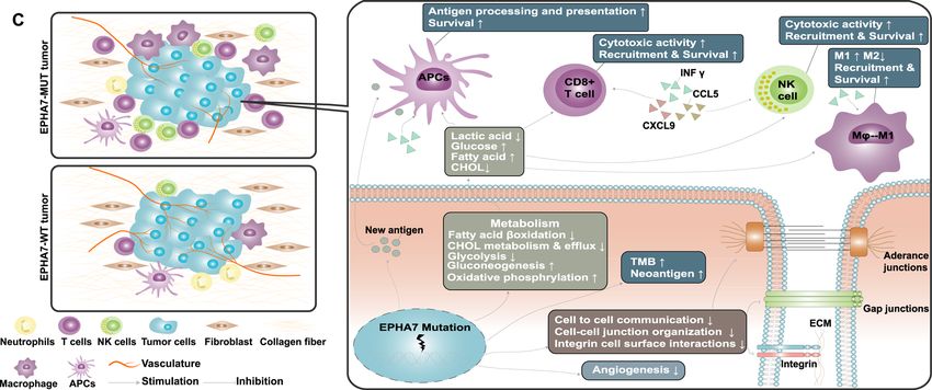
Zhang et al. BMC Medicine        (2021) 19:26                                                                                             Page 11 of 15

 (See figure on previous page.)
 Fig. 5 EPHA7-MUT was associated with enhanced anti-tumor immunity in TCGA cohort. a Violin plot depicting the distribution of TMB, CNA, and
 NAL in EPHA7-MUT and EPHA7-WT tumors. b Boxplot depicting the infiltration of 22 immune cells in EPHA7-MUT and EPHA7-WT tumors.
 CIBERSORT was used to calculate the infiltration degree of these immune cells. Gene expression profiles were uploaded to CIBERSORT web portal,
 and the algorithm was configured with 1000 permutations. CIBERSORT results were recorded in Additional file 5: Table S3. Samples with
 deconvolution P value ≥ 0.05 were excluded (n = 2967) (Mann-Whitney U test; ns, not significant; *P < 0.05, **P < 0.01, ***P < 0.001, ****P < 0.0001).
 c Boxplot depicting the expression level of immune-related genes in EPHA7-MUT and EPHA7-WT groups (Mann-Whitney U test; ns, not
 significant; *P < 0.05, **P < 0.01, ***P < 0.001, ****P < 0.0001). d Heatmap depicting the log2-transformed fold change in the expression level of
 immune-related genes across multiple cancer types (EPHA7-MUT vs EPHA7-WT). Blue indicated downregulation and red indicated upregulation

clinical outcomes in ICI-treated patients across multiple                     progression in various cancers [41, 42]. Survival analysis
cancer types.                                                                 in both non-ICI-treated cohort and TCGA cohort re-
  We found some meaningful changes in biological                              vealed longer median OS in EPHAT-WT instead of
functions of EPHA7-MUT tumors, including intercellu-                          EPHA7-MUT patients, indicating that EPHA7-MUT
lar communication, angiogenesis, and metabolism. First                        might potentially have a worse prognostic impact on
of all, Eph receptors and their ligands (ephrin) have been                    cancer patients. Accordingly, the clinical benefits of
proven essential in the cell communication system [14].                       EPHA7-MUT patients with ICIs should be the result
Hence, it is reasonable to observe a downregulation of                        that the ICI treatment benefits of EPHA7-MUT out-
intercellular communication in EPHA7-MUT tumors in                            weighed its harmful prognostic impact. We then further
our analysis. Previous study showed that inhibiting inter-                    analyzed cancer subgroups in TCGA cohort, which
cellular communication by targeting EPHA10 could                              showed a generally and numerically worse prognosis of
boost anti-tumor immunity by reducing PD-L1 expres-                           EPHA7-MUT patients within each cancer type (Add-
sion [21]. However, we found an upregulation of PD-L1                         itional file 7: Figure S3). Colorectal cancer with EPHA7-
expression in EPHA7-MUT tumors with decreased cell-                           MUT had the worst prognosis in TCGA cohort (HR =
cell contact. This finding suggested that decreased inter-                    5.21 [95% CI, 2.22–12.21], adjusted P < 0.0001). This
cellular communication in EPHA7-MUT tumors may                                could partially explain why EPHA7-MUT colorectal can-
have other underlying mechanisms that enhance anti-                           cer was the only one that presented with worse OS in
tumor immunity rather than depressing PD-L1 expres-                           the validation cohort, because the harmful prognostic
sion. Secondly, Eph-ephrin signaling promotes tumor                           impact of EPHA7-MUT outweighed its ICI treatment
angiogenesis [14]. As expected, we found the angiogen-                        benefits in colorectal cancer. Also, besides the aforemen-
esis pathway was significantly downregulated in EPHA7-                        tioned sample size of the discovery cohort, this assump-
MUT tumors. Angiogenesis and immunosuppression are                            tion could be another reason why only numerical
closely related. Tortuous tumor vasculature causes a                          survival advantage of EPHA7-MUT patients was ob-
hypoxic tumor environment and hinders the infiltration                        served in the discovery cohort, since the harmful prog-
of lymphocytes [39]. Decreased angiogenesis in EPHA7-                         nostic impact of EPHA7-MUT could partly mask the
MUT tumors could promote lymphocyte infiltration. Fi-                         ICI therapy benefits. Notably, these findings supported
nally, metabolic changes in EPHA7-MUT tumors could                            our previous assumption and further stressed the im-
also strengthen anti-tumor immunity. Fatty acids and                          portance of ICI therapy in EPHA7-MUT patients, which
glucose promote the survival of immune cells in the                           could potentially turn the harmful prognostic impact of
tumor microenvironment, while cholesterol and lactate                         EPHA7-MUT patients into an overall survival benefit.
function reversely [40]. Accumulation of fatty acids and                         In the initial screening process, EPHA3 was ruled out
glucose, and depletion of cholesterol and lactate within                      since its FDR adjusted P value of DCB was 0.053. How-
EPHA7-MUT tumor were detected in our analysis,                                ever, given this borderline P value, EPHA3 was worth
which could create a better TIME and enable cytotoxic                         following up. Hence, we have also done the survival ana-
lymphocytes to work more effectively. Overall, EPHA7-                         lysis of EPHA3 in both discovery cohort and validation
MUT tumors are more likely to provide a friendly living                       cohort. Results could be found in the supplementary
environment for those effective immune cells and thus                         material (Additional file 8: Figure S4). However, only nu-
enhance the anti-tumor immunity.                                              merical survival benefits were observed in both discovery
  Ephrin receptors form a large family of receptor tyro-                      and validation cohort for EPHA3-MUT patients. There-
sine kinase and regulate various biological functions.                        fore, EPHA3 might not be as effective as EPHA7 in
Both oncogenic and tumor suppressive roles have been                          predicting the efficacy of immunotherapy in current
reported for specific ephrin receptors [14]. Particularly,                    analysis.
EPHA7 has been previously identified as a tumor                                  In our primary analysis, it is individual EPH gene that
suppressive gene that inhibits tumor growth and                               was evaluated rather than cumulative effects of all 14
Zhang et al. BMC Medicine      (2021) 19:26                                                                                      Page 12 of 15

 Fig. 6 Pathway enrichment analysis in TCGA dataset and possible tumor immune microenvironment in EPHA7-MUT and EPHA7-WT tumors. a
 Differences in pathway activities scored by GSEA between EPHA7-MUT and EPHA7-WT tumors in TCGA dataset. Significant results (P < 0.05 and
 FDR < 0.25) of enrichment analysis were summarized in Additional file 6: Table S4. Pathways which might potentially impact the tumor immune
 microenvironment were presented in a. These pathways were divided into four groups: immune function (blue), intercellular signaling (brown),
 metabolism (green), and other biological functions (gray). b GSEA plot depicting representative pathways identified by GSEA between EPHA-MUT
 and EPHA7-WT tumors, including metabolism, cell communication, immune response, and angiogenesis. c Comparison of possible tumor
 immune microenvironment between EPHA7-MUT and EPHA7-WT tumors. APCs, antigen presenting cells; NK cell, nature killer cell; ECM,
 extracellular matrix

EPH genes. To investigate the combined effects of all 14                 defined as at least one EPH gene has mutation among
EPH genes, we performed further analysis combining all                   14 genes, while EPH-WT was defined as none of EPH
14 genes (Additional file 9: Figure S5). EPH-MUT was                     genes has mutation. Although EPH-MUT patients
Zhang et al. BMC Medicine   (2021) 19:26                                                                                    Page 13 of 15

presented with a higher ORR than EPH-WT patients              researches or ongoing trials co-targeting these pathways
(40.8% vs 24.8%), there were no significant differences       and tumor immunity [18, 40], our study introduces a
between EPH-MUT and EPH-WT patients regarding                 novel angle that Eph receptors might influence the im-
DCB, PFS, and OS. Activation of different Eph receptors       munotherapy response through corresponding biological
can have highly varied impacts on cellular processes, but     functions. Further elucidation of the molecular mechan-
exact function of each Eph receptor has not been fully        ism between Eph receptors and anti-tumor immunity is
understood. Therefore, it would be more reasonable to         warranted to validate the predictive value as well as help
test the combined predictive value of EPH genes that          to identify potential therapeutic targets.
have synergic effect in the future rather than all EPH
genes.                                                        Conclusion
  This retrospective analysis also has several limitations.   Our study demonstrated the robust link between
Firstly, only four out of fourteen EPH genes are included     EPHA7-MUT and better clinical outcomes in ICI-
in the MSK-IMPACT panel. To analyze all EPH genes,            treated cancer patients. Therefore, EPHA7-MUT has the
we only included cohorts with WES data in the discov-         potential to serve as a predictive biomarker for immune
ery cohort. Considering the limited sample size of the        checkpoint blockades across multiple cancer types. Val-
discovery cohort (n = 386), we should not completely ex-      idation of the predictive value in future prospective trials
clude the predictive function of other EPH genes. Sec-        and exploration of the molecular mechanism in further
ondly, mutation rate of EPHA7 in melanoma was nearly          molecular researches are warranted for EPHA7-MUT.
2.5 times higher than in other cancer types. The majority
of EPHA-MUT samples were melanoma (33/38) in the              Supplementary Information
discovery cohort, which is a major confounding factor         The online version contains supplementary material available at https://doi.
causing bias. However, the survival advantages across         org/10.1186/s12916-020-01899-x.
multiple cancers in the validation cohort as well as the
                                                               Additional file 1: Table S1. EPH genes and corresponding clinical
general upregulation of anti-tumor immunity in various
                                                               outcomes in the discovery cohort.
cancers could compensate the bias to some degree. Still,
                                                               Additional file 2: Table S2. Patient characteristics in the discovery
the predictive value of EPHA7 mutation with regard to          cohort stratified by EPHA7 status.
cancer types needs to be verified in future prospective        Additional file 3: Figure S1. Violin plot depicting the distribution of
trials. Additionally, the possible TIME and molecular          TMB and CNA in EPHA7-MUT and EPHA7-WT tumors.
mechanisms of EPHA7-MUT were demonstrated based                Additional file 4: Figure S2. Truncating vs non-truncating EPHA7 mu-
                                                               tation analysis in both discovery and validation cohort. Figure S2 A-D: dis-
on GSEA, which requires further molecular researches
                                                               covery cohort. Figure S2 E: validation cohort.
to validate. Finally, gene expression data has not been in-
                                                               Additional file 5: Table S3. Results of CIBERSORT analysis in TCGA
cluded in both the discovery and validation cohorts.           cohort.
Therefore, combination analysis of EPHA7 and other             Additional file 6: Table S4. Significant pathways detected by gene set
predictive biomarkers (e.g., expression of PD-L1) has not      enrichment analysis. (EPHA7-MUT vs EPHA7-WT tumors).
been performed. Clinical trials with expression data are       Additional file 7: Figure S3. Survival analysis of cancer subgroups in
                                                               the TCGA cohort.
needed to expand our findings and test the added value
                                                               Additional file 8: Figure S4. Survival analysis of EPHA3 in both
of tumor-infiltrating lymphocytes in the survival analysis
                                                               discovery and validation cohort.
of EPHA7-MUT.
                                                               Additional file 9: Figure S5. Association between clinical outcomes
  Importantly, these limitations do not preclude the fa-       and the combination of all 14 EPH genes in the discovery cohort.
vorable clinical outcomes derived from immunotherapy
in EPHA7-MUT patients. Unlike continuous variables            Abbreviations
such as TMB, CNA, or PD-L1 expression, EPHA7-MUT              ACC: Adrenocortical carcinoma; BLCA: Bladder urothelial carcinoma;
are easily detected by NGS and clearly classify patients      BRCA: Invasive breast carcinoma; CHOL: Cholangiocarcinoma; CI: Confidence
                                                              interval; CNA: Copy number alteration; COAD: Colorectal adenocarcinoma;
into two groups that are associated with immunotherapy        CR: Complete response; CRC: Colorectal cancer; CSCC: Cervical squamous cell
response. The scope of EPHA7-MUT falls in compensat-          carcinoma; CTLA-4: Cytotoxic T lymphocyte antigen 4; DCB: Durable clinical
ing the existing biomarkers to detect those patients who      benefit; DLBC: Diffuse large B cell lymphoma; EAC: Esophagogastric
                                                              adenocarcinoma; EC: Endometrial carcinoma; ECA: Endocervical
are most likely to benefit from immunotherapy. Our            adenocarcinoma; ESCC: Esophageal squamous cell carcinoma; FDA: Food
study not only paved the way for precise treatments           and Drug Administration; FDR: False discovery rate; FPKM: Fragments per
tailored to molecular subtypes, but also indicated the        kilobase of exon model per million mapped fragments; GBM: Glioblastoma;
                                                              HCC: Hepatocellular carcinoma; HNSC: Head and neck squamous cell
association between Eph receptor-related TIME and im-         carcinoma; HR: Hazard ratio; ICIs: Immune checkpoint inhibitors; KIRC: Kidney
munotherapy response. Biological functions mediated by        renal clear cell carcinoma; KIRP: Kidney renal papillary cell carcinoma;
Eph receptors, especially tumor angiogenesis, intercellu-     LAML: Acute myeloid leukemia; LGG: Diffuse glioma; Mb: Megabase;
                                                              MESO: Mesothelioma; MSI-H: High microsatellite instability; NAL: Neoantigen
lar contact, and tumor metabolism, should be better           load; NSCLC: Non-small cell lung cancer; ORR: Objective response rate;
characterized in future studies. Although there are some      OS: Overall survival; OV: Ovarian epithelial tumor; PAAD: Pancreatic
Zhang et al. BMC Medicine (2021) 19:26 Page 14 of 15
adenocarcinoma; PCC: Pheochromocytoma; PD: Progression of disease; PD- 8. Ott PA, Bang YJ, Piha-Paul SA, Abdul Razak AR, Bennouna J, Soria JC, et al. T-
(L)1: Programmed cell death (ligand) 1; PFS: Progression-free survival; cell–inflamed gene-expression profile, programmed death ligand 1
PGL: Paraganglioma; PRAD: Prostate adenocarcinoma; PR: Partial response; expression, and tumor mutational burden predict efficacy in patients
RECIST: Response Evaluation Criteria in Solid Tumors; SARC: Sarcoma; treated with pembrolizumab across 20 cancers: KEYNOTE-028. J Clin Oncol.
SD: Stable disease; SKCM: Melanoma; STAD: Stomach adenocarcinoma; 2019;37:318–27.
TCGA: The Cancer Genome Atlas; EPHA7-MUT: EPHA7-mutant; EPHA7- 9. Hakimi AA, Voss MH, Kuo F, Sanchez A, Liu M, Nixon BG, et al.
WT: EPHA7-wildtype; TMB: Tumor mutational burden; UCES: Uterine corpus Transcriptomic profiling of the tumor microenvironment reveals distinct
endometrial carcinoma; UVM: Uveal melanoma; WES: Whole-exome subgroups of clear cell renal cell cancer: data from a randomized phase III
sequencing trial. Cancer Discov. 2019;9:510–25.
10. Ready N, Hellmann MD, Awad MM, Otterson GA, Gutierrez M, Gainor JF,
Acknowledgements et al. First-line nivolumab plus ipilimumab in advanced non-small-cell lung
We would like to thank Prof. Luc G. T. Morris from Memorial Sloan Kettering cancer (CheckMate 568): outcomes by programmed death ligand 1 and
Cancer Center for generously sharing the clinical data of the non-ICI-treated tumor mutational burden as biomarkers. J Clin Oncol. 2019;37:992–1000.
cohort from Samstein et al., and the staff members of TCGA Research Net- 11. Zhou H, Liu J, Zhang Y, Huang Y, Shen J, Yang Y, et al. PBRM1 mutation and
work, the cBioPortal, the UCSC Xena data portal, and the CIBERSORT portal, preliminary response to immune checkpoint blockade treatment in non-
as well as all the authors for making their valuable research data public. small cell lung cancer. NPJ Precis Oncol. 2020;4:4–7.
12. Gonzalez-Cao M, Viteri S, Karachaliou N, Aguilar A, García-Mosquera JJ,
Authors’ contributions Rosell R. Tumor mutational burden as predictive factor of response to
Study concept and design: HL, ZZ, and HW. Acquisition, analysis, or immunotherapy. Transl Lung Cancer Res. 2018;7:S358–61.
interpretation of data: all authors. Drafting of the manuscript: all authors. 13. Funk SD, Orr AW. Ephs and ephrins resurface in inflammation, immunity,
Critical revision of the manuscript for important intellectual content: all and atherosclerosis. Pharmacol Res. 2013;67:42–52.
authors. Study supervision: HL. All authors read and approved the final 14. Pasquale EB. Eph receptors and ephrins in cancer: bidirectional signalling
manuscript. and beyond. Nat Rev Cancer. 2010;10:165–80.
15. Barquilla A, Pasquale EB. Eph receptors and ephrins: therapeutic
Funding opportunities. Annu Rev Pharmacol Toxicol. 2015;55:465–87.
This study was funded by the National Natural Science Foundation of China 16. Song Y, Fu Y, Xie Q, Zhu B, Wang J, Zhang B. Anti-angiogenic agents in
(81930065, 81871985), Natural Science Foundation of Guangdong Province combination with immune checkpoint inhibitors: a promising strategy for
(2014A030312015, 2019A1515011109), and Science and Technology Program cancer treatment. Front Immunol. 2020;11:1–17.
of Guangzhou (201803040019, 202002030208). 17. Taylor MH, Lee CH, Makker V, Rasco D, Dutcus CE, Wu J, et al. Phase Ib/II
trial of lenvatinib plus pembrolizumab in patients with advanced renal cell
carcinoma, endometrial cancer, and other selected advanced solid tumors. J
Availability of data and materials
Clin Oncol. 2020;38:1154–63.
All of the data we used in this study were publicly available as described in
18. Fukuoka S, Hara H, Takahashi N, Kojima T, Kawazoe A, Asayama M, et al.
the “Methods” section.
Regorafenib plus nivolumab in patients with advanced gastric or colorectal
cancer: an open-label, dose-escalation, and dose-expansion phase Ib trial
Ethics approval and consent to participate (REGONIVO, EPOC1603). J Clin Oncol. 2020;38:2053–61.
Ethical approval was waived since we used only publicly available data and 19. Zhu AX, Finn RS, Ikeda M, Sung MW, Baron AD, Kudo M, Okusaka T,
materials in this study. Kobayashi M, Kumada H, Kaneko S, Pracht M, Mamontov K, Meyer T, Mody
K, Kubota T, Dutcus CE, JML K. A phase Ib study of lenvatinib (LEN) plus
Consent for publication pembrolizumab (PEMBRO) in unresectable hepatocellular carcinoma (uHCC).
Not applicable J Clin Oncol. 2020;38(15_suppl):4519 (May 20, 2020).
20. Shiuan E, Chen J. Eph receptor tyrosine kinases in tumor immunity. Cancer
Competing interests Res. 2016;76:6452–7.
The authors declare that they have no competing interests. 21. Yang WH, Cha JH, Xia W, Lee HH, Chan LC, Wang YN, et al. Juxtacrine
signaling inhibits antitumor immunity by upregulating PD-L1 expression.
Received: 13 September 2020 Accepted: 29 December 2020 Cancer Res. 2018;78:3761–8.
22. Rizvi NA, Hellmann MD, Snyder A, Kvistborg P, Makarov V, Havel JJ, et al.
Mutational landscape determines sensitivity to PD-1 blockade in non-small
References cell lung cancer. Science (80- ). 2015;348:124–8.
1. Gutzmer R, Stroyakovskiy D, Gogas H, Robert C, Lewis K, Protsenko S, et al. 23. Snyder A, Makarov V, Merghoub T, Yuan J, Zaretsky JM, Desrichard A, et al.
Atezolizumab, vemurafenib, and cobimetinib as first-line treatment for Genetic basis for clinical response to CTLA-4 blockade in melanoma. N Engl
unresectable advanced BRAFV600 mutation-positive melanoma (IMspire150): J Med. 2014;371:2189–99.
primary analysis of the randomised, double-blind, placebo-controlled, phase 24. Van Allen EM, Miao D, Schilling B, Shukla SA, Blank C, Zimmer L, et al.
3 trial. Lancet. 2020;395:1835–44. Genomic correlates of response to CTLA-4 blockade in metastatic
2. Gandhi L, Rodríguez-Abreu D, Gadgeel S, Esteban E, Felip E, De Angelis F, melanoma. Science (80- ). 2016;352:207–12.
et al. Pembrolizumab plus chemotherapy in metastatic non-small-cell lung 25. Miao D, Margolis CA, Vokes NI, Liu D, Taylor-Weiner A, Wankowicz SM, et al.
cancer. N Engl J Med. 2018;378:2078–92. Genomic correlates of response to immune checkpoint blockade in
3. Marabelle A, Le DT, Ascierto PA, Di Giacomo AM, de Jesus-Acosta A, Delord microsatellite-stable solid tumors. Nat Genet. 2018;50:1271–81.
JP, et al. Efficacy of pembrolizumab in patients with noncolorectal high 26. Hugo W, Zaretsky JM, Sun L, Song C, Moreno BH, Hu-Lieskovan S, Berent-
microsatellite instability/ mismatch repair–deficient cancer: results from the Maoz B, Pang J, Chmielowski B, Cherry G. Genomic and transcriptomic
phase II KEYNOTE-158 study. J Clin Oncol. 2020;38:1–10. features of response to anti-PD-1 therapy in metastatic melanoma. Cell.
4. Sharma P, Hu-Lieskovan S, Wargo JA, Ribas A. Primary, adaptive, and 2016;165(1):35–44.
acquired resistance to cancer immunotherapy. Cell. 2017;168:707–23. 27. Hellmann MD, Nathanson T, Rizvi H, Creelan BC, Sanchez-Vega F, Ahuja A, et al.
5. Havel JJ, Chowell D, Chan TA. The evolving landscape of biomarkers for Genomic features of response to combination immunotherapy in patients
checkpoint inhibitor immunotherapy. Nat Rev Cancer. 2019;19:133–50. with advanced non-small-cell lung cancer. Cancer Cell. 2018;33:843–852.e4.
6. Davoli T, Uno H, Wooten EC, Elledge SJ. Tumor aneuploidy correlates with 28. Miao D, Miao D, Margolis CA, Gao W, Voss MH, Li W, et al. Genomic
markers of immune evasion and with reduced response to immunotherapy. correlates of response to immune checkpoint therapies in clear cell renal
Science. 2017;355(6322). cell carcinoma. Science (80- ). 2018;5951:1–11.
7. Wu HX, Chen YX, Wang ZX, Zhao Q, He MM, Wang YN, et al. Alteration in 29. Samstein RM, Lee CH, Shoushtari AN, Hellmann MD, Shen R, Janjigian YY,
TET1 as potential biomarker for immune checkpoint blockade in multiple et al. Tumor mutational load predicts survival after immunotherapy across
cancers. J Immunother Cancer. 2019;7:1–13. multiple cancer types. Nat Genet. 2019;51:202–6.Zhang et al. BMC Medicine (2021) 19:26 Page 15 of 15
30. Liu J, Lichtenberg T, Hoadley KA, Poisson LM, Lazar AJ, Cherniack AD, et al.
An integrated TCGA Pan-Cancer Clinical Data Resource to drive high-quality
survival outcome analytics. Cell. 2018;173:400–416.e11.
31. Thorsson V, Gibbs DL, Brown SD, Wolf D, Bortone DS, Ou Yang TH, et al.
The immune landscape of cancer. Immunity. 2018;48:812–830.e14.
32. Goldman MJ, Craft B, Hastie M, Repečka K, McDade F, Kamath A, Banerjee A,
Luo Y, Rogers D, Brooks AN. Visualizing and interpreting cancer genomics
data via the Xena platform. Nature Biotechnology. 2020. p. 1–4.
33. Yarchoan M, Hopkins A, Jaffee EM. Tumor mutational burden and response
rate to PD-1 inhibition. N Engl J Med. 2017;377:2500–1.
34. Chalmers ZR, Connelly CF, Fabrizio D, Gay L, Ali SM, Ennis R, et al. Analysis
of 100,000 human cancer genomes reveals the landscape of tumor
mutational burden. Genome Med. 2017;9:1–14.
35. Newman AM, Liu CL, Green MR, Gentles AJ, Feng W, Xu Y, et al. Robust
enumeration of cell subsets from tissue expression profiles. Nat Methods.
2015;12:453–7.
36. Love MI, Huber W, Anders S. Moderated estimation of fold change and
dispersion for RNA-seq data with DESeq2. Genome Biol. 2014;15:1–21.
37. Yu G, Wang LG, Han Y, He QY. ClusterProfiler: an R package for comparing
biological themes among gene clusters. Omi A J Integr Biol. 2012;16:284–7.
38. Gao J, Chang MT, Johnsen HC, Gao SP, Sylvester BE, Sumer SO, et al. 3D
clusters of somatic mutations in cancer reveal numerous rare mutations as
functional targets. Genome Med. 2017;9:1–13.
39. Khan KA, Kerbel RS. Improving immunotherapy outcomes with anti-
angiogenic treatments and vice versa. Nat Rev Clin Oncol. 2018;15:310–24.
40. Li X, Wenes M, Romero P, Huang SCC, Fendt SM, Ho PC. Navigating
metabolic pathways to enhance antitumour immunity and immunotherapy.
Nat Rev Clin Oncol. 2019;16:425–41.
41. Oricchio E, Nanjangud G, Wolfe AL, Schatz JH, Mavrakis KJ, Jiang M, et al.
The Eph-receptor A7 is a soluble tumor suppressor for follicular lymphoma.
Cell. 2011;147:554–64.
42. Li S, Wu Z, Ma P, Xu Y, Chen Y, Wang H, et al. Ligand-dependent EphA7
signaling inhibits prostate tumor growth & progression. Cell Death Dis.
2017;8:e3122.
Publisher’s Note
Springer Nature remains neutral with regard to jurisdictional claims in
published maps and institutional affiliations.You can also read