Total Recall: Lateral Habenula and Psychedelics in the Study of Depression and Comorbid Brain Disorders - MDPI
←
→
Page content transcription
If your browser does not render page correctly, please read the page content below
International Journal of
            Molecular Sciences

Review
Total Recall: Lateral Habenula and Psychedelics in the
Study of Depression and Comorbid Brain Disorders
Matas Vitkauskas 1           and Ajay S. Mathuru 1,2,3, *
 1    Yale-NUS College, Singapore 637551, Singapore; matas.vitkauskas@u.yale-nus.edu.sg
 2    Institute of Molecular and Cell Biology (IMCB), Singapore 637551, Singapore
 3    Department of Physiology, Yong Loo Lin School of Medicine, NUS, Singapore 637551, Singapore
 *    Correspondence: ajay.mathuru@yale-nus.edu.sg

 Received: 23 July 2020; Accepted: 4 September 2020; Published: 7 September 2020

 Abstract: Depression impacts the lives and daily activities of millions globally. Research into the
 neurobiology of lateral habenula circuitry and the use of psychedelics for treating depressive states
 has emerged in the last decade as new directions to devise interventional strategies and therapies.
 Several clinical trials using deep brain stimulation of the habenula, or using ketamine, and psychedelics
 that target the serotonergic system such as psilocybin are also underway. The promising early results
 in these fields require cautious optimism as further evidence from experiments conducted in animal
 systems in ecologically relevant settings, and a larger number of human studies with improved
 spatiotemporal neuroimaging, accumulates. Designing optimal methods of intervention will also
 be aided by an improvement in our understanding of the common genetic and molecular factors
 underlying disorders comorbid with depression, as well as the characterization of psychedelic-induced
 changes at a molecular level. Advances in the use of cerebral organoids offers a new approach for
 rapid progress towards these goals. Here, we review developments in these fast-moving areas of
 research and discuss potential future directions.

 Keywords: lateral habenula; psilocybin; depression; comorbid brain disorders; cerebral organoids

1. Introduction
     Depression affects millions of people worldwide. Analysis of the data collated by the World Health
Organisation suggests that depression as a leading cause of disability has grown worldwide and in 2017
had more than 250 million sufferers, making it one of the most common mental ailments [1]. Depression has
often been observed to be comorbid with other neuropsychiatric ailments and brain disorders.
Epidemiological evidence suggests a co-occurrence with nicotine dependence [2,3], substance use
disorders [3,4], anxiety disorders [5], insomnia, and chronic pain [6]. Further, patients with comorbid
disorders have comparatively worse therapeutic outcomes in treatments [5,7,8]. The neurobiological,
genetic, and circuit-level commonalities between these conditions are anticipated but are not well
understood at present [6]. Unraveling these commonalities in molecular mechanisms and brain circuits
is thus likely to give us an insight into both depression as a brain disorder as well as into the potential
therapeutic routes to take for intervention.
     In recent times, there has also been an increasing appreciation for the dynamic nature of neural
circuitry under duress and the potential role of neuroplasticity that becomes maladaptive in conditions
like major depressive disorder (MDD) [9]. This interpretation associates the clinical symptoms of
depression with dysfunctional molecular and cellular changes in critical neural circuits in the brain
that regulate emotion. One region of the brain that has received an unusual amount of interest in
the past few years in this context is the habenula complex [10–15]. The habenula complex has been
variously described as a “conductor hidden in the orchestra” [16], and as a structure at the “crossroads

Int. J. Mol. Sci. 2020, 21, 6525; doi:10.3390/ijms21186525                         www.mdpi.com/journal/ijms
Int. J. Mol. Sci. 2020, 21, 6525 2 of 17
between basal ganglia and the limbic system”, vital for decision making in vertebrates [17]. One reason
to describe the habenula using these metaphors is the regulatory control it exerts in the release of
several neuromodulators—serotonin, dopamine, noradrenaline, and histamine among others [18,19].
The habenula complex has two subnuclei, which are biochemically, histologically, transcriptionally,
and functionally distinct. Both the lateral and medial habenulae malfunctions have been implicated in
several neuropsychiatric disorders. Among the two, the lateral habenula has primarily been associated
with depression, while the medial with substance use disorders [11,20,21]. There is growing enthusiasm
for the possibility that the lateral habenula is a key player in MDD based on a large number of studies
in rodent models of depression [11,14,22–26]. Alongside this body of research, the lateral habenula has
become the anatomical target for testing antidepressants such as ketamine infusions in animals [13,14]
and its action in humans [27]. Clinical studies of Deep Brain Stimulation (DBS) targeting the habenula
in humans [28–33] are also underway based on favorable results in rodents [34–36]. As promising
as this direction of research is, it is still early days and there is a need for cautious optimism as not
every observation is fully explained by current theory about how habenula dysfunction could result in
depressive states [10].
Interestingly, a very different approach from that of targeting a specific brain region has reemerged
in parallel in the search for an appropriate antidepressant treatment regimen [37]. This renaissance
in the use of psychedelics in psychiatry for pharmaco-resistant depression has come to prominence
because of dramatically different treatment procedures and effects documented compared to other
pharmacological agents [38–41]. In one account, a single dose of psilocybin appears to have lowered
the severity of an MDD with effects lasting for 5-weeks or longer [38]. The antidepressant effects of
psilocybin are proposed to be brought about by a “reset” of the default mode network of the brain in
this study [38]. While how such a long-term change in circuits spanning the entire brain occurs at a
physiological and molecular level is not fully understood [37,42], clues exist about its action via the
serotonergic system. It is interesting to note that psilocybin has also shown promise in the treatment of
addiction [43,44]. The co-occurrence of the two disorders and the potential for a common treatment
that is effective for both is suggestive of shared underlying affected mechanisms [3].
In this article, we review recent developments in these two fast-moving areas of research and
discuss the potential future directions that can bridge progress in both.
2. Lateral Habenula, “Negative-Reward”, and the Relation to Depression
The revival of interest in the habenula complex in the context of neuropsychiatric ailments has been
fueled by the seminal work conducted by Matsumoto, Hikosaka, and colleagues that demonstrated
the reward-prediction error function of lateral habenula [45]. That is, the lateral habenula neurons fire
when expected rewards are omitted, and are silenced with unexpected rewards. Their results [46–50]
in a behaving monkey have helped contextualize older observations of indirect modulation of the
substantia nigra and ventral tegmental areas by the habenula, primarily resulting in the inhibition of
dopaminergic neurons [51,52], and to the serotonergic dorsal raphe nucleus, resulting in the regulation
of serotonin release [15]. As these immediately downstream regions play a critical role in reward
processing and reinforcement learning, these discoveries, in addition to the discovery of functional
conservation of this complex in the vertebrate lineage, have sparked a resurgence in the interest in the
habenula complex [18,53–55].
Malfunction in the reward processing and reinforcement behaviors are closely tied to many
brain disorders including addiction and MDD [25]. Consequently, animal models where the
neurobiology of such behaviors can be studied have also emerged as powerful tools for the study of
depression [11,34,55–58]. A recent study, for instance, examined passive coping behavior in zebrafish
while applying brain-wide imaging techniques that allow high-speed imaging of the entire brain at the
resolution of single neurons [59]. Passive coping (or learned helplessness) occurs when animals are
exposed to a prolonged behavioral challenge, in this case, an inescapable shock regiment. Such coping
has been equated with despair in human patients, a common feature of MDD. Analysis of theInt. J. Mol. Sci. 2020, 21, 6525 3 of 17
whole-brain Ca2+ responses of neurons revealed that a passive coping strategy was associated with
progressive activation of neurons in the ventral habenula (equivalent to the lateral habenula in humans).
Conversely, activation of ventral-habenula neurons using optogenetic manipulation, resulted in both
behavioral passivity and suppression of the serotonergic neurons in the raphe [59], thus connecting the
dots between the neurophysiological response of the habenula neurons and the behavioral outcome.
Congenitally learned helpless (cLH) rats and mice, where a similar rationale was applied, have also
been used to examine the response of the habenula under conditions of normalcy and coping [14].
A whole-cell patch-clamp in this study showed that 23% percent of lateral habenula neurons in cLH
rats display a bursting type activity while the percentage of such firing was only 7% in controls.
Moreover, the number of spikes in each burst was also higher at 43% to 7% in controls [14]. A series
of experiments from several other groups of researchers has added vastly to our knowledge of the
habenula neurophysiology in this context in the last few years [58,60–62]. Together, these studies
suggest that a hyper or overactivation of the lateral habenula is involved in the animal phenotypes
associated with depression.
3. Targeting the Hyperactivity of Lateral Habenula Provides Antidepressant Effects in Animals
Preclinical animal models provide a valuable avenue to dissect a complex phenomenon such
as depression and discover potential antidepressant treatments. In rodents, several phenotypes
in specific behavioral tests have been used to model human depression, though not all of them
may have a face, predictive, and construct validity, and the interpretation of the animal’s behavior
in these tests has changed over time [63]. Among them, time of immobility in forced swim tests,
sucrose consumption, exploration in an open field, and participation in reinforcement behavior
associated with rewards, alongside measurement of neuromodulator concentration, have been used
as readouts to quantify coping abilities, the effectiveness of antidepressants, and depressive-like
behaviors. One therapeutic angle being tested using these readouts is to find the exact anatomical
location for implanting electrodes that deliver deep brain stimulation (DBS) [36]. High-frequency
stimuli ranging between 100 and 200 Hz applied either intermittently, chronically over days, or acutely
up to an hour before the behavioral tests are conducted have been found to be effective in alleviating
readouts that are considered depressive in these animals [28]. Studies show that DBS applied to the
lateral habenula of rats over 28 days improved the exploration of open field and sucrose consumption
in chronically stressed rats [34]. A study examining maternal separation-induced depressive-like
behaviors in rodents also found that a high-frequency DBS to the lateral habenula attenuates the
behavioral symptoms [60]. The authors, in this case, also demonstrated that the DBS procedure reduces
the lateral habenula hyperactivity [60]. Further, in support of the hypothesis that high-frequency
DBS likely works by reducing the hyperactivity, one study showed that low-frequency stimulation
conversely activates lateral habenula and produces an increase in depressive-like behaviors in rats [35].
The exact mechanism of DBS action is still not clearly understood, but could occur either because of a
change in the presynaptic neurotransmitter release mechanisms [22,24,34] or due to the downstream
effects [52]. The behavioral symptom relief in most studies is correlated with an elevation in serotonin,
dopamine, and norepinephrine levels [34].
Another approach to target the lateral habenula hyperactivity has been the use of pharmacological
aids. The two most notable examples are the administration of protein-phosphotase 2A (PP2A)
inhibitors and ketamine. Mice experiencing inescapable shock develop depressive-like phenotypes
and exhibit neuronal hyperactivity in the lateral habenula [61]. The authors found that one reason for
the hyperactivity was a reduction in the surface expression of a G protein—gated inwardly rectifying
potassium channels and GABAB receptors, regulated by the enzyme PP2A. An increase in the PP2A
activity due to the shock paradigm increases the internalization of these proteins. Inhibition of PP2A,
specifically in the lateral habenula, on the other hand, restores the currents from these channels and
reverses both the effects on neuronal excitability, and the depressive-like behaviors exhibited [61].Int. J. Mol. Sci. 2020, 21, 6525 4 of 17
A pair of studies from the Hailan Hu lab has also charted out another physiological dysfunction
in the lateral habenula and how ketamine rapidly reverses this deficit in rodent models to produce
antidepressant effects [12,14]. Ketamine is known to be an NMDA receptor antagonist and has been
reported to produce rapid antidepressant effects even when applied systemically [27]. The authors
reasoned that ketamine could be acting at the lateral habenula given that the brain region receives
glutamatergic afferents and controls neuromodulators implicated in depression [64]. A direct infusion
of ketamine converted the bursting activity seen in a subset of lateral habenula neurons of cLH mice to
the tonic firing more common in control animals [14]. The group further discovered that the bursting
firing pattern in the habenula neurons was dependent on a change in the resting membrane potential
of these neurons, which in turn was occurring due to a change in the expression levels of the channel
Kir4.1 in the astrocytes that wrap these neurons [12]. Another recent study also suggests the same
site of action for ketamine but attributes the effects to the function of mu-opioid receptors (MOR)
and NMDA receptors (NMDAR), both of which are highly expressed in the lateral habenula [13].
Ketamine could be acting on many other neurons in the brain with NMDAR and MOR expression,
but these observations together suggest a potential explanation for the rapid antidepressant effects of
ketamine in comparison to alternatives like SSRIs since the site of action is upstream and can directly
correct the putative monoamine imbalance linked with depressive states.
Targeting the hyperactivity of the lateral habenula, either through DBS or chemogenetic
inhibitors [60], or by using drugs [11,36], has been highly effective in animals in reducing depressive-like
behaviors, but the suitability of applying these directly to humans raises both ethical and technological
considerations. Only a handful of clinical trials of DBS with a small number of subjects have been
initiated in humans until now [33,65]. The initial findings are promising, but further studies with
larger numbers of subjects will be needed to understand how individual variability impacts humans,
before a more complete picture can be constructed. New findings such as the possibility of using
non-invasive light therapy delivered through the retina to modulate lateral habenula activity open
additional avenues to combine with existing approaches for clinical applications in humans that need
further exploration [66].
4. Lateral Habenula Activity in Human Mdd Patients
Human habenula studies have generally been in agreement with the findings in animals and
non-human primates. Habenula activation was reported when there was a punishment prediction
error, and in aversive events rather than in neutral or rewarding events [67–69]. Functional studies of
the lateral habenula in humans, however, are difficult to perform in general in humans as anatomically,
both medial and lateral habenula together occupy a volume of approximately 30 mm3 , accounting for
only a few voxels in an fMRI image. This makes delineating the responses of these two distinct nuclei
quite difficult [68,70]. Studies that examined MDD patients compared to matched controls have yielded
mixed results [10,71]. Contrary to expectations, MDD patients do not show a noticeable increase in
baseline activity or hyperactivity in depressive states. In these neuroimaging studies, BOLD signals
were measured during Pavlovian conditioning when the association between symbols that indicate the
probability of obtaining monetary incentives or disincentives, and shock, were being made. One of
these studies paralleled findings from animal studies that the habenula activity increased in control
subjects when a negative event, the chance of receiving a shock, was high. In the MDD patients for the
same task, the habenula activity decreased instead, which is difficult to interpret [10]. A second study
also reported no significant difference between the activities of habenulae in healthy controls and MDD
patients [71]. However, the second study found an overall positive correlation between the positive
prediction error-related habenula activity and the frequency of depressive episodes in the participants.
What these results mean in the broader context of the research in this area needs further analysis.
As the authors of these studies point out, caution is required when interpreting human studies of
this kind and when comparing the results from animal studies. To draw robust conclusions on
the role of the lateral habenula activity in MDD patients, at least four methodological refinementsInt. J. Mol. Sci. 2020, 21, 6525 5 of 17
are necessary. First, fMRI resolution for structures as small as the habenula can be faulty even
after careful quality control during registration. An exploration of new methods, such as those
adapting computational geometry to compare spatiotemporal dissimilarities, may be needed for
disambiguation [72]. Better methods are also needed to differentiate the medial and the lateral
habenula activity as their activity could be divergent in these tasks and may further complicate
interpretation [10]. Larger sample sizes or a meta-analysis of standardized experimental designs
are also necessary to increase confidence. Fourth, research groups may also need to evaluate and
correct for the possibility of workflow-dependent observational and interpretational bias that has been
recently described as being a common issue among functional studies [73]. Apart from these technical
considerations, there are other possibilities for the differences between human and animal studies.
The BOLD signal recorded in the fMRI study is an abstraction of complex neural activity—of afferents,
local inhibition, and spiking efferents [74]. Therefore, an average response is difficult to untangle.
The lateral habenula has a complex local circuitry [24], which makes deconvolving, and interpreting
the BOLD signal from this region even more difficult. As a large number of neuron types have been
reported in the habenula [75], it may also be overly simplistic to interpret an average activity when
comparing depressive and non-depressive states. Another reason, as pointed out previously [10],
is the possibility that although the metabolic activity in the habenula may be inversely correlated to
depressive states, the extent to which this holds may depend on chronic conditions and the state of
remission of the subject [32,33], and is therefore dynamic. Finally, the experimental design for human
experiments that can reveal the type of bursting activity in a subset of neurons of the lateral habenula
observed in MDD rodent models is also not easy to implement and requires further refinement.
5. Perils of Reliance on Single Models to Understand the Neurobiology of Habenula Function in
Depression
The use of animal models to dissect the underpinnings of human brain disorders is
irreplaceable [76], yet studies have to fulfill a tall order of requirements where differences in cellular
physiology, species differences, and the differences due to the complexity of the circuitry need to be
taken into account while generating meaningful, transferable knowledge. The studies on MOR and
NMDAR described above both used cLH rodents [13,64]. While this model effectively captures many
aspects of a depressive state in rodents, whether it adequately represents the cellular and physiological
changes associated with MDD in humans in its entirety is debatable. Further, as noted by many
researchers, certain natural behaviors such as freezing, escaping, or fighting are altered significantly in
animals reared in laboratories for prolonged periods [77,78]. Even if it is argued that relative differences
are being examined between two conditions, the generalizability of the findings can be impacted
as such alterations are subject to “floor” and “ceiling” effects. Expanding the types of phenotypes
examined, and testing many more species broadly, in ecologically naturalistic settings, can overcome
the concerns of studying species-specific evolutionary trajectories in singular animal models. In the
process, as discussed in a recent review on this topic, it will also deliver more generalizable knowledge
and conditions applicable to humans [77]. Comparing and contrasting results across a plurality of
animal systems and experimental designs that incorporate behaviors animals normally exhibit in the
wild, such as social isolation, social defeat, and dominance hierarchy-dependent stresses will further
improve the predictive validity alongside the construct validity of systems used to model depressed
states [11]. Some studies have also begun to explore other paradigms, such as symptoms resulting
from maternal separation in rodents [60]. More such diverse paradigms are necessary to improve our
understanding of the transferability of the accompanying neurobiological changes to the biological
changes associated with psychiatric disorders in humans. We acknowledge that starting completely
new research programs is neither feasible nor practical. However, adapting already existing models
that emulate ecologically naturalistic settings, in tandem with the assays standardized on lab-reared
animal models, is both warranted, and plausible. For instance, prairie voles form long-term social
bonds with mating companions and display many depressive-like behaviors when separated from theirInt. J. Mol. Sci. 2020, 21, 6525 6 of 17
partner [79]. Studying the underlying factors modulating such natural behaviors will be a beneficial
complement to current studies using animal systems and will go some distance in addressing the
concerns of overinterpretation of animal behavioral data.
The second point of caution is with respect to the interpretation of results from the standard
practice of activating or inactivating neurons to understand how their (in)operation affects specific
aspects of a given behavior [80]. Such studies in recent times have been bolstered by the use of
optogenetics. They have also been crucial in the study of the habenula function in depression [57].
The ability to control neuronal activity with light is a powerful tool. However, researchers need to be
cautious and avoid the trappings of faulty logic [81]. For instance, a claim that “neuron X is sufficient
to elicit behavior Y” has the danger of treating the neuron X in isolation, and suggests the highly
unlikely possibility that neuron X is the only neuron required in the entire brain for the execution
of the behavior [81]. Though this is a universal problem and may appear to be hair-splitting of the
implied meaning, it becomes a weak scaffold for future studies reliant on these findings. The same is
also applicable to studies employing such manipulation to study depression [14]. Bursting patterns
of neural activity could trigger depressive-like behaviors in most mice in this study; however, as the
activity of other regions or other neurons is not monitored, the exact physiological conditions of
the brain that may result in MDD in an individual are difficult to circumscribe even from such a
convincing demonstration of immediate effect. How the dynamics of the homeostatic processes and
compensatory changes operate continuously in these neurons and the downstream circuits in response
to the increase in the frequency of bursting type pattern of electrical activity is unclear. How that
impacts the overall manifestation of depressive states becomes the next question that needs further
investigation. More studies, in other animals and humans, and with a variety of experimental designs,
will increase the confidence in the interpretation that the pattern of the electrical activity of the lateral
habenula neurons is the central player in triggering a depressive state.
6. Comorbidity of Depression with Neuropsychiatric Disorders: An Underexplored Avenue of
Research
While such detailed investigations at the physiological level are essential to devise pharmacological
interventions for depression, a complementary approach practiced to a lesser extent is to conceptualize
the phenomena and related deficits more broadly. Depressive behaviors have been noted to be
often comorbid with a few neuropsychiatric disorders [2,3,5,6]. Smoking in youths in the past year,
for example, is highly predictive of ongoing major depressive episodes [2,82]. Moreover, depressed
youths are more likely to transition from experimenting to lifetime tobacco use than non-depressed
individuals are [83]. Whether the anxiety relief experienced by many when smoking is the primary
reason for such associations, or whether there are more fundamental, deeper biological reasons for the
co-occurrence, is unclear. In general, psychiatric diagnostics depend on the psychological and somatic
symptoms reported by the individual. It is, therefore, possible that the diagnosis of two psychiatric
disorders being comorbid could in actuality be a misclassification due to the presence of overlapping
cognitive symptoms, artificially inflating the rate of comorbidity. For example, the diagnosis of
generalized anxiety disorder (GAD) has been shown to be comorbid with major depressive disorder
(MDD), ranging from 40 to 98% of the time [84]. Even though the criteria used for each diagnosis are
different, there are a number of overlaps. Overlapping psychopathological criteria include fatigue,
sleep disturbances, diminished ability to concentrate, and increased levels of agitation [84]. One way
to distinguish if there is indeed shared pathophysiology between conditions is to improve diagnostic
criteria with an emphasis on criteria that are unique to a disorder. Irrespective of these necessary
improvements in categorization, the co-occurrence of these symptoms itself is indicative of shared
psychopathological maladaptations and dysfunction in common molecular or genetic players in these
disorders. Therefore, interventional strategies that take into account such possibilities and a broader
perspective are highly relevant.Int. J. Mol. Sci. 2020, 21, 6525 7 of 17
One commonality between symptoms of different conditions could be the neuromodulator system
impacted. A common substrate altered in depression, chronic pain, and insomnia for instance is the
dopaminergic function. Its role in arousal has been hypothesized as the potential common factor for the
high frequency of the concurrence of these three disorders, each likely exacerbating the symptoms of the
other conditions [6]. Anxiety and depression in a similar manner may have the GABAergic system and
the GABAB receptors as the common substrate whose function is compromised [85,86]. GABAB receptor
antagonists in general show antidepressant effects, while temporal lobe epilepsy patients with depression
and anxiety show an increased expression of GABAB receptors as determined by autoradiography
studies [86]. The role of this receptor in reward perception, especially that of nicotine reward, is also well
studied [86,87], which suggests it to be one point of molecular convergence between addictive disorders,
anxiety disorders, and depressive states. Interestingly, hyperactive lateral habenula might also have a role
in the manifestation of comorbid disorders. A study investigating the maladaptation of the glial glutamate
transporter GLT-1 in changing the activity of lateral habenula neurons in an alcohol-withdrawal rat
model found that systemic administration of ceftriaxone, an antibiotic known to increase GLT-1 expression,
normalized the hyperactivity of lateral habenula neurons in slices, and reversed depression-like and
anxiety-like behaviors in rats undergoing alcohol-withdrawal [88]. The lateral habenula could also be
the neural circuit link that connects the Alcohol Use Disorders (AUD) comorbidity to depression and
anxiety. Research into the circuits where such molecular convergences could play out is anticipated and
has been theorized, but is still in its infancy [89]. The physiological conditions that differ between the
susceptible and the resistant to comorbid disorders have not yet been fully elaborated. As the study of
alcohol withdrawal rats shows [88], new insights and novel roles for molecular players can be discovered
when studies directly focus on comorbidity. These may go amiss and remain unanticipated when studies
regard each disorder independently.
Recognition of conditions where mental illnesses are comorbid with depression is also critical to
improve the overall clinical outcomes. Preventive measures to counsel subjects who are at high risk for
a second disorder when presenting symptoms of the first among comorbid disorders could enhance
treatment objectives and subject well-being. Treatment of one of the comorbid diseases may have
positive effects on the treatment of other diseases if a common element is targeted. For example, in a
study where Cognitive Behavioral Therapy (CBT) was used to alleviate symptoms of social anxiety
disorder, a decrease in depressive mood episodes was also reported [5]. Similarly, the reduction in
symptoms of depression comorbid with insomnia was observed in treatment with antidepressant
medication escitalopram in an initial evaluation study of combining pharmacotherapy with CBT [7].
More such studies alongside animal studies where these related phenomena are modeled together
rather than as separate ailments are needed for this line of investigation to bear fruit.
7. Psychedelics—A “New” Approach to Antidepressant Treatment
In the context of studying depressive states holistically, psychedelic research has resurfaced in
the past few years as an important avenue to seek solutions, after decades of research attempts being
stifled by regulatory constraints. The modern era of psychedelic research can be traced back to the
well-documented discovery of the properties of the synthetic psychedelic lysergic acid diethylamide
(LSD) in 1938 [37]. After an initial period of exploration post the discovery, due to the abuse of
the drug and an increase in the societal stigma associated with its use, LSD and several other
psychedelics were placed under the controlled Schedule I category of substances, making research
with it difficult to conduct in most parts of the world until recently [37]. Despite these impediments,
and aided more recently by a relaxation of conditions, a large body of studies have now accumulated,
which together suggest that psychedelic use alongside traditional behavioral and cognitive therapies
can radically transform the treatment of many mental ailments including addiction to substances,
generalized anxiety, clinical depression, and post-traumatic stress disorders [37]. A new generation of
behavioral health researchers has begun to re-examine the safety concerns and the efficacy of these
substances when administered under medical supervision. Overall, their findings suggest that theseInt. J. Mol. Sci. 2020, 21, 6525 8 of 17
substances may provide the long-sought breakthrough in the treatments available for mental illnesses.
These developments, along with recent advancements and the promise of psychedelic use in the
treatment of brain disorders, have been reviewed in a special issue of Neuropharmacology titled
“Psychedelics: New Doors, Altered Perceptions” [90].
An important step has been the recognition of the potential of psychedelics in treatments beyond
their use as a medication to reduce the despair and suffering of patients in palliative care coping with a
terminal illness [91]. For example, a recent study examined the resting-state functional connectivity
of the brain after a single, high-dose of psilocybin administered to 19 treatment-resistant depression
patients. The team examined symptoms and collected fMRI scans 1-day post-treatment to examine the
“after-glow” that subjects report experiencing after the administration of psychedelics, and 5 weeks
post-treatment to examine the long-term impacts [38]. While it is impressive that psilocybin treatment
showed antidepressant effects at the 1-day post-treatment time point itself (in comparison to SSRIs that
can take 4–6 weeks on an average), remarkably, the effects were seen to persist 5-weeks later in most
subjects. All but one subject of the 19 showed statistically significant changes 5 weeks post-treatment in
the mood on a 16-item Quick Inventory of Depressive Symptoms (QIDS-SR16) survey [38]. The increase
in the cerebral blood flow was similar to that observed during LSD and Ayahuasca treatments [92,93];
however, the functional connectivity or the integrity of the default mode network (the DMN) was
increased compared to pre-administration scans and can be categorized as being normalized to healthy
volunteers. This increase remained at the 5 weeks timepoint for returning subjects. The authors
interpret this as a “reset” of the DMN. Wherein, the activity of participating modules disintegrates
initially on the administration of the psychedelics, then it is reintegrated, allowing the resumption
of “normal function”, unlike their operation in a depressive state [38]. Initial reports from a different
cohort of subjects suggest the effects may last for up to 6 months, a highly unusual long-lasting effect
when compared to other antidepressants [40].
Each psychedelic has a different profile of effects with a fair degree of overlap [39,92–94]. The reason
for the reset effect described best for psilocybin, could be the increased globular connectivity that has
been described as the experience of “expansion of the mind” under the influence of some psychedelics.
LSD, for instance, has also been shown to decrease modular connectivity between neural networks while
simultaneously increasing global connectivity between high level-cortical regions [95]. This change
correlates with the self-reported experience of “ego-dissolution” [95]. Psilocybin, in a similar fashion,
increases global communication, which has also been described as the overall increase in the between
community (of brain regions) communication rather than within community communication [96].
The “entropic brain” hypothesis encapsulates the phenomenon in the most understandable form,
where the entropic metrics are related to both richness-of-content and the diversity of the subject’s
experience. These experiences increase under psychedelic use [94]. Thus, greater globular connectivity
might mean that the subject is having conscious experiences that engage a larger number of brain
regions in a manner not recruited normally [94]. Exactly why this expanded consciousness experience
has the desired therapeutic effects is not completely clear. One proposal is that, in some mental
illnesses, the networks engage in a particular manner and this becomes rigid and resistant to change.
These high-level priors may be “relaxed” under psychedelic use. This decomposition of the existing
network connectivity allows for a recalibration of these networks, providing relief in a range of
psychiatric ailments where the priors or “beliefs” interfere with normal function [94]. The “reset”
mechanism outlined in the study on psilocybin as an antidepressant is consistent with this proposal [38].
In support of such a proposal being a plausible explanation, this study also found that the self-reported
intensity of the mystical experience was predictive of the antidepressant effect experienced [38].
While the research on potential mechanisms and symptom-level explanations is being investigated
actively, a new animal study coincidentally connects these findings in humans to the lateral habenula.
A recent study that modeled treatment-resistant depression in rats used optogenetic manipulation
to silence the activity of the lateral habenula and examined the rodent DMN using high-resolution
rodent fMRI [97]. This perturbation in cLH rats, that the authors categorize as “negative cognitiveInt. J. Mol. Sci. 2020, 21, 6525 9 of 17
state” rats, resulted in an overall reduction in the connectivity of the DMN in the rats, similar to the
initial effects observed after administration of psychedelics [92,95]. The authors of this study speculate
that the anterior cingulate cortex, in the anterior DMN involved in monitoring negative reward, may
be receiving indirect input from the lateral habenula via the ventral tegmental area. More importantly,
as the authors of the rodent study discuss, recent high-resolution fMRI imaging studies suggest that the
lateral habenula is functionally connected to the DMN [98,99]. Therefore, perturbations to its activity
can impact the functionality of the DMN (Figure 1). These findings thus connect depression-related
studies
Int. J. Mol.at two
Sci. different
2020, 20, x FOR scales—at
PEER REVIEW the scale of the physiological activity of habenula neurons9and of 16
at the network-level scale of psychedelic effect. Understanding of a circuit-level explanation of the
is critical and
immediate andinsights
long-term intoeffects
it are just beginning toisemerge
of psychedelics critical [37,90,94].
and insightsAnother
into it topic of beginning
are just great interest
to
for its application for treatments at present is also to understand the molecular mechanisms
emerge [37,90,94]. Another topic of great interest for its application for treatments at present is also to by which
psychedelicsthe
understand bring about their
molecular effect. by which psychedelics bring about their effect.
mechanisms
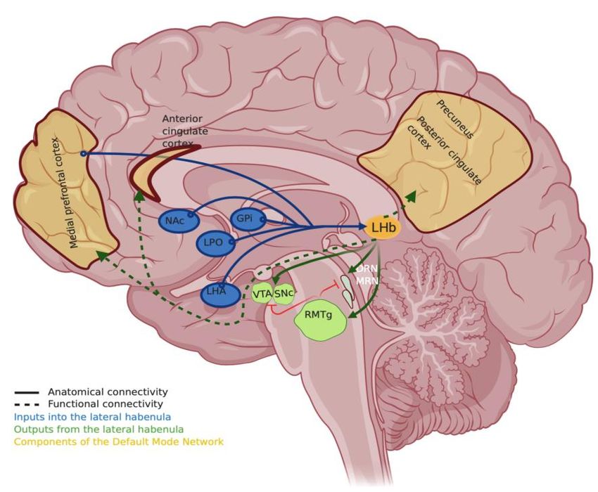
Figure1.
Figure 1. The cartoon
cartoon shows
shows aa few
few major
major inputs
inputs (in
(in blue)
blue) and
and outputs
outputs (in
(in green)
green) of
of the
the lateral
lateral habenula
habenula
basedon
based on[11]—NAc,
[11]—NAc,Nucleus
NucleusAcumbmens;
Acumbmens;GPi,GPi,globus
globuspallidus
pallidusinternus;
internus;LHA,
LHA, lateral
lateral hypothalamic
hypothalamic
area;LPO,
area; LPO,lateral
lateralpreoptic
preoptic area;
area; MRNMRNandand
DRN, DRN,
medialmedial
rapheraphe nucleus
nucleus or dorsal
or dorsal raphe nucleus;
raphe nucleus; RMTg,
RMTg, rostromedial
rostromedial tegmentaltegmental
nucleus;nucleus; SNc, substantia
SNc, substantia nigracompacta;
nigra pars pars compacta; VTA, ventral
VTA, ventral tegmental
tegmental area.
area. Components
Components of Default
of Default Mode Network
Mode Network (DMN, in(DMN,
golden)inwhose
golden) whose connectivity
connectivity and gene
and gene expression
expression
profile changesprofile changes byinclude
by psychedelics psychedelics include precuneus/posterior
precuneus/posterior cingulate
cingulate cortex (PCC), cortex
medial (PCC),
prefrontal
medial(mPFC)
cortex prefrontal
andcortex (mPFC)
the anterior and thecortex.
cingulate anteriorThe
cingulate cortex. is
DMN cartoon The DMN
based oncartoon
[42,93]. isSolid
based on
lines
show major
[42,93]. Solidanatomical
lines showconnectivity described
major anatomical from animal
connectivity and human
described from studies,
animal andwhile dashed
human lines
studies,
show
whilefunctional
dashed linesconnectivity based on
show functional recent human
connectivity basedstudies [97–99].
on recent human Created with
studies BioRender.com.
[97–99]. Created with
BioRender.com.
8. Molecular Mechanisms of Psychedelic Action and the Future of Antidepressant Discovery
8. Molecular Mechanisms
Pharmacologically, ofPsilocybin
both Psychedelic Action
and LSD, and
and the Future
other of Antidepressant
molecules Discovery
that act via the serotonergic
system have been categorized
Pharmacologically, as psychedelics
both Psilocybin [94,100–102].
and LSD, and otherTheir function,
molecules thatprimarily
act via theasserotonergic
agonists of
serotonin receptors (5-HTR) has been confirmed in a remarkable study that used multi-modal
system have been categorized as psychedelics [94,100–102]. Their function, primarily as agonists imaging to of
simulate serotonin receptor activity in the whole brain, generating the closest match
serotonin receptors (5-HTR) has been confirmed in a remarkable study that used multi-modal to the brain on LSD
to date [102].
imaging They achieved
to simulate thisreceptor
serotonin by overlaying Positron
activity in the Emission Tomography
whole brain, generating (PET)
the imaging-derived
closest match to
maps of serotonin receptor density distribution on fMRI images of healthy
the brain on LSD to date [102]. They achieved this by overlaying Positron Emission volunteers on Tomography
LSD. Several
other
(PET)studies also point maps
imaging-derived to serotonin receptors,
of serotonin particularly
receptor density to 5-HT2a , to on
distribution be fMRI
the molecular
images oftarget of
healthy
psychedelics [94]. As pointed out by the author Carhart-Harris, it is indeed an extraordinary
volunteers on LSD. Several other studies also point to serotonin receptors, particularly to 5-HT2a, to finding that
abe
single moleculartarget
the molecular targetof
could underlie the
psychedelics [94].profound andout
As pointed varied subjective
by the experience of psychedelics.
author Carhart-Harris, it is indeed
an extraordinary finding that a single molecular target could underlie the profound and varied
subjective experience of psychedelics. What happens downstream to the receptor activation is not
fully understood yet. Given that a single treatment with psilocybin can have sustained antidepressant
effects even 5-weeks after the treatment [38], it has been hypothesized that long term changes could
include transcriptional changes and activation of specific immediate early genes (IEGs) in theInt. J. Mol. Sci. 2020, 21, 6525 10 of 17
What happens downstream to the receptor activation is not fully understood yet. Given that a single
treatment with psilocybin can have sustained antidepressant effects even 5-weeks after the treatment [38],
it has been hypothesized that long term changes could include transcriptional changes and activation
of specific immediate early genes (IEGs) in the receiving neurons [42]. As IEGS are expressed within
minutes after an intense or unusual neural activity, they are not only used to identify active neurons but
are also the starting point to decipher the cascade of events after exposure to a substance. Brain-derived
neurotrophic factor (BDNF) and activity-regulated cytoskeletal protein (arc) have been associated with
psychedelic exposure; however, a large gap still exists in the characterization of the downstream signaling
pathways involved as the circuitry activated is complex [42].
One way forward for this research is to use the “mini-brains” or cortical organoids that
show complex cellular organization and physiological responses [103]. Cerebral organoids have
an exceptional potential to help bridge the gap between animal studies and human trials. Organoids
are 3-D cellular structures that show realistic micro-anatomy and that are capable of self-renewal
and self-organization. Recent studies show that cerebral organoids derived from human pluripotent
cells progress through similar developmental trajectories and can develop to features resembling a
10-week old human brain [104,105]. Organoids allow for a more controlled environment to perform
investigations of neuropsychiatric illnesses at a molecular and cellular level compared to animal models
or ex-vivo primary cultures. Importantly, as they are directly derived from human pluripotent cells,
it circumvents questions raised about species-specific differences in the brain tissue. Organoids have
become a useful platform to study cellular interactions between different cell types that are not as easy
to conduct in-vivo; for example, for studies on glial–neuron interactions important in the pathology
of neuropsychiatric diseases [12,106]. Findings such as the development of microglia important for
the synapse formation innately within cerebral organoids support the possibility of using cerebral
organoids for such research [107]. Moreover, cerebral organoids maturing over a period of months
show many complex features with dendritic spines and spontaneously active neuronal networks,
thus allowing for the study of a “circuit” level analysis of the effects of antidepressants [108].
The use of the cerebral organoids to uncover the molecular mechanisms of psychedelic action
overcomes many limitations imposed by the lack of appropriate animal models and regulatory
restrictions [109–111]. Detailed studies with single-cell level “omics” analyses can be performed
to characterize the transcriptomic and proteomic profile after different periods of exposure to a
psychedelic. A recent study demonstrated the capability of such an approach by examining the effects
of 5-methoxy-N, N-dimethyltryptamine (5-MeO-DMT) on the proteome of cells in human cerebral
organoids. Dimethyltryptamines (DMTs) are naturally occurring psychoactive agents present in
brews and Ayahuasca used in Shamanic religious practices for their antidepressant properties [112].
Using mass spectrometry to perform unbiased proteome analysis, the study found that a single
exposure for 24 h modulated several signaling pathways. Signaling cascades indicative of increased
anti-inflammatory effects and modulation of proteins implicated in spine morphogenesis and function,
such as those participating in protrusion, microtubule dynamics, cytoskeletal reorganization, and Long
Term Potentiation (LTP), were detected [111]. In addition, the study also found inhibition of pathways
associated with cell death, thus providing some very new insights into the potential molecular
mechanism by which DMT likely produces its effects. Another direction for the use of this approach is
to simulate the effect of psychoactive agents and stimulants in utero or on the developing fetus [106].
Such molecular characterization of drug responses to psychedelics could yield equally exciting insights
into their effect on different sub-populations of neurons and their effects on synaptic plasticity and
neural proliferation.
Another promising direction anticipated in the antidepressant research is to derive organoids
for use in precision medicine type studies and examine the potential effects of antidepressants in
the context of individual risk variants and known genetic susceptibilities [113]. They may also be
particularly well suited to study the genetics of a combination of alleles that each have a small effect
on the phenotype, and are difficult to recreate in animal models. For instance, it has been shownInt. J. Mol. Sci. 2020, 21, 6525 11 of 17
that only approximately 50% of patients respond to SSRIs the first time, and less than one-third go
into remission [114]. Failure to respond to the initial course of treatment can mean an increased risk
of suicide, distress, and loss of productivity among the effected [115]. Predictive biomarkers that
can help stratify potential responders can dramatically reduce such risk [116]. However, the clinical
applications of such biomarker-based selection can be hampered due to small samples and effect
sizes [117]. Patient-derived cerebral organoids offer a potential way to compliment the knowledge of
current biomarkers with tests to examine the potential efficacy of the treatment. Assays that screen for
multiple drugs and their combinations could improve throughput and arrive at the appropriate course
of action faster [113,118]. For such applications to work, however, issues such as the time taken to
grow mature cerebral organoids (4 to 6 weeks) and the cost-effectiveness of the strategy have to be
taken into account. Despite these considerations, the organoids technology provides a new dimension
in psychedelic research and an exciting prospect for the discovery of therapeutic aids for MDD and
treatment-resistant depressive states.
9. Conclusions
Lateral habenula is one of the most intensely researched brain regions currently to find remedies for
treating depression and several comorbid neuropsychiatric disorders. Methodological refinements in
human studies, alongside an expansion of the animal systems used to model depression, are necessary
to understand the role of this brain region in these disorders. Studies that address the sparsity of models
investigating the common underlying molecular factors in comorbid disorders have the potential
for discovering new therapeutic approaches. At the same time, understanding the antidepressant
mechanism of psilocybin and psychedelics warrants scientific priority as the use of these agents has
the possibility of providing profound insights not only into how to “rewire” a brain in depression but
also into the normal operation of brains. We think it will also reveal the exact nature of the connections
between these two areas of research that appear to be emerging (Figure 1).
Funding: This research was funded by Yale-NUS College grant numbers R-607-265-225-121 and R-607-000-452-114
417 to ASM.
Conflicts of Interest: The authors declare no conflict of interest. The funders had no role in the design of the
study; in the collection, analyses, or interpretation of data; in the writing of the manuscript, or in the decision to
publish the results.
References
1. Liu, Q.; He, H.; Yang, J.; Feng, X.; Zhao, F.; Lyu, J. Changes in the global burden of depression from 1990 to 2017:
Findings from the Global Burden of Disease study. J. Psychiatr. Res. 2020, 126, 134–140. [CrossRef] [PubMed]
2. McKenzie, M.; Olsson, C.A.; Jorm, A.F.; Romaniuk, H.; Patton, G.C. Association of adolescent symptoms of
depression and anxiety with daily smoking and nicotine dependence in young adulthood: Findings from a
10-year longitudinal study. Addiction 2010, 105, 1652–1659. [CrossRef] [PubMed]
3. Mathew, A.R.; Hogarth, L.; Leventhal, A.M.; Cook, J.W.; Hitsman, B. Cigarette smoking and depression
comorbidity: Systematic review and proposed theoretical model. Addiction 2017, 112, 401–412. [CrossRef]
[PubMed]
4. Vanyukov, M.M.; Tarter, R.E.; Kirisci, L.; Kirillova, G.P.; Maher, B.S.; Clark, D.B. Liability to substance
use disorders: 1. Common mechanisms and manifestations. Neurosci. Biobehav. Rev. 2003, 27, 507–515.
[CrossRef] [PubMed]
5. Fracalanza, K.; McCabe, R.E.; Taylor, V.H.; Antony, M.M. The effect of comorbid major depressive disorder or
bipolar disorder on cognitive behavioral therapy for social anxiety disorder. J. Affect. Disord. 2014, 162, 61–66.
[CrossRef] [PubMed]
6. Finan, P.H.; Smith, M.T. The comorbidity of insomnia, chronic pain, and depression: Dopamine as a putative
mechanism. Sleep Med. Rev. 2013, 17, 173–183. [CrossRef]Int. J. Mol. Sci. 2020, 21, 6525 12 of 17
7. Manber, R.; Edinger, J.D.; Gress, J.L.; San Pedro-Salcedo, M.G.; Kuo, T.F.; Kalista, T. Cognitive behavioral
therapy for insomnia enhances depression outcome in patients with comorbid major depressive disorder
and insomnia. Sleep 2008, 31, 489–495. [CrossRef] [PubMed]
8. Möller, H.-J. Occurrence and treatment of depressive comorbidity/cosyndromality in schizophrenic psychoses:
Conceptual and treatment issues. World J. Biol. Psychiatry 2005, 6, 247–263. [CrossRef]
9. Krishnan, V.; Nestler, E.J. The molecular neurobiology of depression. Nature 2008, 455, 894–902. [CrossRef]
10. Lawson, R.P.; Nord, C.L.; Seymour, B.; Thomas, D.L.; Dayan, P.; Pilling, S.; Roiser, J.P. Disrupted habenula
function in major depression. Mol. Psychiatry 2017, 22, 202–208. [CrossRef]
11. Hu, H.; Cui, Y.; Yang, Y. Circuits and functions of the lateral habenula in health and in disease. Nat. Rev.
Neurosci. 2020, 21, 277–295. [CrossRef] [PubMed]
12. Cui, Y.; Yang, Y.; Ni, Z.; Dong, Y.; Cai, G.; Foncelle, A.; Ma, S.; Sang, K.; Tang, S.; Li, Y.; et al. Astroglial Kir4.1
in the lateral habenula drives neuronal bursts in depression. Nature 2018, 554, 323–327. [CrossRef] [PubMed]
13. Klein, M.E.; Chandra, J.; Sheriff, S.; Malinow, R. Opioid system is necessary but not sufficient for antidepressive
actions of ketamine in rodents. Proc. Natl. Acad. Sci. USA 2020, 117, 2656–2662. [CrossRef] [PubMed]
14. Yang, Y.; Cui, Y.; Sang, K.; Dong, Y.; Ni, Z.; Ma, S.; Hu, H. Ketamine blocks bursting in the lateral habenula to
rapidly relieve depression. Nature 2018, 554, 317–322. [CrossRef]
15. Amat, J.; Sparks, P.D.; Matus-Amat, P.; Griggs, J.; Watkins, L.R.; Maier, S.F. The role of the habenular complex
in the elevation of dorsal raphe nucleus serotonin and the changes in the behavioral responses produced by
uncontrollable stress. Brain Res. 2001, 917, 118–126. [CrossRef]
16. Lecourtier, L.; Kelly, P.H. A conductor hidden in the orchestra? Role of the habenular complex in monoamine
transmission and cognition. Neurosci. Biobehav. Rev. 2007, 31, 658–672. [CrossRef]
17. Hikosaka, O. The habenula: From stress evasion to value-based decision-making. Nat. Rev. Neurosci. 2010,
11, 503–513. [CrossRef]
18. Stephenson-Jones, M.; Floros, O.; Robertson, B.; Grillner, S. Evolutionary conservation of the habenular
nuclei and their circuitry controlling the dopamine and 5-hydroxytryptophan (5-HT) systems. Proc. Natl.
Acad. Sci. USA 2012, 109, E164–E173. [CrossRef]
19. Grillner, S.; von Twickel, A.; Robertson, B. The blueprint of the vertebrate forebrain—With special reference
to the habenulae. Semin. Cell Dev. Biol. 2018, 78, 103–106. [CrossRef]
20. Lee, H.W.; Yang, S.H.; Kim, J.Y.; Kim, H. The Role of the Medial Habenula Cholinergic System in Addiction
and Emotion-Associated Behaviors. Front. Psychiatry 2019, 10, 100. [CrossRef]
21. Mathuru, A.S. A little rein on addiction. Semin. Cell Dev. Biol. 2018, 78, 120–129. [CrossRef] [PubMed]
22. Li, K.; Zhou, T.; Liao, L.; Yang, Z.; Wong, C.; Henn, F.; Malinow, R.; Yates, J.R., 3rd; Hu, H. βCaMKII in lateral
habenula mediates core symptoms of depression. Science 2013, 341, 1016–1020. [CrossRef] [PubMed]
23. Shabel, S.J.; Wang, C.; Monk, B.; Aronson, S.; Malinow, R. Stress transforms lateral habenula reward responses
into punishment signals. Proc. Natl. Acad. Sci. USA 2019, 116, 12488–12493. [CrossRef] [PubMed]
24. Shabel, S.J.; Proulx, C.D.; Piriz, J.; Malinow, R. Mood regulation. GABA/glutamate co-release controls habenula
output and is modified by antidepressant treatment. Science 2014, 345, 1494–1498. [CrossRef] [PubMed]
25. Proulx, C.D.; Hikosaka, O.; Malinow, R. Reward processing by the lateral habenula in normal and depressive
behaviors. Nat. Neurosci. 2014, 17, 1146–1152. [CrossRef] [PubMed]
26. Lipina, T.V.; Khrapova, M.V.; Serykh, A.; Dubrovina, N.I.; Petrova, E.S.; Mikhnevich, N.; Starostina, M.V.;
Amstyslavskaja, T.G. The increased density of the habenular neurons, high impulsivity, aggression and
resistant fear memory in Disc1-Q31 L genetic mouse model of depression. Behav. Brain Res. 2020, 392, 112693.
27. Carlson, P.J.; Diazgranados, N.; Nugent, A.C.; Ibrahim, L.; Luckenbaugh, D.A.; Brutsche, N.; Herscovitch, P.;
Manji, H.K.; Zarate, C.A., Jr.; Drevets, W.C. Neural correlates of rapid antidepressant response to ketamine in
treatment-resistant unipolar depression: A preliminary positron emission tomography study. Biol. Psychiatry
2013, 73, 1213–1221. [CrossRef]
28. Dandekar, M.P.; Fenoy, A.J.; Carvalho, A.F.; Soares, J.C.; Quevedo, J. Deep brain stimulation for
treatment-resistant depression: An integrative review of preclinical and clinical findings and translational
implications. Mol. Psychiatry 2018, 23, 1094–1112. [CrossRef]
29. Sartorius, A.; Henn, F.A. Deep brain stimulation of the lateral habenula in treatment resistant major depression.
Med. Hypotheses 2007, 69, 1305–1308. [CrossRef]
30. Drobisz, D.; Damborská, A. Deep brain stimulation targets for treating depression. Behav. Brain Res. 2019,
359, 266–273. [CrossRef]You can also read