Impact of Gastrointestinal Symptoms in COVID-19: a Molecular Approach - Alijansa ...
←
→
Page content transcription
If your browser does not render page correctly, please read the page content below
SN Comprehensive Clinical Medicine (2020) 2:2658–2669
https://doi.org/10.1007/s42399-020-00619-z

    COVID-19

Impact of Gastrointestinal Symptoms in COVID-19:
a Molecular Approach
Charu Sonkar 1 & Dharmendra Kashyap 1 & Nidhi Varshney 1 & Budhadev Baral 1 & Hem Chandra Jha 1

Accepted: 25 October 2020 / Published online: 4 November 2020
# Springer Nature Switzerland AG 2020

Abstract
The pandemic of novel coronavirus disease (COVID-19) caused by the Severe Respiratory Syndrome Coronavirus-2 (SARS-
CoV-2) creates an immense menace to public health worldwide. Currently, the World Health Organization (WHO) has recog-
nized the novel coronavirus as the main cause of global pandemic. Patients infected with this virus generally show fever, nausea,
and respiratory illness, while some patients also manifest gastrointestinal symptoms such as abdominal pain, vomiting, and
diarrhea. Traces of SARS-CoV-2 RNA have been found in gastrointestinal cells. Further angiotensin converting enzyme 2
(ACE2) the known receptor for the virus is extensively expressed in these cells. This implies that gastrointestinal tract can be
infected and can also present them as a replication site for SARS-CoV-2, but since this infection may lead to multiple organ
failure, therefore identification of another receptor is a plausible choice. This review aims to provide comprehensive information
about probable receptors such as sialic acid and CD147 which may facilitate the virus entry. Several potential targets are
mentioned which can be used as a therapeutic approach for COVID-19 and associated GI disorders. The gut microbiomes are
responsible for high levels of interferon-gamma which causes hyper-inflammation and exacerbates the severity of the disease.
Briefly, this article highlights the gut microbiome’s relation and provides potential diagnostic approaches like RDT and LC-MS
for sensitive and specific identification of viral proteins. Altogether, this article reviews epidemiology, probable receptors and put
forward the tentative ideas of the therapeutic targets and diagnostic methods for COVID-19 with gastrointestinal aspect of
disease.

Keywords COVID-19 . SARS-CoV-2 . Gastrointestinal disorders . Drugs . Diagnostic methods

Introduction                                                          challenge because of exponential increase in patients, rate of
                                                                      infectivity, scarcity of resources, poor prognosis of disease,
COVID-19 caused by novel SARS-CoV-2 is known to arrive                and ambiguity regarding disease pathogenesis [3].
from Wuhan wet market as an etiological agent leading to a               Furthermore, SARS-CoV-2 is known to attack the lower
global pandemic (2019–2020) [1]. According to the WHO as              respiratory system and cause viral pneumonia; however, it
of September 17, 2020, this infection has more than 29 million        may also affect the gastrointestinal system [4]. Reports sug-
active cases and about 0.58 million reported deaths in the form       gest that patients infected with nCoV showing digestive
of viral pneumonia and affecting about 214 countries world-           symptoms like diarrhea, vomiting may be among presenting
wide [2]. The pandemic of novel coronavirus is a great                features of disease [5]. However, the prognosis of COVID-19
                                                                      patients with gastrointestinal symptoms is mostly unknown.
Dharmendra Kashyap and Nidhi Varshney contributed equally to this     In about 50% of COVID-19 cases, there is the presence of
work.
                                                                      SARS-CoV-2 in fecal samples and its viral RNA has also
This article is part of the Topical Collection on Covid-19            been identified in intestinal mucosa suggesting that GI tract
                                                                      may be a probable route of infection [6]. Based on the studies
* Hem Chandra Jha                                                     in SARS-CoV-2, its receptor (ACE2) is known to be a critical
  hemcjha@iiti.ac.in
                                                                      component of gastric mucosa and gastrointestinal cells as
1                                                                     well, due to which gastric mucosa or gastrointestinal tract
     Infection Bioengineering Group, Department of Biosciences and
     Biomedical Engineering, Indian Institute of Technology Indore,   may be considered as vulnerable site for SARS-CoV-2 infec-
     Simrol, Madhya Pradesh 453552 Indore, India                      tion [7, 8]. The lasting presence of virus in gastric mucosa may
SN Compr. Clin. Med. (2020) 2:2658–2669                                                                                         2659

indicate the possibility of gastric glandular epithelial cells as   gut. Although infection results in diffuse alveolar damage, the
an incubation site of virus. This can be further consolidated by    changes in the gut are more precise which may include trans-
the presence of viral nucleocapsid protein in the cytoplasm of      migration of intestinal bacteria and more lipopolysaccharide
gastrointestinal tract days after its clearance from respiratory    (LPS) permeability in the intestine. LPS causes an increase in
sputum [9].                                                         the production of tumor necrosis factor (TNF), interleukin-1
   Since this virus arises from the family of Coronaviridae, it     (IL-1), and IL-6, thus resulting in aggravation of disease [20].
belongs to betacoronavirus genera. Moreover, the collectively       Furthermore, studies showed by Leung et al., states the SARS
known host receptor utilized by the SARS-CoV-2 is ACE2,             replication in the cells of small and large intestine of patients
sialic acids, and CD147 for the host cell infection [10–12].        with accumulation of higher viral titer inside the endoplasmic
Human leukocyte antigen (HLA) is an antigen-presenting fac-         reticulum (ER) and viral particles might leave from the apical
tor for viral peptide and may be prioritized for the development    membrane of the enterocytes. While the report suggests that
of a vaccine against SARS-CoV-2 [13]. Both sialic acids and         there is only minimal disruption of intestinal cells caused by
HLA are known to be important for Helicobacter pylori               the virus despite the tropism, thus, diarrhea associated with
(H. pylori) mediated gastritis and gastric cancer (GC) respec-      SARS infection may be more related to proteins or toxins
tively [14, 15]. Thus, they played an important role in causing     produced during viral replication than malabsorption or in-
gastrointestinal symptoms associated with COVID-19, with the        flammation [21].
emergence of the gastrointestinal tract as an important site for        Middle East respiratory syndrome coronavirus (MERS-
SARS-CoV-2 pathogenesis. A possible mechanism of viral in-          CoV), another virus of the coronaviridae family, known to
teraction and pathology is not yet completely known. In this        cause human respiratory infections and gastrointestinal symp-
review, we summarize the association of gastrointestinal disor-     toms. Zhou et al. illustrated that intestinal organoids and hu-
ders with SARS-CoV-2, their association from the other              man primary intestinal epithelial cells were immensely sus-
coronaviruses along with the receptors which play a crucial role    ceptible to MERS-CoV and can sustain vigorous viral repli-
in facilitating the virus entry. Briefly, we also highlighted the   cation [22]. Apart from the respiratory tract, the human ali-
gut microbe association in enhancing the infection, potential       mentary tract may be analyzed as a potential site for viral
targets used as therapeutics and drugs which can be repurposed      entry. Enteric viruses (adenovirus, rotavirus) and some non-
for the COVID-19 patients with gastric co-morbidities. This         enteric viruses (adenovirus A12) can bypass the physical bar-
article highlights potential diagnostic approaches like RDT         riers and infect susceptible cells in the alimentary tract [23].
and LC-MS for sensitive and specific identification of viral        The gastrointestinal symptoms are generally seen in most hu-
proteins. Taken all together, this article reviews epidemiology,    man coronaviruses [24], so far, seven human coronaviruses
probable receptors and put forward the tentative ideas of the       have been known as the causative agents of mild or severe
therapeutic targets, their drugs, and diagnostic tool for COVID-    respiratory infections.
19 with gastrointestinal aspect of disease.

                                                                    Epidemiology and Gastric Disorder as Clinical
Coronavirus and Gastrointestinal Symptoms:                          Predictor of SARS-CoV-2
a Long-Back Association
                                                                    SARS-CoV-2 shows the most common symptoms of the dis-
The coronaviruses comprise large, enveloped, positive-              ease include pneumonia along with cough, sore throat, myal-
stranded RNA viruses. Coronavirus caused a broad spectrum           gia, diarrhea, nausea, vomiting, and fatigue [25, 26]. In adults,
of diseases in animals and humans [16]. Human coronaviruses         most common symptoms are anorexia (39.9–50.2%) and di-
(HCoV) can be classified into two serogroups with HCoV-             arrhea (2–49.5%) while children suffered most from vomiting
229E and HCoV-NL63 included in serogroup one and                    (6.5–66.7%). Furthermore, 34.3% of COVID-19 patients are
HCoV-OC43 and HCoV-HKU1 falling in serogroup 2 [17].                having digestive symptoms that contribute to their delayed
The first two human coronaviruses, HCoV-229E and HCoV-              recovery, unlike the remaining patients who recover early
OC43, are correlated with upper respiratory tract infections.       [27], since the incubation time of a virus can range from 1 to
Furthermore, HCoV-OC43 and HCoV-HKU1 cause gastroin-                24 days, screening the patients becomes a tough task to handle
testinal symptoms such as diarrhea, vomiting, nausea, and           [27, 28]. Moreover, several reports showed that viral RNA
abdominal pain in up to 57% and 38% of infected people              was detected in stool samples from 48.1% patients even in
respectively [18, 19]. Therefore, gastrointestinal symptoms         stool collected after the respiratory samples tested negative
can be treated as evident as respiratory symptoms in corona-        [27, 29]. This suggests that the virus utilized fecal-oral route
virus colds, often designated “gastric flu.”                        for its transmission. Additional evidence of the association of
   Another coronavirus is the severe acute respiratory syn-         SARS-CoV-2 and gut came when it was found in the endos-
drome virus (SARS) which is also well known to affect the           copy sample of a COVID-19 patient [9]. Wang et al. have
2660 SN Compr. Clin. Med. (2020) 2:2658–2669
cultured the SARS-CoV-2 from four different patients’ stool patients from Wuhan has reported that there is an increased
samples and found live virions in two samples through mi- level of neutrophil, Interleukin-6 (IL-6), chemokine’s IP-10,
croscopy [30], although there are also reports that viruses are MCP-1, MIP-1A, tumor necrosis factor-alpha (TNFα), and
transmitted through fomites [31]. However, more research is less expression of lymphocytes [42, 43]. Here we precisely
required to confirm if this virus is viable in the stool and also explain the downstream signaling from the aforementioned
to analyze the level of transmission through the fecal-oral factors concerning gastrointestinal disorders and COVID-19
route [32]. taking SARS-CoV as a reference.
Importantly, multiple studies have found that the suscepti- ACE2 is largely found in the gastrointestinal tract [44]. The
ble population for COVID-19 includes elderly mostly > messenger RNA of the ACE2 receptor is highly expressed and
70 years of age, individuals with underlying disease, or weak- stabilized by neutral amino acid transporter B0AT1(SLC6A19)
ened immune systems [33]. Furthermore, in a meta-analysis in the gastro-intestinal system [45]. SARS-CoV-2 utilizes spike
by Men and colleagues, 10% of patients were showing only protein for binding to its receptor ACE2, upon binding the
gastrointestinal features upon infection. This may be due to plasma membrane fusion occurs and releases viral RNA.
delayed diagnosis which may lead to potential problems in the Further, viral RNA is detected as pathogen associated molecu-
patient and the person who comes in contact with the patient. lar pattern (PAMP’s) by pattern recognition receptor (mostly
Another study suggests that about 3% of COVID-19 cases toll like receptor) [46]. The RNA released is recognized by the
showed only digestive symptoms and no respiratory symp- viral RNA receptor retinoic-acid inducible gene I (RIG-I), cy-
toms [8, 34]. H. pylori is a well-established gastrointestinal tosolic receptor melanoma differentiation-associated gene5
pathogen associated with multiple gastric disorders like chron- (MDA5), nucleotidyltransferase cyclic GMP-AMP synthase
ic gastritis and gastric cancer. According to reports, SARS- (cGAS), and stimulator of interferon genes (STING) [40, 47].
CoV-2 and H. pylori infection are more likely to occur in Furthermore, this binding recruits TIR-domain-containing
patients with blood group A, thus increases the risk of gastro- adaptor protein along with mitochondrial antiviral-signaling
intestinal infection [35]. Furthermore, the patient having the protein (MAVS) and induces downstream signaling which in-
history of H. pylori infection may become more susceptible to cludes activation of nuclear factor-κB (NF-κB), interferon
oral-fecal route transmission. (IFN), and series of pro-inflammatory (IL-6) and antiviral cy-
Nearly one-fifth of COVID-19 patients have reported with tokines (Fig. 1) [48, 49]. This viral entry can also be through
gastro-intestinal symptoms [36]. About 70% of patients with endocytosis by clathrin dependent or independent pathway
viral RNA shedding through gastrointestinal tract were report- which may be used as a mechanism to avoid host detection
ed which lasted for about 10 weeks after the symptom onset (Fig. 1) [50, 51]. The upregulation of RIG-I and MDA-5 me-
[31]. Pathophysiological reports have suggested that no con- diated through retinoic acid-inducible gene I-like receptors
siderable damage was observed in the mucosal epithelium of (RLRs) and toll-like receptors is also associated with gastric
the esophagus, stomach, and duodenum tissues. However, adenocarcinoma cells [52]. The role of ACE is controversial;
major infiltration of lymphocytes is observed in squamous it is known to have a significant role in gastric ulcer healing and
epithelium of lamina propria of the stomach, duodenum, and can be related to virus mediated diarrhea [53]. Hence, studying
rectum which may cause abdominal pain [37]. Furthermore, ACE2 related signaling with gastric disorders and SARS-CoV-
abdominal pain is highly associated with COVID-19 severity 2 would provide better insight in determining therapeutic
along with nausea and vomiting which are comparatively less targets.
frequent. Considering the association of gut anomalies and its Reports suggest that through in-silico analysis of viral pep-
association with COVID-19 some of the symptoms like ab- tides of major histocompatibility complex class I gene (MHC)
dominal pain and diarrhea can also be considered COVID-19 ([HLA] A, B, C), HLA may present highly conserved SARS-
symptoms or can be used as clinical predictors [36]. CoV-2 peptides which suggest its ability to activate cross-
protective T cell mediated immunity. These findings suggest
that severity of SARS-CoV-2 may be affected by genetic var-
Receptor-Mediated Signaling iability of [HLA] A, B, and C [54]. Downregulation of HLA is
with Gastrointestinal Disorders a probable cause for poor prognosis in gastric and esophageal
cancer. Though epigenetic and oncogenic studies of HLA are
It is well established that ACE2 is a receptor for SARS-CoV- still ambiguous, reports suggest that MAPK and AKT (HER2)
2. Moreover, sialic acid, CD147 can also act as a receptor [10, signaling regulates the expression of HLA in gastric and
38]. However, the human coronavirus HKU1 and OC43 in- esophageal cancer [55]. Considering the importance of HLA
cludes human leukocyte antigen (HLA) as its attachment fac- and MAPK in the signaling pathway, they appear to be im-
tor and sialic acid as its receptor respectively [39, 40]. In portant targets for therapeutic use.
SARS-CoV-2 too, HLA may present the viral peptides [41]. Sialic acid which is responsible for regulating various phys-
Moreover, another study by Ming et al. (2020) on COVID-19 iological and pathological processes is composed of aSN Compr. Clin. Med. (2020) 2:2658–2669 2661 Fig. 1 Potential colliding targets, their signaling, and inhibitors: SARS- cytokines. Moreover, commercially available drugs and the targets with CoV-2 RNA enters the cells through plasma membrane and endocytosis no known drugs have also been shown. Here ACE2, TMPRSS2, RIG-I, followed by recognition by RIG-I and MDA5 which binds to MAVs. MDA5, IL-6, and ADAM-17 may serve as important targets concerning Furthermore, this leads to the secretion of IL-6, IFN, and antiviral gastric disorder as well diversified family of acidic sugar [56]. This can be used for Menicagli et.al., one of the hypotheses states that sialic acid internalization of bacteria like H. pylori in gastric mucosa strengthens the capacity of diffusion which relies on the varied through sialic acid binding adhesin (SabA) for subsequent per- number of glycoproteins present on the COVID-19 capsule sistence of infection. H. pylori infection is considered a crucial [62]. Additionally, there are certain sialic acids present on the risk factor in gastric carcinogenesis where only a subset of host cell surface which act as additional receptors for binding individuals develops tumor [57]. In healthy conditions, gastric sites of the S protein of SARS-CoV-2 [63], hence playing a role mucosa mostly expresses neutral fucosylated glycans whose in the pathogenicity and epidemiology of the associated disease glycophenotype is modified by H.pylori infection which leads [63]. to overexpression of β3-N-acetylglucosaminyltransferase-5 CD147 also known as Basigin or EMMPRIN is a trans- (β3GnT5) which is followed by increased biosynthesis of membrane glycoprotein known to bind to spike protein of sialyl-Lewis x [58]. This increased biosynthesis of sialyl- SARS-CoV-2 and mediate in virus invasion and infection to Lewis x further leads to successful strengthening of gastric other cells [11, 38]. Recently, a research team by Zhinan et al. epithelial attachment to H.pylori for efficient colonization, after conducting surface Plasmon resonance analysis and hence increasing the risk of gastric disorder. It has also been competitive inhibition experiment found that CD147 anti- found that high levels of α 2,3 sialic acid residues were linked body competitively inhibited binding of CD147 and S protein to GC cells invasion and metastasis [59]. Thus, the role of sialic [64]. Hence, it can be the potent target for therapeutics for acid in gastric comorbidities is evident; however, its role in COVID-19 patients. Moreover, CD147 is required for malaria SARS-CoV-2 is also emerging. The SARS-CoV-2 has very parasite, Plasmodium falciparum invasion, which can explain high infectivity due to its structure which contains various the infection of SARS-CoV-2 in red blood cells [65]. groups of terminal sialic acid [60, 61]. According to However, it is also associated with gastric cancer invasion,
2662 SN Compr. Clin. Med. (2020) 2:2658–2669
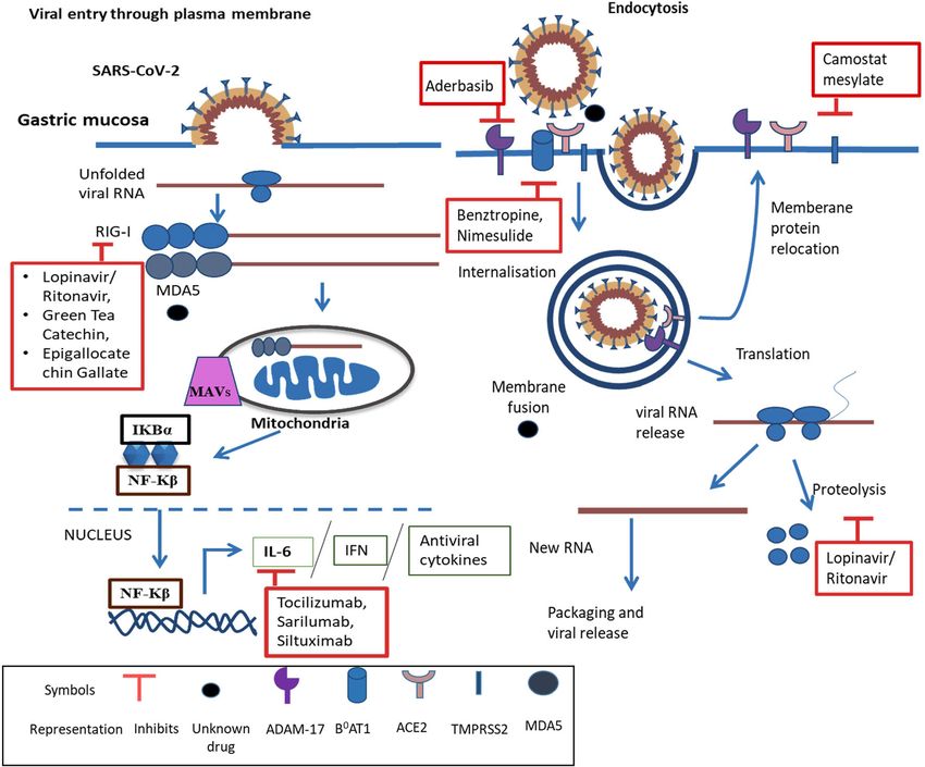
Fig. 2 Analysis of SARS-CoV-2 proteins, mutations, and post-translational modifications through LCMS followed by ESI/MS for effective therapeutics
metastasis and might be utilized for prognosis and indicator of SARS-CoV-2 spike protein binds to the ACE-2 receptor
tumor recurrence [66]. which may cause activation of p38 mitogen-activated protein
Increased level of IL-6 is often related to respiratory failure kinase (MAPK), upregulate ADAM-17, and stimulate ROS
and acute respiratory distress syndrome (ARDS). It plays a formation [69, 70]. The Spike-ACE-2 complex is proteolyti-
crucial role in aggravating cytokine storms in COVID-19. It cally processed by type 2 transmembrane protease
mainly follows two pathways cis and trans. In cis, IL-6 binds (TMPRSS2) at its S1/S2 junction to release S2 subunit, which
with membrane bound IL-6 receptor (mIL-6R) and gp130 further facilitates viral and cell membrane fusion [71, 72].
which activates Janus kinases (JAKs) and signal transducer Hence, to facilitate virus entry, TMPRSS2 and cathepsin L/
and activator of transcription 3 (STAT3) pathway. This JAK/ B primes the S-protein of SARS-CoV-2 [71]. The commer-
STAT3 pathway then activates innate and acquired immunity cially available drug for TMPRSS2 is camostat mesylate and
causing cytokine release syndrome (CRS). In trans pathway, nafamostat (Table 1), which is a clinically proven protease
IL-6 binds to its soluble receptor (sIL-6R) which again acti- inhibitor and it can also improve reflux esophagitis, dyspepsia
vates JAK-STAT3 signaling. Further, it leads to secretion of and inhibit SARS-CoV-2 infection [73–76]. Cathepsin L/B
vascular endothelial growth factor (VEGF), monocyte which also plays a crucial role in gastric cancer can be
chemoattractant protein–1 (MCP-1), IL-8 and reduced E- inhibited by cysteine protease inhibitor E64d (Table 1) [71,
cadherin expression on endothelial cells hence aggravating 99]. CD147 helps in P. falciparum invasion by binding to
cytokine storm [67]. IL-6 R can also be used as a prognostic reticulocyte-binding protein 5 (Rh5) [95]. CD147 can also
marker in gastric cancer. Hence, IL-6 can be a potent thera- bind to the spike protein of SARS-CoV-2 for entry into the
peutic target for COVID-19 patients suffering from gastric host cell [11, 38]. Targeting of CD147 through the
cancer [68]. meplazumab monoclonal antibody could be a possible poten-
tial therapy against COVID-19 disease (Table 1) [96].
STING (stimulator of interferon genes) is encoded by
Potential Colliding Targets and Associated TMEM173, considered as a key adaptor molecule which links
Drugs to the identification of cytosolic DNA leading to production
of interferons (IFNs) and NFkB. STING can also identify
The potential targets like ACE-2, TMPRSS2, Cathepsin L/B, infections by some RNA viruses. There are arguments that
CD147, STING, RIG-I, MDA5, P38 MAPK, ADAM-17, si- suggest the polymorphisms of the STING pathway could be
alic acid B0AT1, and IL-6 along with drugs can be used as involved in the pathogenesis of COVID-19 [97]. It is also
therapeutics in COVID-19 and associated gastrointestinal dis- reported that decreased STING is associated with poor prog-
orders have been briefly explained. nosis of gastric cancer patients (Table 1) [98]. RIG-I preciselySN Compr. Clin. Med. (2020) 2:2658–2669 2663
Table 1 Potential colliding
targets and probable drugs with S. Target Gastrointestinal Drug/phaseI/II-(PI) Drug activity References
their phase I/II/III trials with their No disorder Phase II/III-(PII)
activity association
1 ACE2 Gastric ulcer PII-APN01 ACE2 inhibitor [7–10, 44,
healing, virus 53, 69,
mediated 70]
diarrhea
2 TMPRSS2 Dyspepsia, Camostat mesylate Protease Inhibitor [71–76]
reflux PII-NCT04352400
esophagitis PII-nafamostat
3 (RIG-I) H.pylori Lopinavir/ritonavir, green Protease Inhibitor [47, 77–81]
infection, tea catechin,
gastric epigallocatechin gallate
adenocarci- PI-NCT03065023
noma
4 MDA5 Gastric [47]
adenocarci-
noma
5 [HLA]A Gastric cancer, No known commercial drug [13–15, 41,
esophageal available 54, 55]
cancer
6 Sialic acid H.pylori Egg whites [10–12, 14,
containing infection 39,
56–63,
82,
83, 79]
7 ADAM-17 Gastric acid Aderbasib (INCB7839) ADAM-17/TACE [69, 70, 84,
secretion inhibitor 85]
8 P38 MAPK Gastric cancer PI-Ralimetinib P38 MAPK [69, 70, 85,
(LY2228820) inhibitor 86]
9 B0AT1 Benztropine, nimesulide B0AT1 Inhibitor, [45, 87, 88]
(SLC6- Cyclooxygena-
A19) se Inhibitor
10 IL-6 Chronic gastritis Tocilizumab, sarilumab Interleukin-6 [20, 39, 42,
receptor 43, 48,
antagonist 49, 67,
68,
89–94]
12 CD147 Gastric cancer (PII) anti-CD147 [11, 38,
NCT04275245/meplazu- antibody 64–66,
mab 95, 96]
13 Stimulator of Gastric cancer [47, 97, 98]
interferon
genes
(STING)
14 Cathepsin B Digestive E64d Cysteine protease [71, 99]
cancer inhibitor
identifies the intracellular double-stranded viral RNA bearing Sialic acid also plays a key role in eradicating H.pylori
5′ triphosphate and invites molecules to activate antiviral sig- infection by inhibiting ROS production and NF-kB activation
naling [77]. Hence, antiviral drugs would be useful to target its [82]. Hence, sialic acid mediated inhibitors may provide po-
activity such as lopinavir with combination with ritonavir, tent treatment to patients. Soluble macromolecules containing
along with them natural products like green tea catechin, epi- sialic acid can act as decoy receptors and competitively inhibit
gallocatechin gallate are also been found to be effective [78], the receptor binding such as α2-macroglobulin, umifenovir,
though the administration of lopinavir /ritonavir is often asso- and other natural inhibitors include egg white which can be
ciated with drug-related diarrhea [79, 80]. Green tea catechin useful in COVID-19 patients having gastric disorders
is considered a beneficial and effective way to prevent gastro- (Table 1) [82, 83]. B0AT1 is an amino acid transporter, abbre-
intestinal disorders (Table. I) [81]. viation of major apical neutral amino acid transport system B02664 SN Compr. Clin. Med. (2020) 2:2658–2669
and it belongs to the solute carrier family 6 (SLC6A19) [87]. It Generally, the microbiota can enhance chronic phase protein
is the major Na+-dependent transporter for neutral amino acids and interferon signaling in lung cells to protect it from viral
in the small intestine and kidney [87]. The approved drug for infection [110]. However, in the case of SARS-CoV-2 infec-
B0AT1 inhibitor is Nimesulide (cyclooxygenase inhibitor) tion, the body’s response to infection changes the scenario.
and Benztropine (Table 1) [88]. Increased secretion of pro- Occasionally, COVID-19 patients’ immune response against
inflammatory cytokines like Interleukin-6 (IL-6) is a common the virus results in a cytokine storm ultimately leading to
factor in patients with gastritis and SARS-CoV-2 infection. hyper-inflammation and multi-organ failure [111, 112], so
IL-6 being a pro-inflammatory cytokine is linked to increased far, a cytokine profile associated with COVID-19 severity
inflammation in chronic acute gastritis [89, 90]. IL-6 may act has been characterized by increased interferon-γ inducible
as a prognostic marker for gastric cancer and a potential bio- protein and many other cytokines. There is a lack of clinical
marker for COVID-19 progression [68, 91]. Hence, toci- evidence supporting the modulation of the gut microbiota that
lizumab and sarilumab are FDA-approved drugs which is an may have therapeutic value in COVID-19 patients, subjected
interleukin-6 receptor antagonist can be a potent therapeutic to further research. From the current understanding, it can be
drug (Table 1) [92, 93]. Siltuximab is a monoclonal antibody speculated that the host cytokine molecular pathways, micro-
against IL-6 (Table 1) [94]. biota components, in association with cytokine responses, can
be used as novel microbiome-based therapeutic approaches
for SARS-CoV-2 infection [113].
Gut Dysbiosis: Interaction of Microbiota
with SARS-CoV-2
Diagnostic Approaches
The human gut consists of 1014 microorganisms like bacteria,
fungi, archaebacteria, and viruses [100]. These gut microbiomes Due to the growing COVID-19 pandemic, there is a shortage
have an important role in maintaining the health of the individual. of molecular testing capacity. Therefore, there is a need for
The microbiomes and host have a symbiotic association in that new point of care immunodiagnostic tests for fast and accurate
the earlier gets food and shelter and in turn, helps the later in testing of the disease. However, they can only be used in
regulating physiological functioning like dietary digestion, and research purposes and cannot be used for clinical decision
imparting protective immunity against pathogens [101]. making. There are 2 types of new point-of-care immunodiag-
Alterations of gut microbiota are known as “gut dysbiosis” which nostic tests for antigen detection and host antibody detection.
is associated with several diseases and disorders such as type 2
diabetes, IBD, cardiovascular disease, and depression [102–104].
Moreover, the COVID-19 treatment regime includes sets of Rapid Diagnostic Tests Based on Antigen
drugs that have negative impacts on different organelles and Detection
may cause gut dysbiosis also [105].
Intriguingly, the pulmonary health is also affected through COVID-19 virus proteins (antigens) present in a sample in-
a vital cross-talk between the lungs and the gut microbiota fected person are detected by rapid diagnostic test (RDT). If
known as the “gut-lung axis” [106]. This axis is bidirectional, the concentration of target antigen is sufficient in the sample,
i.e., microbial metabolites and the endotoxins can modulate the specific antibodies fixed to a paper strip enclosed in a
the lung through blood, and during inflammation in the lung, plastic casing will bind to the target protein. This will generate
it affects the gut microbiota [107]. This interdependency a visually detectable signal, usually within 30 min. Actively
boosts a striking possibility that SARS-CoV-2 may affect replicating a virus expresses the antigen; hence, this test can be
the gut microbiota. Numerous reports have pointed out that used for early detection of infection. However, the tests de-
alteration in the composition of the gut microbiota is correlat- pend on various factors such as virus concentration, quality of
ed with respiratory infections [108]. Moreover, severe clinical sample, and precise formulation of reagents. Hence, its accu-
outcomes in SARS-CoV-2 infected patients are associated racy for SARS-CoV-2 virus can range from 34 to 80% [114].
with immune-compromisation and aging. Therefore, it is
tempting to speculate the probable cross-talk between the
gut microbiota and the lung in COVID-19 which may further Rapid Diagnostic Tests Based on Host
influence the clinical manifestation. Antibody Detection
One hypothesis regarding gut dysbiosis is microbiomes’
impacts on cytokines. Type II interferon (interferon-γ) is This RDT detects the antibodies present in the blood of the
one such cytokine that plays important roles in antiviral re- patients believed to be infected COVID-19 with body pro-
sponses [109]. Furthermore, microbial metabolic processes in duced in response to the infection [115]. The strength of anti-
the gut strongly impact the production of cytokines [101]. bodies produced depends on various factors such as severitySN Compr. Clin. Med. (2020) 2:2658–2669 2665
of disease, age, nutritional status, and certain medication different structural proteins such as spike and nucleocapsid
against infections like HIV [116]. Furthermore, most antibod- that are encoded by SARS-CoV were identified by Krokhin
ies are produced in the second week of infection or may be et al. through the MS technique [126]. Intriguingly, Zeng et al.
generated in the recovery state of patients. However, one of first time identified the four structural protein and cytosol and
the drawbacks of this test is that it may provide false-positive nucleus fractions of SARS-CoV infected vero E6 cells and
results by interacting with antibodies generated for other in- also from the crude virion with shotgun strategy with 2D-
fections [114]. LC-MS/MS followed by ESI-MS/MS or by one dimensional
electrophoresis followed by ESI-MS/MS [127]. Post-
translational modifications (PTMs) of viral proteins interfere
Proteomics of SARS-CoV-2 Infected Host Cells with host cell signaling, cellular machinery hijacking and en-
Reveal Their Potential Targeted Therapy hancing infectivity [128]. Thus, viruses like influenza, SARS-
CoV, and SARS-CoV-2 utilize these PTM for enhancing the
Reaction towards SARS-CoV-2 outbreak through expedi- replication of their genome and for virion production.
tious, fast, and specific testing widely recognized as critical. Moreover, the novel phosphorylation of structural proteins
Nowadays, most qRT-PCR–based methods are used for the of SARS-CoV has been identified by this approach fig. 2
testing of SARS-CoV-2, while the non-MS (mass spectrome- [129]. Heavy glycosylation of spikes may facilitate viral at-
try) methods such as enzyme-linked immunosorbent assays tachment, membrane fusion and critically stimulate the host
(ELISAs), western blots, and protein arrays depend on anti- immune response. There are about 22 potential N-glycosyla-
bodies that were more successful during the outbreak of tion sites in S1 and S2 subunits of spike proteins. Shajahan
SARS-CoV in 2003 [117, 118]. Considering the immense et al. mapped the glycosylation sites of spike protein subunits
variability in antibody production, the liquid chromatography S1 and S2 which are expressed on human cells through reso-
coupled to mass spectrometry (LCMS) is an alternative attrac- lution MS [130]. Moreover, they have quantitatively charac-
tive diagnostic approach for the identification of small mole- terized the N-glycosylation sites. Intriguingly, they have ob-
cules such as peptides and proteins in clinical settings with served the unpredicted O-glycosylation modifications on the
consistent results [119, 120]. RBD domain of S1 subunit, spike protein. This is the first
These techniques measure the quantity of intact or proteo- report where they have shown O-glycosylation on S1 subunit.
lytically digested proteins with specificity, speed, sensitivity Thus, this study might play their role in vaccine development
and in resolution up to the femto-gram [121]. The most of the through elucidation of the glycan attachment on spike protein
LCMS techniques recruit tandem MS [122]. Furthermore, the of SARS-CoV-2 [130]. The limitations of LCMS in its med-
measurement of fragment ions that is formed in tandem MS ical setup, complex matrices, trace level analytes, and time-
has their clinical significance due to its higher specificity and consuming sample preparations [131].
lesser chances of false-positive results [123]. Ihling et al. iden-
tified the SARS-CoV-2 virus nucleoproteins from diluted gar-
gle solution of COVID-19 patients through the precipitation Conclusion
of protein followed by the proteolytic digestion through MS
[124]. Study shows that the expression of ACE-2 receptor is We conclude that SARS-CoV-2 is a causative agent of COVID-
high in heart tissue through the tandem-MS via tandem mass 19 and its association with the GI tract is well known from earlier
tag (TMT)-labeling and correlated with the higher heart fail- coronaviruses leaving a long-lasting impact on patients. In fact,
ure. Bojkova et al. have isolated the SARS-CoV-2 and infect- the severity will increase in patients having GI disorders. The
ed the human Caco-2 cell line which is a human colon epithe- attribute of gastrointestinal symptoms existing in COVID-19 is
lial cell and used proteome and translatome mass spectrometry more subtle than the respiratory symptoms; hence, they are easily
to perform the cellular response [125]. Furthermore, they were ignored. However, during the entire course of COVID-19, pa-
identified as the key casualties of the host cell retaliation to tients might have only gastrointestinal symptoms and may shed
infection. The above finding revealed the potential key mole- the virus in feces, even though their respiratory samples tests
cules as a drug target for the SARS-CoV-2 infection [125]. negative. Thus, it is pivotal to observe these gastrointestinal
symptoms with caution in the early stage of COVID-19.
Furthermore, dynamic monitoring of the digestive system and
One Dimensional and Two Dimensional cytokines is also required during clinical practice to decrease the
Liquid Chromatography ESI/MS chances of the complications and mortality of COVID-19 pa-
and Quantification of Virions tients. Moreover, the detection of SARS-CoV-2 in fecal samples
is essential for clinical practice along with routine testing, partic-
Although it is easy to identify the genome sequences of ularly for patients with atypical symptoms before leaving the
SARS-CoV, but the recognition of protein is difficult. Two hospital to confirm viral clearance. The ACE2 receptor is2666 SN Compr. Clin. Med. (2020) 2:2658–2669
ubiquitously found on the surface of the GI tract; thus, it is a References
potential replication site for SARS-CoV-2. Another receptor si-
alic acid is used by SARS-CoV-2 for its entry and HLA as its 1. Jin Z, Du X, Xu Y, Deng Y, Liu M, Zhao Y, et al. Structure of
Mpro from SARS-CoV-2 and discovery of its inhibitors. Nature.
attachment factor. We have mentioned several potential targets
2020;582:289–93.
that could be used as possible therapeutics. COVID-19 patients 2. WHO Coronavirus Disease (COVID-19) Dashboard. https://
have suffered from hyper-inflammation due to which gut mi- covid19.who.int. Accessed 17 Sep 2020.
crobes will further exacerbate the infection. In addition to inflam- 3. Endeman H, van der Zee P, van Genderen ME, van den Akker
mation, the current treatment regimens can also negatively affect JPC, Gommers D. Progressive respiratory failure in COVID-19: a
hypothesis. Lancet Infect Dis. 2020; S1473309920303662.
gut microbiota and cause digestive complications. We have also
4. Chen Y, Chen L, Deng Q, Zhang G, Wu K, Ni L, et al. The
aimed to provide insight techniques like RDT, LC-MS which presence of SARS-CoV-2 RNA in the feces of COVID-19 pa-
can be used for diagnosis and target the viral proteins with high tients. J Med Virol. 2020; 92:833–840. https://doi.org/10.1002/
sensitivity. Hence, this review intends to provide comprehensive jmv.25825.
information on SARS-CoV-2 concerning GI disorders. 5. Pan L, Mu M, Yang P, Sun Y, Wang R, Yan J, et al. Clinical
characteristics of COVID-19 patients with digestive symptoms in
Hubei, China: a descriptive, cross-sectional, multicentre study.
Acknowledgments We are thankful to CSIR for a scholarship to Charu Am J Gastroenterol. 2020;115: 766–773.
Sonkar, Dharmendra Kashyap, University Grants Commission, and DST-
6. Ng SC, Tilg H. COVID-19 and the gastrointestinal tract: more
inspire for fellowship to Budhadev Baral and Nidhi Varshney respective-
than meets the eye. Gut. 2020;69:973–4.
ly in the form of research stipend. We appreciate our lab colleagues for
7. Xu J, Fan J, Wu F, Huang Q, Guo M, Lv Z, et al. The
insightful discussions and advice. We gratefully acknowledge the Indian
ACE2/angiotensin-(1–7)/mas receptor axis: pleiotropic roles in
Institute of Technology Indore for providing facilities and support.
cancer. Front Physiol. 2017;8. https://doi.org/10.3389/fphys.
2017.00276.
Authors’ Contributions HCJ: supervision and reviewing and editing; CS: 8. Mao R, Qiu Y, He JS, Tan JY, Li XH, Liang J, et al.
conceptualization, methodology, data curation, writing—original draft Manifestations and prognosis of gastrointestinal and liver involve-
preparation, and reviewing and editing; DK: writing—original draft prep- ment in patients with COVID- 19: a systematic review and meta-
aration, reviewing and editing, and data curation; NV: methodology, analysis. Lancet Gastroenterol Hepatol. 2020; 5:667–678
original draft preparation, reviewing and editing, and data curation; BB:
9. Xiao F, Tang M, Zheng X, Liu Y, Li X, Shan H. Evidence for
visualization and reviewing and editing.
gastrointestinal infection of SARS-CoV-2. Gastroenterology.
2020;158:1831–1833.e3.
Funding This study is funded by the Council of Scientific and Industrial 10. Morniroli D, Giannì ML, Consales A, Pietrasanta C, Mosca F.
Research grant no 37(1693)/17/EMR-II, Department of Science and Human Sialome and coronavirus disease-2019 (COVID-19) pan-
Technology as Ramanujan fellowship grant no. SB/S2/RJN-132/20/5. demic: an understated correlation? Front Immunol. 2020;11:1480.
The funding organization has not played any role in the study design or 11. Ulrich H, Pillat MM. CD147 as a target for COVID-19 treatment:
the preparation of the manuscript. suggested effects of azithromycin and stem cell engagement. Stem
Cell Rev Rep. 2020;16:434–40.
Compliance with Ethical Standards 12. Shi Y, Wang Y, Shao C, Huang J, Gan J, Huang X, et al. COVID-
19 infection: the perspectives on immune responses. Cell Death
Conflict of Interest The authors declare that they have no conflict of Differ. 2020;27:1451–4.
interest. 13. Chan CM, Lau SKP, Woo PCY, Tse H, Zheng B-J, Chen L, et al.
Identification of major histocompatibility complex class I C mol-
ecule as an attachment factor that facilitates coronavirus HKU1
Abbreviations (COVID-19), Coronavirus disease 2019; (SARS-CoV-
spike-mediated infection. JVI. 2009;83:1026–35.
2), Severe acute respiratory syndrome coronavirus 2; (SARS), Severe
acute respiratory syndrome virus; (GC), Gastric cancer; (H. pylori), 14. Unemo M, Aspholm-Hurtig M, Ilver D, Bergström J, Borén T,
Helicobacter pylori; (IFN) (STING), Stimulator of interferon; (HLA), Danielsson D, et al. The sialic acid binding SabA adhesin of
Human leukocyte antigen; (TGEV), Transmissible gastroenteritis virus; Helicobacter pylori is essential for nonopsonic activation of hu-
(LPS), lipopolysaccharide; (TNF), Tumor necrosis factor; (IL-1), man neutrophils. J Biol Chem. 2005;280:15390–7.
Interleukin-1; (IL-6), Interleukin-6; (MERS-CoV), Middle East respira- 15. Xu D-P, Shi W-W, Zhang T-T, Lv H-Y, Li J-B, Lin A, et al.
tory syndrome coronavirus; (PAMP’s), Type II interferon (interferon-γ), Elevation of HLA-G-expressing DC-10 cells in patients with gas-
pathogen associated molecular pattern; (RIG-I), Retinoic-acid inducible tric cancer. Hum Immunol. 2016;77:800–4.
gene I; (MDA5), Melanoma differentiation-associated gene5; (RLRs), 16. Zhou J, Li C, Zhao G, Chu H, Wang D, Yan HH-N, et al. Human
Retinoic acid-inducible gene I-like receptors; (cGAS), cyclic GMP- intestinal tract serves as an alternative infection route for Middle
AMP synthase; (TMPRSS2), Type 2 transmembrane protease; (MHC), East respiratory syndrome coronavirus. Sci Adv. 2017;3:
Major histocompatibility complex class I gene; (β3GnT5), β3-N- eaao4966.
acetylglucosaminyltransferase-5; (9-OAc-Sia), 9-O-acetylated sialic acid; 17. Esper F, Ou Z, Huang YT. Human coronaviruses are uncommon in
(SLC6A19), Solute carrier family 6; (JAKs), Janus kinases; (STAT3), patients with gastrointestinal illness. J Clin Virol. 2010;48:131–3.
Signal transducer and activator of transcription 3; (RDT), Rapid diagnos- 18. Kanwar A, Selvaraju S, Esper F. Human coronavirus-HKU1 in-
tic test; (ELISAs), Enzyme-linked immunosorbent assays; (LCMS), fection among adults in Cleveland. Ohio Open Forum Infect Dis.
Liquid chromatography coupled to mass spectrometry; (MS), Mass spec- 2017;4:ofx052. https://doi.org/10.1093/ofid/ofx052.
trometry; (TMT), Tandem mass tag; (PTMs), Post-translational modifi- 19. Kumthip K, Khamrin P, Ushijima H, Maneekarn N. Enteric and
cations; (AT1), Angiotensin II receptor type 1 non-enteric adenoviruses associated with acute gastroenteritis in
pediatric patients in Thailand, 2011 to 2017. PLoS One. 2019;14:
e0220263. https://doi.org/10.1371/journal.pone.0220263.SN Compr. Clin. Med. (2020) 2:2658–2669 2667
20. To K, Tong JH, Chan PK, Au FW, Chim SS, Allen Chan K, et al. protein. preprint. Microbiology. 2020. https://doi.org/10.1101/
Tissue and cellular tropism of the coronavirus associated with 2020.03.14.988345.
severe acute respiratory syndrome: anin-situ hybridization study 39. Pöhlmann S, Gramberg T, Wegele A, Pyrc K, van der Hoek L,
of fatal cases. J Pathol. 2004;202:157–63. Berkhout B, et al. Interaction between the spike protein of human
21. Leung WK, To K, Chan PKS, Chan HLY, Wu AKL, Lee N, et al. coronavirus NL63 and its cellular receptor ACE2. In: Perlman S,
Enteric involvement of severe acute respiratory syndrome- Holmes KV, editors. The Nidoviruses. Boston: Springer; 2006. p.
associated coronavirus infection1 1The authors thank Man-yee 281–4. https://doi.org/10.1007/978-0-387-33012-9_47.
Yung, Sara Fung, Dr. Bonnie Kwan, and Dr. Thomas Li for their 40. Li X, Geng M, Peng Y, Meng L, Lu S. Molecular immune path-
help in retrieving patient information. Gastroenterology. ogenesis and diagnosis of COVID-19. J Pharm Anal. 2020;10:
2003;125:1011–7. 102–8.
22. Li H, Liu SM, Yu XH, Tang SL, Tang CK. Coronavirus disease 41. Zahn LM. HLA genetics and COVID-19. Science. 2020;368:
2019 (COVID-19): current status and future perspectives. Int J 841.2–841.
Antimicrobial Agents. 2020; 55:105951 42. Ni M, Tian F, Xiang D, Yu B. Characteristics of inflammatory
23. Verity R, Okell LC, Dorigatti I, Winskill P, Whittaker C, Imai N, factors and lymphocyte subsets in patients with severe COVID-
et al. Estimates of the severity of coronavirus disease 2019: a 19. J Med Virol. 2020. https://doi.org/10.1002/jmv.26070.
model-based analysis. Lancet Infect Dis. 2020;20:669–77. 43. Costela-Ruiz VJ, Illescas-Montes R, Puerta-Puerta JM, Ruiz C,
https://doi.org/10.1016/S1473-3099(20)30243-7. Melguizo-Rodríguez L. SARS-CoV-2 infection: The role of cyto-
24. Portes SAR, Volotão E d M, Rocha MS, Rebelo MC, Xavier M d kines in COVID-19 disease. Cytokine Growth Factor Rev. 2020;
PTP, de Assis RM, et al. A non-enteric adenovirus A12 gastroen- 54: 62–75
teritis outbreak in Rio de Janeiro, Brazil. Mem Inst Oswaldo Cruz. 44. Sungnak W, Huang N, Bécavin C, Berg M, Queen R, Litvinukova
2016;111:403–6. M, et al. SARS-CoV-2 entry factors are highly expressed in nasal
25. Tian Y, Rong L, Nian W, He Y. Review article: gastrointestinal epithelial cells together with innate immune genes. Nat Med.
features in COVID-19 and the possibility of faecal transmission. 2020;26:681–7.
Aliment Pharmacol Ther. 2020;51:843–51. 45. Jiang Y, Rose AJ, Sijmonsma TP, Bröer A, Pfenninger A, Herzig
26. Li X, Zai J, Wang X, Li Y. Potential of large “first generation” S, et al. Mice lacking neutral amino acid transporter B0AT1
human-to-human transmission of 2019-nCoV. J Med Virol. (Slc6a19) have elevated levels of FGF21 and GLP-1 and im-
2020;92:448–54. proved glycaemic control. Molec Metab. 2015;4:406–17.
27. Cheung KS, Hung IFN, Chan PPY, Lung KC, Tso E, Liu R, et al. 46. Kawai T, Akira S. The role of pattern-recognition receptors in
Gastrointestinal manifestations of SARS-CoV-2 infection and vi- innate immunity: update on toll-like receptors. Nat Immunol.
rus load in fecal samples from the Hong Kong cohort and system- 2010;11:373–84.
atic review and meta-analysis. Gastroenterology. 2020; 159:81–95 47. Essig M, Matt M, Massy Z. The COVID-19 outbreak and the
angiotensin-converting enzyme 2: too little or too much?
28. Lee I-C, Huo T-I, Huang Y-H. Gastrointestinal and liver manifes-
Nephrol Dial Transplant. 2020;35:1073–5. https://doi.org/10.
tations in patients with COVID-19. J Chin Med Assoc. 2020;83:
1093/ndt/gfaa113.
521–3. https://doi.org/10.1097/JCMA.0000000000000319.
48. Pawlik MW, Kwiecien S, Ptak-Belowska A, Pajdo R, Olszanecki
29. Xu Y, Li X, Zhu B, Liang H, Fang C, Gong Y, et al.
R, Suski M, et al. The renin-angiotensin system and its vasoactive
Characteristics of pediatric SARS-CoV-2 infection and potential
metabolite angiotensin-(1-7) in the mechanism of the healing of
evidence for persistent fecal viral shedding. Nat Med. 2020;26:
preexisting gastric ulcers. The involvement of mas receptors, nitric
502–5.
oxide, prostaglandins and proinflammatory cytokines. J Physiol
30. Wang W, Xu Y, Gao R, Lu R, Han K, Wu G, et al. Detection of
Pharmacol. 2016;67:75–91.
SARS-CoV-2 in different types of clinical specimens. JAMA.
49. Fujita M, Hayashi I, Yamashina S, Fukamizu A, Itoman M,
2020; 323:1843–1844
Majima M. Angiotensin type 1a receptor signaling-dependent in-
31. Xing Y-H, Ni W, Wu Q, Li W-J, Li G-J, Wang W-D, et al. duction of vascular endothelial growth factor in stroma is relevant
Prolonged viral shedding in feces of pediatric patients with coro- to tumor-associated angiogenesis and tumor growth.
navirus disease 2019. J Microbiol Immunol Infect. 2020;53:473– Carcinogenesis. 2005;26:271–9.
80. https://doi.org/10.1016/j.jmii.2020.03.021. 50. Wang H, Yang P, Liu K, Guo F, Zhang Y, Zhang G, et al. SARS
32. Gupta S, Parker J, Smits S, Underwood J, Dolwani S. Persistent coronavirus entry into host cells through a novel clathrin- and
viral shedding of SARS-CoV-2 in faeces – a rapid review. Color caveolae-independent endocytic pathway. Cell Res. 2008;18:
Dis. 2020;22:611–20. 290–301.
33. Pk C, Da W, Lk T, Sm I, Ac L, Cs L, et al. Viral shedding patterns 51. Kuba K, Imai Y, Ohto-Nakanishi T, Penninger JM. Trilogy of
of coronavirus in patients with probable severe acute respiratory ACE2: a peptidase in the renin-angiotensin system, a SARS re-
syndrome. Lancet. 2004;363:1699–700. ceptor, and a partner for amino acid transporters. Pharmacol Ther.
34. Ma C, Cong Y, Zhang H. COVID-19 and the digestive system. 2010;128:119–28.
Am J Gastroenterol. 2020; 115:1003–1006 52. Qu J, Hou Z, Han Q, Zhang C, Tian Z, Zhang J. Poly(I:C) exhibits
35. Uno Y. Why does SARS-CoV-2 invade the gastrointestinal epi- an anti-cancer effect in human gastric adenocarcinoma cells which
thelium? Gastroenterology. 2020;159:1622–3. is dependent on RLRs. Int Immunopharmacol. 2013;17:814–20.
36. Henry BM, de Oliveira MHS, Benoit J, Lippi G. Gastrointestinal 53. D’Amico F, Baumgart DC, Danese S, Peyrin-Biroulet L. Diarrhea
symptoms associated with severity of coronavirus disease 2019 during COVID-19 infection: pathogenesis, epidemiology, preven-
(COVID-19): a pooled analysis. Intern Emerg Med. 2020;15: tion, and management. Clin Gastroenterol Hepatol. 2020;18:
857–9. https://doi.org/10.1007/s11739-020-02329-9. 1663–72. https://doi.org/10.1016/j.cgh.2020.04.001.
37. Camargo SMR, Singer D, Makrides V, Huggel K, Pos KM, 54. Nguyen A, David JK, Maden SK, Wood MA, Weeder BR,
Wagner CA, et al. Tissue-specific amino acid transporter partners Nellore A, Thompson RF. Human leukocyte antigen susceptibil-
ACE2 and collectrin differentially interact with hartnup muta- ity map for severe acute respiratory syndrome coronavirus 2. J
tions. Gastroenterology. 2009;136:872–882.e3. Virol 2020; 94: e00510–20
38. Wang K, Chen W, Zhou Y-S, Lian J-Q, Zhang Z, Du P, et al. 55. Qu J-L, Qu X-J, Zhao M-F, Teng Y-E, Zhang Y, Hou K-Z, et al.
SARS-CoV-2 invades host cells via a novel route: CD147-spike Gastric cancer exosomes promote tumour cell proliferation2668 SN Compr. Clin. Med. (2020) 2:2658–2669
through PI3K/Akt and MAPK/ERK activation. Dig Liver Dis. 75. Sai JK, Suyama M, Kubokawa Y, Matsumura Y, Inami K,
2009;41:875–80. Watanabe S. Efficacy of camostat mesilate against dyspepsia as-
56. Jun L, Yuanshu W, Yanying X, Zhongfa X, Jian Y, Fengling W, sociated with non-alcoholic mild pancreatic disease. J
et al. Altered mRNA expressions of sialyltransferases in human Gastroenterol. 2010;45:335–41.
gastric cancer tissues. Med Oncol. 2012;29:84–90. 76. Yamamoto M, Matsuyama S, Li X, Takeda M, Kawaguchi Y,
57. Pinho SS, Carvalho S, Marcos-Pinto R, Magalhães A, Oliveira C, Inoue J, et al. Identification of nafamostat as a potent inhibitor of
Gu J, et al. Gastric cancer: adding glycosylation to the equation. Middle East respiratory syndrome coronavirus S protein-mediated
Trends Mol Med. 2013;19:664–76. membrane fusion using the split-protein-based cell-cell fusion as-
58. Marcos NT, Magalhães A, Ferreira B, Oliveira MJ, Carvalho AS, say. Antimicrob Agents Chemother. 2016;60:6532–9.
Mendes N, et al. Helicobacter pylori induces β3GnT5 in human 77. Ranjith-Kumar CT, Murali A, Dong W, Srisathiyanarayanan D,
gastric cell lines, modulating expression of the SabA ligand sialyl– Vaughan R, Ortiz-Alacantara J, et al. Agonist and antagonist rec-
Lewis x. J Clin Invest. 2008;118:2325–36. ognition by RIG-I, a cytoplasmic innate immunity receptor. J Biol
59. Wang F-L, Cui S-X, Sun L-P, Qu X-J, Xie Y-Y, Zhou L, et al. Chem. 2009;284:1155–65.
High expression of α 2, 3-linked sialic acid residues is associated 78. Ranjith-Kumar CT, Lai Y, Sarisky RT, Cheng KC. Green tea
with the metastatic potential of human gastric cancer. Cancer catechin, epigallocatechin gallate, suppresses signaling by the
Detect Prev. 2009;32:437–43. dsRNA innate immune receptor RIG-I. PLoS One. 2010;5:
60. Robson B. Bioinformatics studies on a function of the SARS- e12878. https://doi.org/10.1371/journal.pone.0012878.
CoV-2 spike glycoprotein as the binding of host sialic acid gly- 79. Cvetkovic RS, Goa KL. Lopinavir/ritonavir. Drugs. 2003;63:769–
cans. Comput Biol Med. 2020;122:103849. 802.
61. Fantini J, Di Scala C, Chahinian H, Yahi N. Structural and mo- 80. Chandwani A, Shuter J. Lopinavir/ritonavir in the treatment of HIV-1
lecular modelling studies reveal a new mechanism of action of infection: a review. Ther Clin Risk Manag. 2008;4:1023–33.
chloroquine and hydroxychloroquine against SARS-CoV-2 infec- 81. Koo MWL, Cho CH. Pharmacological effects of green tea on the
tion. Int J Antimicrob Agents. 2020;55:105960. gastrointestinal system. Eur J Pharmacol. 2004;500:177–85.
62. Menicagli R, Limodio M. COVID-19 solution. Int J Prev Med. 82. Matrosovich M, Klenk H-D. Natural and synthetic sialic acid-
2020;11(6):11–73. containing inhibitors of influenza virus receptor binding. Rev
63. Devaux CA, Rolain J-M, Colson P, Raoult D. New insights on the Med Virol. 2003;13:85–97.
antiviral effects of chloroquine against coronavirus: what to expect 83. Leneva IA, Burtseva EI, Yatsyshina SB, Fedyakina IT, Kirillova
for COVID-19? Int J Antimicrob Agents. 2020;55:105938. ES, Selkova EP, et al. Virus susceptibility and clinical effective-
64. The second key protein of SARS-CoV-2 infection, CD147 - ness of anti-influenza drugs during the 2010–2011 influenza sea-
CLOUD-CLONE CORP.(CCC). http://www.cloud-clone.com/ son in Russia. Int J Infect Dis. 2016;43:77–84.
topic/The-second-key-protein-of-SARS-CoV-2-infection%2D% 84. Murumkar PR, Ghuge RB, Chauhan M, Barot RR, Sorathiya S,
2DCD147.html. Accessed 16 Jul 2020. Choudhary KM, et al. Recent developments and strategies for the
65. Muramatsu T. Basigin: a multifunctional membrane protein with discovery of TACE inhibitors. Expert Opin Drug Discov.
an emerging role in infections by malaria parasites. Expert Opin 2020;15:779–801
Ther Targets. 2012;16:999–1011. 85. Smolka AJ, Backert S. How Helicobacter pylori infection controls
66. Chu D, Zhu S, Li J, Ji G, Wang W, Wu G, et al. CD147 gastric acid secretion. J Gastroenterol. 2012;47:609–18.
Expression in human gastric cancer is associated with tumor re- 86. Vergote I, Heitz F, Buderath P, Powell M, Sehouli J, Lee CM,
currence and prognosis. PLoS ONE. 2014;9:e101027. et al. A randomized, double-blind, placebo-controlled phase 1b/2
67. COVID-19 and the cytokine storm the crucial role of IL-6 - Enzo study of ralimetinib, a p38 MAPK inhibitor, plus gemcitabine and
Life Sciences. https://www.enzolifesciences.com/science-center/ carboplatin versus gemcitabine and carboplatin for women with
technotes/2020/april/covid-19-and-the-cytokine-storm-the- recurrent platinum-sensitive ovarian cancer. Gynecol Oncol.
crucial-role-of-il-6/. Accessed 16 Jul 2020. 2020;156:23–31.
68. Simondurairaj C, Krishnakumar R, Sundaram S, Venkatraman G. 87. Bröer S, Gether U. The solute carrier 6 family of transporters. Br J
Interleukin-6 receptor (IL-6R) expression in human gastric carcinoma Pharmacol. 2012;167:256–78.
and its clinical significance. Cancer Investig. 2019;37:293–8. 88. Cheng Q, Shah N, Bröer A, Fairweather S, Jiang Y, Schmoll D, et al.
69. Grimes JM, Grimes KV. p38 MAPK inhibition: a promising thera- Identification of novel inhibitors of the amino acid transporter B0AT1
peutic approach for COVID-19. J Mol Cell Cardiol. 2020;144:63–5. (SLC6A19), a potential target to induce protein restriction and to treat
70. Gwathmey TM, Pendergrass KD, Reid SD, Rose JC, Diz DI, type 2 diabetes. Br J Pharmacol. 2017;174:468–82.
Chappell MC. Angiotensin-(1-7)-angiotensin-converting enzyme 89. Bauditz J, Ortner M, Bierbaum M, Niedobitek G, Lochs H,
2 attenuates reactive oxygen species formation to angiotensin II Schreiber S. Production of IL-12 in gastritis relates to infection
within the cell nucleus. Hypertension. 2010;55:166–71. with Helicobacter pylori. Clin Exp Immunol. 1999;117:316–23.
71. Gheblawi M, Wang K, Viveiros A, Nguyen Q, Zhong J-C, Turner 90. Huang M, Dong D, Qi D, Ling C, Zhang D. Hp infection and
AJ, et al. Angiotensin-converting enzyme 2: SARS-CoV-2 recep- expression of TGF-βR II,IL-6 and TNF-α in patients with chronic
tor and regulator of the renin-angiotensin system. Circ Res. atrophic gastritis. Chin J Immunol. 2018;34:751–6.
2020;126:1456–74. https://doi.org/10.1161/CIRCRESAHA.120. 91. Ulhaq ZS, Soraya GV. Interleukin-6 as a potential biomarker of
317015. COVID-19 progression. Med Mal Infect. 2020;50:382–3.
72. Rabi FA, Al Zoubi MS, Kasasbeh GA, Salameh DM, Al-Nasser 92. Tu Y-F, Chien C-S, Yarmishyn AA, Lin Y-Y, Luo Y-H, Lin Y-T,
AD. SARS-CoV-2 and coronavirus disease 2019: what we know et al. A review of SARS-CoV-2 and the ongoing clinical trials. Int
so far. Pathogens. 2020;9:231. J Mol Sci. 2020;21:2657.
73. McKee DL, Sternberg A, Stange U, Laufer S, Naujokat C. 93. Fu B, Xu X, Wei H. Why tocilizumab could be an effective treat-
Candidate drugs against SARS-CoV-2 and COVID-19. ment for severe COVID-19? J Transl Med. 2020;18:164.
Pharmacol Res. 2020;157:104859. 94. van Rhee F, Fayad L, Voorhees P, Furman R, Lonial S, Borghaei
74. Kono K, Takahashi A, Sugai H, Umekawa T, Yano T, Kamiyasu H, et al. Siltuximab, a novel anti-interleukin-6 monoclonal anti-
K, et al. Oral trypsin inhibitor can improve reflux esophagitis after body, for Castleman’s disease. J Clin Oncol. 2010;28:3701–8.
distal gastrectomy concomitant with decreased trypsin activity. 95. van Ooij C. Basigin opens the door to malaria. Nat Rev Microbiol.
Am J Surg. 2005;190:412–7. 2012;10:3–3.You can also read