Case Report Malignant Transformation of Temporal Bone Schneiderian Papilloma Associated with HPV-6
←
→
Page content transcription
If your browser does not render page correctly, please read the page content below
Hindawi
Case Reports in Otolaryngology
Volume 2021, Article ID 6684254, 5 pages
https://doi.org/10.1155/2021/6684254

Case Report
Malignant Transformation of Temporal Bone Schneiderian
Papilloma Associated with HPV-6

          O. Marzouk,1 F. Brasch,2 I. Todt,1 P. K. C. Goon,3 and H. Sudhoff                               1

          1
            Department of Otorhinolaryngology, Head and Neck Surgery, Medical Faculty OWL, Bielefeld University, Campus Klinikum,
            Bielefeld, Germany
          2
            Department of Pathology, Academic Teaching Hospital Bielefeld, Bielefeld, Germany
          3
            Department of Dermatology, Peterborough City Hospital, North West Anglia NHS Foundation Trust, Peterborough, UK

          Correspondence should be addressed to H. Sudhoff; holger.sudhoff@rub.de

          Received 6 December 2020; Revised 4 January 2021; Accepted 13 January 2021; Published 25 January 2021

          Academic Editor: M. Tayyar Kalcioglu

          Copyright © 2021 O. Marzouk et al. This is an open access article distributed under the Creative Commons Attribution License,
          which permits unrestricted use, distribution, and reproduction in any medium, provided the original work is properly cited.

          Introduction. Temporal bone Schneiderian papillomas (TBSPs) rarely present as a primary tumors arising from the middle ear and
          mastoid process. The clinical findings and imaging of TBSPs are not specific. Therefore, diagnosis can only reliably be definitively
          established by histopathology. Objective. To report a novel case of a malignant transformation of TBSP associated with HPV-6 and
          to present its management. Case Report. A 68-year-old woman presented with conductive hearing loss and recurrent right-sided
          otorrhoea. Initially, we performed a lateral temporal bone resection and obliteration with abdomen fat. Early histology described
          TBSP associated with HPV-6. Follow-up detected malignant transformation of the Schneiderian papillomatous variant. Post-
          operative radiotherapy combined with extended temporal bone resection resulted in a disease-free 17-month period of follow-up.
          Discussion. TBSPs are not very specific, and the diagnosis can only reliably be established by histopathology. There is a risk of
          malignant transformation, and due to the absence of reliable prognostic markers, strict postoperative follow-up is mandatory and
          should consist of regular otoscopy, nasal endoscopy, and imaging. This case also supports the importance of extended temporal
          bone resections as salvage surgery, combining radical surgery with radiotherapy for improved survival rates.

1. Introduction                                                        iatrogenic implantation of tumor cells during a surgical
                                                                       procedure within the sinonasal tract [2, 6, 7]. A malignant
Temporal bone inverted papillomas (Schneiderian-type                   transformation of SPs can occur in about 10% of cases
papillomas, SPs) can rarely present as a primary tumor                 [1, 2]. The typical symptoms of SPs in the temporal bone
rising from the middle ear and mastoid process. Addi-                  are otalgia, otorrhoea, and hearing loss [5].
tionally, this benign middle ear lesion may originate from
the sinonasal tract, accounting for 5% of all tumors [1].
The etiology is unclear, but human papilloma virus
                                                                       2. Case Presentation
(HPV) is considered a risk factor [1, 2]. Inverted papil-              A 68-year-old female Caucasian patient was referred to our
lomas can also occur in the oral cavity, nasopharynx,                  clinic for a conductive hearing loss and recurrent right-sided
lacrymal sac, and temporal bone [3–5]. There is a debate               middle ear infections presenting as episodes of painless
in the literature on whether temporal bone Schneiderian                otorrhoea in January 2019. Otoscopy of the right ear revealed
papilloma (TBSP) involves the same pathology as sino-                  a completely occluded livid-coloured mass with mucopur-
nasal SP [1, 3]. The involvement of the temporal bone is               ulent secretions. Anterior rhinoscopy revealed no pathol-
extremely rare. It can occur either with direct tumor                  ogies. Pure tone audiogram of the right side revealed a
spread via the eustachian tube or originating from ectopic             moderate to severe mixed hearing loss. There was no medical
mucosa. Another possible mechanism is related to the                   history of any previous pathology or surgery of the sinonasal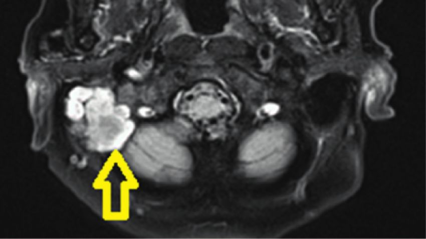
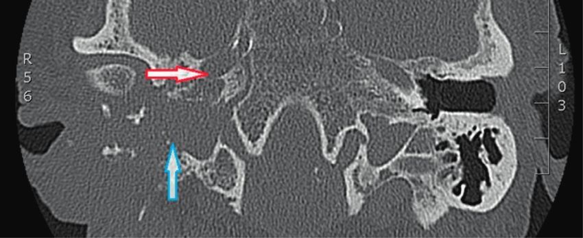
2                                                                                                      Case Reports in Otolaryngology

Figure 1: Axial view of the initial temporal CT showed complete opacification of the external auditory canal, middle ear, and mastoid on the
right side indicating a soft tissue mass.

Figure 2: Initial T2-weighted magnetic resonance imaging revealing a mass occupying the right middle ear and mastoid (yellow arrow).

tract. An outpatient biopsy of the right external auditory             respiratory epithelium with invagination throughout the
canal revealed an SP. CT scan and magnetic resonance                   stroma and small cysts in the epithelial lining containing
imaging (MRI) of the temporal bone showed a mass that                  cellular debris and neutrophils. The patient was discharged
filled the area of the right external auditory canal with               after 14 days. Subsequently, the patient was monitored by
penetration into the mastoid cavity with involvement of the            the referring an ENT doctor.
dura of the right temporal lobe (Figures 1–3). Subsequently,               A follow-up CT scan of the temporal bone due to new
a combined transmastoidal and transcanal surgical approach             right-sided facial palsy and complete deafness after 5 months
of the middle ear was performed, with exenteration of the              demonstrated a progressive and destructive lesion of the
mastoid cells resulting in a lateral temporal bone resection           right mastoid and middle ear adjacent to the carotid canal
without any evidence for residual disease after immediate              and jugular bulb, erosion of the cochlea, styloid process, and
postoperative CT scans. The polypoid tissue found in the               partially involved the temporal mandibular joint in July 2019
external ear canal, middle ear, and mastoid was completely             (Figure 4).
excised in February 2019. Postoperatively, the patient had no              The patient subsequently underwent a right-sided
facial palsy or vertigo. A temporary otoliquorrhea was                 mastoid exploration, and frozen sections confirmed a
successfully managed with conservative therapy. Histopa-               squamous cell carcinoma. Subsequently, an extended tem-
thology diagnosed a Schneiderian-type papilloma with low-              poral bone resection with sacrifice of the facial nerve, lab-
risk HPV infection (HPV-6 polymerase chain reaction                    yrinth, and cochlea including a duraplasty with abdominal
positive), p16 positive immunohistochemistry without any               fat obliteration was performed (Figure 5). Pathology reports
malignant signs. It exhibited aggregates of hyperplastic               confirmed a moderately differentiated nonkeratinizing
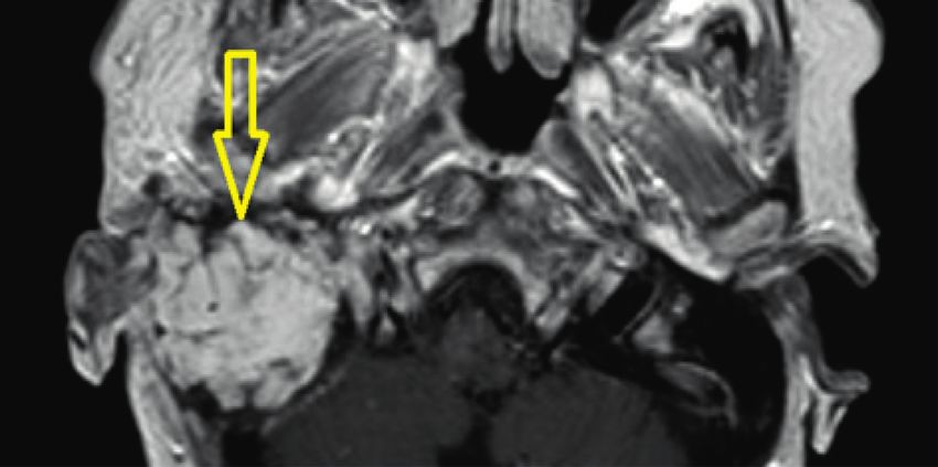
Case Reports in Otolaryngology                                                                                                           3

Figure 3: Initial coronal MRI demonstrating a heterogenous tissue mass in the right middle ear mastoid up to the dura of the middle cranial
dura (white arrow).

Figure 4: Axial view of temporal CT 5 months after the initial surgery showing complete opacification and massive bone erosion of the
middle ear and mastoid on the right side indicating an extended soft tissue mass adjacent to the carotid canal (red arrow) and jugular bulb
(blue arrow) and partially obliterating the temporal mandibular joint.

Figure 5: Axial T1-weighted MRI with gadolinium revealed obliteration with abdominal fat (yellow arrow) without any residual tumor 16
months after the extended temporal bone resection.
4 Case Reports in Otolaryngology
(a) (b)
(c) (d)
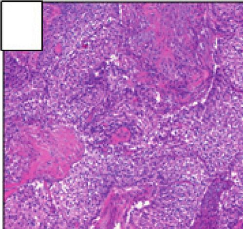
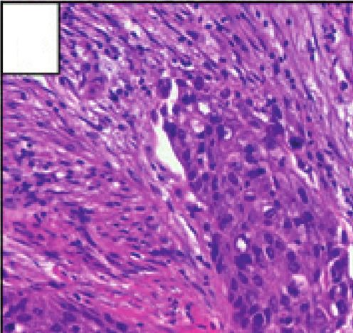
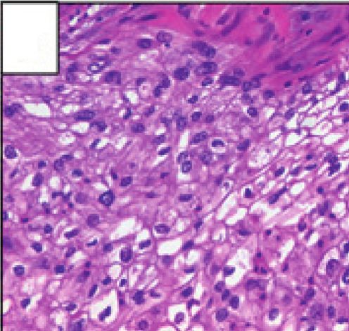
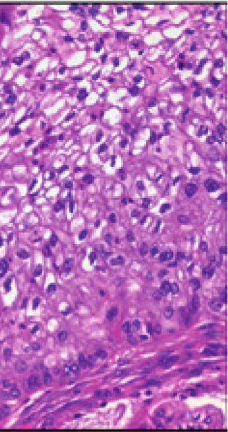
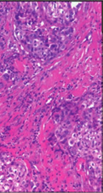
Figure 6: Histopathology revealing parts of the surgical specimen widely occupied by extensions of the temporal bone Schneider papilloma
after complete temporal bone resection. Cellular atypia and increased mitoses demonstrate the transition to an invasive, moderately
differentiated, nonkeratinizing squamous cell carcinoma.
squamous cell carcinoma on a background of SP that had subsequently still demonstrated malignant transformation.
been diagnosed earlier (Figure 6). A postoperative radio- HPV-6 and 11 have been rarely associated with malignant
therapy combined with an extended temporal bone resection transformation in the upper respiratory tract [16–18]. Un-
resulted in a disease-free 17-month follow-up. usually, the malignant transformation and extensive tumor
growth with the destruction of the surrounding structures
occurred in a relatively short time from the first surgical
3. Discussion
intervention in February 2019 to July 2019 along with facial
To our knowledge, up to the present day, only 33 patients palsy and deafness. So far, the majority of the biological
with TBSP have been reported [8]. Of note, in 18 patients, studies have been focused on E6 and E7 from HPV-16 and
including this current case, the tumor arose as a primary, HPV-18, since they are the most frequent types detected in
isolated lesion in the temporal bone, representing a true head and neck cancer. Possibly, an altered immune deviation
primary TBSP [8]. In another 14 reported cases, the tem- could have contributed to this rapid malignant transfor-
poral bone localisation represented an extension from pri- mation [19].
mary sinonasal SP, and in one case, sinonasal involvement is Stone and coworkers reported an inverted papilloma
not clearly described [1, 2, 9]. Despite its benign nature, confined to the temporal bone in 1987 [18]. So far, only 7
inverted Schneiderian papillomas have a high propensity for reported cases have exhibited malignant transformations
local recurrence and may undergo malignant transformation [8]. Patients with temporal bone inverted papilloma are
into squamous cell carcinomas [10]. Approximately 20% of probably more susceptible to recurrence due to the biology
these tumors may present with different severities of epi- of TBSP. This case highlights that the postoperative follow-
thelial dysplasia, which then convey different malignant up period is crucial, not only to detect recurrences but also to
potentials to the inverted Schneiderian papilloma [11, 12]. monitor a potential transformation to squamous cell car-
The possibility of a coincidence of the simultaneous presence cinoma [19]. This case also supports the importance of
of squamous cell carcinoma and Schneider’s papilloma extended temporal bone resections as salvage surgery after
could be excluded by serial sections of the investigated preoperative staging and assessment, combining radical
samples. A malignant transformation of the TBSP is ex- surgery with adjuvant radiotherapy for better survival rates
ceedingly rare. It has been demonstrated that HPV-6 and 11 [20–22].
(low risk viruses) are the most commonly isolated HPV
subtypes in inverted papillomas in the sinonasal tract [13]. 4. Conclusions
On the other hand, HPV-16 and 18 (high risk) are strongly
associated with squamous cell carcinoma [14, 15]. Temporal bone localisation of Schneiderian papilloma may
Interestingly, our case was associated with HPV-6 but represent a primary tumor or an extension from the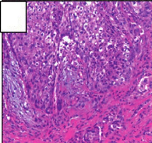
Case Reports in Otolaryngology                                                                                                              5

sinonasal tract; both are rare. The clinical presentation and             [12] P. A. Suarez, K. Adler-Storthz, M. A. Luna, A. K. El-Naggar,
radiology of TBSPs are not very specific, and the diagnosis                     F. W. Abdul-Karim, and J. G. Batsakis, “Papillary squamous
can only reliably be determined by histopathology. There is a                  cell carcinomas of the upper aerodigestive tract: a clinico-
risk of malignant transformation, and due to the absence of                    pathologic and molecular study,” Head & Neck, vol. 22, no. 4,
                                                                               pp. 360–368, 2000.
reliable prognostic markers, strict postoperative follow-up is
                                                                          [13] G. Marioni, G. Altavilla, G. Busatto, S. Blandamura,
mandatory and should consist of regular otoscopy, nasal
                                                                               C. De Filippis, and A. Staffieri, “Detection of human papil-
endoscopy, and imaging. Extended temporal bone resection                       lomavirus in temporal bone inverted papilloma by poly-
as salvage surgery plus the combination of extended tem-                       merase chain reaction,” Acta Oto-Laryngologica, vol. 123,
poral bone resection with radiotherapy seems to improve                        no. 3, pp. 367–371, 2003.
patient survival.                                                         [14] E. Gaio, G. Marioni, S. Blandamura, and A. Staffieri, “Inverted
                                                                               papilloma involving the temporal bone and its association
                                                                               with squamous cell carcinoma: critical analysis of the liter-
Data Availability                                                              ature,” Expert Review of Anticancer Therapy, vol. 5, no. 2,
                                                                               pp. 391–397, 2005.
The data for this case were accessed through the electronic
                                                                          [15] S. M. Caruana, N. Zwiebel, R. Cocker, S. A. McCormick,
medical record. They are not available for readers to review                   R. C. Eberle, and P. Lazarus, “p53 alteration and human
as they contain confidential patient health information.                        papilloma virus infection in paranasal sinus cancer,” Cancer,
                                                                               vol. 79, no. 7, pp. 1320–1328, 1997.
Conflicts of Interest                                                     [16] J. Nudell, S. Chiosea, and L. D. R. Thompson, “Carcinoma ex-
                                                                               Schneiderian papilloma (malignant transformation): a clini-
The authors declare that there are no conflicts of interest.                    copathologic and immunophenotypic study of 20 cases
                                                                               combined with a comprehensive review of the literature,”
                                                                               Head and Neck Pathology, vol. 8, no. 3, pp. 269–286, 2014.
References                                                                [17] M. Re, F. M. Gioacchini, A. Bajraktari et al., “Malignant
                                                                               transformation of sinonasal inverted papilloma and related
 [1] S. Mirza, P. J. Bradley, A. Acharya, M. Stacey, and N. S. Jones,          genetic alterations: a systematic review,” European Archives of
     “Sinonasal inverted papillomas: recurrence, and synchronous               Oto-Rhino-Laryngology, vol. 274, no. 8, pp. 2991–3000, 2017.
     and metachronous malignancy,” The Journal of Laryngology &           [18] D. M. Stone, R. E. Berktold, C. Ranganathan, and R. J. Wiet,
     Otology, vol. 121, no. 9, pp. 857–664, 2007.                              “Inverted papilloma of the middle ear and mastoid,” Oto-
 [2] S. J. Ramey, J. K. Russo, J. M. Condrey 3rd., B. Coulter, and             laryngology-Head and Neck Surgery, vol. 97, no. 4, pp. 416–
     A. K. Sharma, “Synchronous bilateral inverted papilloma of                418, 1987.
     the temporal bone: case report and review of the literature,”        [19] S. Smola, “Immune deviation and cervical carcinogenesis,”
     Head & Neck, vol. 35, no. 8, pp. 240–245, 2013.                           Papillomavirus Research, vol. 7, pp. 164–167, 2019.
 [3] H. S. Kaddour and C. J. Woodhead, “Transitional papilloma of         [20] E. Avallone, T. Lenarz, and K. Willenborg, “Primäre felsen-
     the middle ear,” The Journal of Laryngology & Otology,                    bein schneider papillom mit beteiligung der schädelba-
     vol. 106, no. 7, pp. 628-629, 1992.                                       sis—fallbericht und review der literatur,” Laryngo-Rhino-
 [4] B. M. Wenig, “Schneiderian-type mucosal papillomas of the                 Otologie, vol. 98, no. S2, p. S277, 2019.
     middle ear and mastoid,” Annals of Otology, Rhinology &              [21] M. Lionello, P. Stritoni, M. C. Facciolo et al., “Temporal bone
     Laryngology, vol. 105, no. 3, pp. 226–233, 1996.                          carcinoma. Current diagnostic, therapeutic, and prognostic
 [5] N. Schaefer, J. Chong, A. Griffin, A. Little, P. Gochee, and                concepts,” Journal of Surgical Oncology, vol. 110, no. 4,
     N. Dixon, “Schneiderian-type papilloma of the middle ear: a               pp. 383–392, 2014.
     review of the literature,” International Surgery, vol. 100, no. 6,   [22] D. A. Moffat, P. Grey, R. H. Ballagh, and D. G. Hardy,
     pp. 989–993, 2015.                                                        “Extended temporal bone resection for squamous cell car-
 [6] H. W. Lin, J. D. Richmon, K. S. Emerick et al., “Malignant                cinoma,” Otolaryngology-Head and Neck Surgery, vol. 116,
     transformation of a highly aggressive human papillomavirus                no. 6, pp. 617–623, 1997.
     type 11-associated recurrent respiratory papillomatosis,”
     American Journal of Otolaryngology, vol. 31, no. 4, pp. 291–
     296, 2010.
 [7] J. Shen, F. Baik, M. F. Mafee, M. Peterson, and Q. T. Nguyen,
     “Inverting papilloma of the temporal bone,” Otology &
     Neurotology, vol. 32, no. 7, pp. 1124–1133, 2011.
 [8] L. van der Putten, E. Bloemena, P. Merkus, and E. F. Hensen,
     “Schneiderian papilloma of the temporal bone,” BMJ Case
     Reports, vol. 2013, Article ID bcr2013201219, 2013.
 [9] S. Blandamura, G. Marioni, C. de Filippis, L. Giacomelli,
     P. Segato, and A. Staffieri, “Temporal bone and sinonasal
     inverted papilloma,” Archives of Otolaryngology-Head & Neck
     Surgery, vol. 129, no. 5, pp. 553–556, 2003.
[10] J. E. Smith and Y. Ducic, “Inverting papilloma of the base of
     tongue with malignant transformation,” Otolaryngology-Head
     and Neck Surgery, vol. 130, no. 1, pp. 142–144, 2004.
[11] J. G. Batsakis and P. Suarez, “Schneiderian papillomas and
     carcinomas: a review,” Advances in Anatomic Pathology,
     vol. 8, no. 2, pp. 53–64, 2001.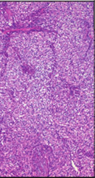
You can also read