Splenic Angiosarcoma with Bone Marrow Involvement Initially Diagnosed as Systemic Mastocytosis: A Case Report - Cureus
←
→
Page content transcription
If your browser does not render page correctly, please read the page content below
Open Access Case
Report DOI: 10.7759/cureus.5804
Splenic Angiosarcoma with Bone Marrow
Involvement Initially Diagnosed as
Systemic Mastocytosis: A Case Report
Paul Plantinga 1 , Sadaf Rahman 2 , Kamilia Rizkalla 3 , Jessica G. Shepherd 4 , Chai W. Phua 5
1. Pathology and Laboratory Medicine, Schulich School of Medicine & Dentistry, Western University,
London, CAN 2. Medicine, Schulich School of Medicine & Dentistry, Western University, London, CAN 3.
Pathology and Laboratory Medicine, London Health Sciences Centre, London, CAN 4. Pathology and
Laboratory Medicine, London Health Science Centre, London, CAN 5. Hematology, London Health
Sciences Centre, London, CAN
Corresponding author: Paul Plantinga, paulplantinga@gmail.com
Disclosures can be found in Additional Information at the end of the article
Abstract
We describe the case of a 67-year-old female patient presenting with constitutional symptoms
and rapid decline. Two bone marrow core biopsies were performed, with spindled cells
identified and thought to represent marrow involvement by systemic mastocytosis on the first
biopsy. A diagnosis of metastatic vascular malignancy with sarcomatoid features was favored
on the second core biopsy. The patient rapidly deteriorated and passed away. The post-mortem
examination revealed a splenic angiosarcoma with metastasis to the liver and bone marrow.
Splenic angiosarcoma is a rare, aggressive entity, with bone marrow metastasis even more
uncommon. This report perceives this as a diagnostic consideration on bone marrow biopsies
with spindled cells and explores the diagnostic dilemma and overlapping features of systemic
mastocytosis and angiosarcoma.
Categories: Internal Medicine, Oncology, Pathology
Keywords: splenic angiosarcoma, bone marrow, autopsy
Introduction
Primary splenic angiosarcoma is a rare, aggressive vascular malignancy with a dismal prognosis.
It is composed of malignant endothelial cells with variable morphology, and can often be
spindled [1-4]. The use of immunohistochemistry is required to make the diagnosis; however,
unexpected immunohistochemical staining patterns can lead to diagnostic difficulty. We
describe the case of metastatic primary splenic angiosarcoma masked by preliminary diagnoses
Received 03/08/2019 of idiopathic myelofibrosis and systemic mastocytosis. This report explores the diagnostic
Review began 03/25/2019 dilemma and overlapping features of systemic mastocytosis and angiosarcoma.
Review ended 09/27/2019
Published 09/30/2019
© Copyright 2019 Case Presentation
Plantinga et al. This is an open
A previously healthy 67-year-old female presented to a community hospital with a four-month
access article distributed under the
history of rapidly progressive decline with early satiety, weight loss, and night sweats. There
terms of the Creative Commons
Attribution License CC-BY 3.0., which was no significant family medical history. Physical examination revealed palpable
permits unrestricted use, distribution, hepatosplenomegaly, bilateral pitting edema of the lower limbs, scleral jaundice, and pallor.
and reproduction in any medium, Additional workup was significant for hepatosplenomegaly (liver was 25 cm and spleen 18 cm
provided the original author and
on CT), and innumerable hypodense lesions of liver, spleen, and bones. Peripheral blood
source are credited.
demonstrated leukoerythroblastic features and mild thrombocytopenia. She underwent an
How to cite this article
Plantinga P, Rahman S, Rizkalla K, et al. (September 30, 2019) Splenic Angiosarcoma with Bone Marrow
Involvement Initially Diagnosed as Systemic Mastocytosis: A Case Report. Cureus 11(9): e5804. DOI
10.7759/cureus.5804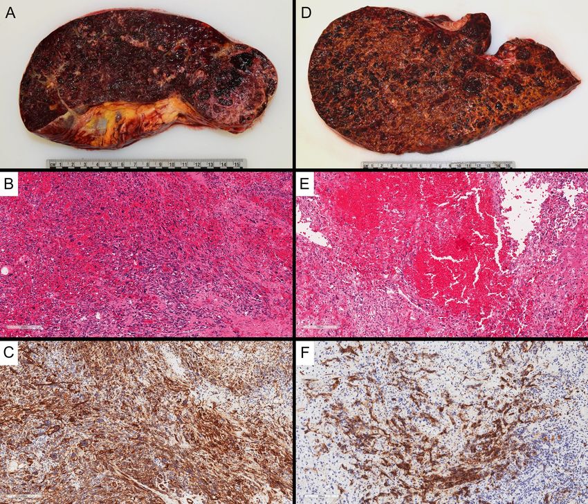
urgent bone marrow biopsy, which reported a preliminary diagnosis of idiopathic myelofibrosis.
                                   To confirm the diagnosis of myelofibrosis, the case was referred to a tertiary center for
                                   evaluation, including a review of the bone marrow aspirate and core biopsy, and testing for
                                   driver mutations associated with primary myelofibrosis. An urgent outpatient hematology-
                                   oncology referral was also made.

                                   Unfortunately, the patient deteriorated quickly with progressive liver dysfunction, anasarca,
                                   and transfusion-dependent bicytopenia. She was then transferred to a tertiary care center and
                                   admitted to the hematology-oncology ward for further management. Working diagnosis at that
                                   time included primary myelofibrosis with significant extramedullary hematopoiesis causing
                                   hepatosplenomegaly. Laboratory investigations at presentation in our center were pertinent for
                                   the following (normal reference ranges given in parentheses): total bilirubin: 72.7 μmol/L (3-22
                                   μmol/L); direct: bilirubin 21.4 μmol/L (0-5 μmol/L); hemoglobin: 69 g/L (115-160 g/L); total
                                   leukocytes: 9 x 10⁹/L (4-10 x 109/L); platelets: 21 x 109/L (150-400 x 109/L); nucleated red
                                   blood cells: 10-20/100 leukocytes (0/100 leukocytes); INR (international normalized ratio): 3.1
                                   (0.9-1.1); fibrinogen: 0.33 g/L (1.7-5.9 g/L); ALT (alanine aminotransferase): 104 U/L (9-52 U/L);
                                   AST (aspartate aminotranferase): 241 U/L (14-26 U/L); ALP (alkaline phosphatase): 171 U/L
                                   (38-126 U/L); LDH (lactate dehydrogenase): 750 U/L (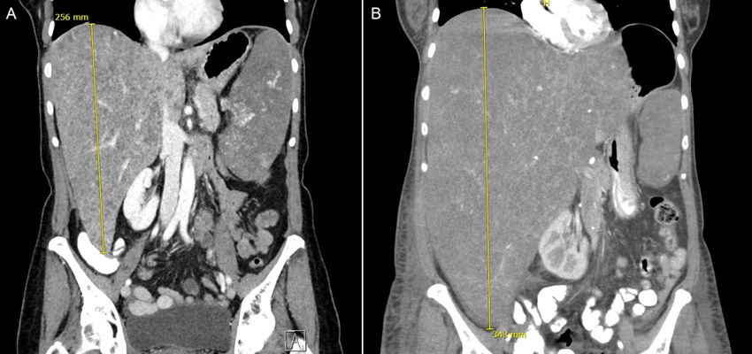
The patient clinically deteriorated further and, on the sixth day of admission, was transferred to
the intensive care unit for hypotension requiring vasopressors and multi-organ failure.
Unfortunately, she passed away within 24 hours of her transfer. Pathologic results from her
subsequent bone marrow biopsy and autopsy are reviewed below.
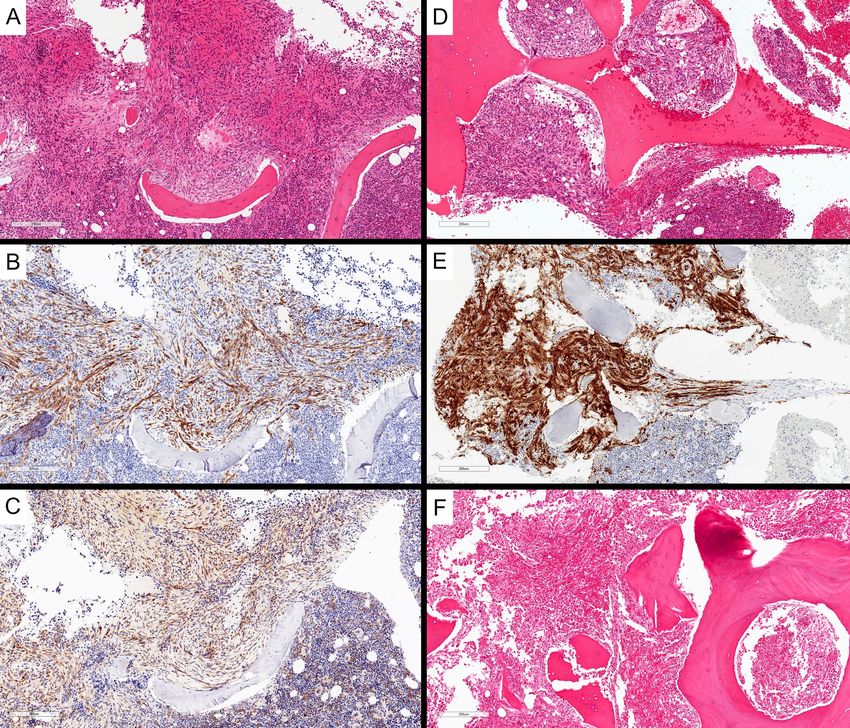
Intriguingly, the secondary review of her initial bone marrow biopsy revealed a hypercellular
marrow together with infiltration of spindled cells (Figure 2A) that were uniform and bland,
admixed with relatively normal bone marrow elements. Immunohistochemistry stains showed
positivity for CD34 and CD117 (c-KIT) (Figure 2B-C). Staining for CD2 and tryptase was not
available through our lab, and CD68R and FLI-1 staining was not completed. The cells were
negative for cytokeratin CK AE1/AE3. These spindled cells were interpreted as spindled mast
cells due to morphologic features and CD117 positivity. Given the findings of multifocal dense
infiltrates of presumed mast cells, of which greater than 25% were spindled, the diagnosis of
systemic mastocytosis by WHO 2016 criteria was suggested. The subsequent bone marrow core
biopsy (Figure 2D-E) was performed shortly before the patient's death and was reported after
her death. On this biopsy, there were occasional foci of infiltrative vascular neoplasm with
sarcomatoid features. There were hyperchromasia and pleomorphism in the spindled cells, and
vascular spaces were identified. The neoplastic cells were positive for vascular markers CD34
and CD31, along with vimentin. Interestingly, in this biopsy, CD117 appeared negative.
Reticulin fibers were increased within the infiltrative metastatic foci.
FIGURE 2: Bone marrow involvement by angiosarcoma. Initial
bone marrow core biopsy showed spindled cells within the
marrow, interpreted as atypical spindled mast cells (A, H&E,
100x magnification), with patchy CD34 staining (B, 100x
2019 Plantinga et al. Cureus 11(9): e5804. DOI 10.7759/cureus.5804 3 of 7magnification) and expressing weak CD117 (C, 100x
magnification). Subsequent bone marrow core biopsy again
showed atypical spindled cells (D, H&E, 100x magnification)
with stronger CD34 expression (E, 100x magnification).
Vertebral bone marrow from post-mortem examination
demonstrated involvement by angiosarcoma (F, H&E, 100x
magnification)
H&E: Hematoxylin and eosin
Macroscopic autopsy findings included massive hepatosplenomegaly (liver: 4,674 g, spleen:
1,364 g), with the parenchyma of both liver and spleen almost entirely replaced by diffuse,
infiltrative, hemorrhagic neoplasm (Figure 3A, D). No definite discrete mass was identified.
There was 650 mL of sanguineous fluid in the peritoneal cavity and diffuse hemorrhage in the
retroperitoneum. There were no significant abnormalities in the remaining abdominal and
pelvic organs. Microscopy revealed a poorly differentiated angiosarcoma. This was diffusely
involving the spleen (Figure 3B-C) and liver (Figure 3E-F), with patchy involvement of
vertebral bone marrow sections (Figure 2F). The lesion was composed of atypical, mitotically
active, spindled cells, forming vascular channels with abundant background hemorrhage. The
lesional cells were positive for vascular immunohistochemical markers CD34 and CD31 and
negative for cytokeratin CK AE1/AE3. The eventual diagnosis was favored to represent primary
splenic angiosarcoma with metastasis to the liver and bone marrow.
2019 Plantinga et al. Cureus 11(9): e5804. DOI 10.7759/cureus.5804 4 of 7FIGURE 3: Post-mortem examination of the spleen (A) and liver
(D) showed diffuse involvement by angiosarcoma, with
minimal residual normal parenchyma, and diffuse enlargement
of both organs. The diffuse involvement by malignant spindled
cells is demonstrated histologically in the spleen (B) and liver
(E) (H&E, 100x magnification), with diffuse positivity for CD34
by immunohistochemistry [C (spleen) and F (liver), 100x
magnification]
H&E: Hematoxylin and eosin
Discussion
Primary splenic angiosarcoma is a rare, highly aggressive vascular malignancy comprising
about 2.6% of angiosarcomas, with an incidence of approximately 0.14-0.25 cases per million
individuals [1,5]. It is often metastatic to the liver, lungs, lymph nodes, and bone and has a poor
prognosis [1-4]. Bone marrow metastasis from primary splenic angiosarcoma is very rare, with
only a few reports in the literature [5]. The lesion is composed of abnormal, malignant
endothelial cells, with variable histology between cases. The malignant endothelial cells can be
round, polygonal, spindled, or epithelioid, forming vascular channels in well-differentiated
areas, which are less defined in more aggressive diseases. Papillary projections into the vascular
lumen and multilayering of the malignant cells can occur. In more poorly differentiated areas,
there may be sheets of malignant cells, with hemorrhage and necrosis [1].
Immunohistochemistry is vital in making the diagnosis of angiosarcoma, typically with
endothelial marker expression, including CD34, CD31, factor VIII related antigen, vascular
endothelial growth factor (VEGF), and FLI-1 [6]. Occasionally, angiosarcomas can express
cytokeratins, leading to difficulty in distinguishing from a poorly differentiated carcinoma [1].
The expression of CD117 has also been demonstrated in angiosarcoma [7,8]. It is unclear as to
why CD117 expression differed between the two bone-marrow core biopsy samples. It could
have been due to a sampling issue, localized expression in parts of the tumour, or related to the
staining procedure.
Angiosarcoma involving the bone marrow can be mistakenly interpreted as systemic
mastocytosis with bone marrow involvement. Mastocytosis is characterized by the
accumulation and aggregation of neoplastic mast cells within one or more organ systems. The
bone marrow is involved in nearly all cases [9]. Atypical morphology of the mast cells,
particularly spindling, is included in the criteria for systemic mastocytosis. Neoplastic mast
cells typically are immunoreactive for CD117, with an activating point mutation of KIT [9,10].
Systemic mastocytosis typically involves the bone marrow, whereas angiosarcoma involving the
bone marrow is rare. Though these diseases are not classically within the same differential
diagnosis, both can show spindled morphology, and both can express CD117 by
immunohistochemistry. However, CD117 positivity is more typically seen in systemic
mastocytosis and is less frequent in angiosarcoma. In addition, immunohistochemical staining
patterns for both CD34 and CD117 can be challenging to interpret in the context of bone
marrow biopsies, as blasts and immature precursors can show positivity for these markers.
CD34 expression is typically absent in neoplastic mast cells but can be displayed on their
2019 Plantinga et al. Cureus 11(9): e5804. DOI 10.7759/cureus.5804 5 of 7precursor neoplastic stem cells [9]. Notably, CD31, while typically positive in angiosarcoma, has
been found to be negative in systemic mastocytosis [10]. A summary of pathological findings of
systemic mastocytosis compared with splenic angiosarcoma is given below (Table 1).
Systemic mastocytosis Splenic angiosarcoma
Bone marrow, spleen, skin, liver, Spleen, with metastasis to liver, lungs, lymph
Sites involved
gastrointestinal tract nodes, and bone, rarely to bone marrow
Clusters of mast cells, which may show
Variable pleomorphic, malignant nuclei: may be
cytologic atypia, including spindling and poly-
Morphology rounded, polygonal, fusiform, or epithelioid;
lobed nuclei; may show associated
vascular spaces in well-differentiated cases
eosinophilia
Positive for vascular markers CD31, CD34, FLI-
Positive for CD117, CD2, CD25, tryptase;
Immunohistochemistry 1, VEGF; rarely CD117 positive; epithelioid
CD31 and CD34 usually negative
variants may be cytokeratin positive
TABLE 1: Summary and comparison of pathological findings in systemic
mastocytosis and splenic angiosarcoma
Conclusions
This case underscores the diagnostic dilemma that can arise in differentiating angiosarcoma
from systemic mastocytosis, both uncommon diseases, based on initial bone marrow biopsy.
Spindled lesions involving the bone marrow, with an unusual morphology and
immunohistochemical staining patterns, can present significant diagnostic challenges, and
rare entities should be considered and thoroughly investigated.
Additional Information
Disclosures
Human subjects: Consent was obtained by all participants in this study. Conflicts of interest:
In compliance with the ICMJE uniform disclosure form, all authors declare the following:
Payment/services info: All authors have declared that no financial support was received from
any organization for the submitted work. Financial relationships: All authors have declared
that they have no financial relationships at present or within the previous three years with any
organizations that might have an interest in the submitted work. Other relationships: All
authors have declared that there are no other relationships or activities that could appear to
have influenced the submitted work.
References
1. Young RJ, Brown NJ, Reed MW, Hughes D, Woll PJ: Angiosarcoma. Lancet Oncol. 2010,
11:983-91. 10.1016/S1470-2045(10)70023-1
2. Neuhauser TS, Derringer GA, Thompson LD, Fanburg-Smith JC, Miettinen M, Saaristo A,
Abbondanzo SL: Splenic angiosarcoma: a clinicopathologic and immunophenotypic study of
28 cases. Mod Pathol. 2000, 13:978-87. 10.1038/modpathol.3880178
3. Falk S, Krishnan J, Meis JM: Primary angiosarcoma of the spleen: a clinicopathologic study of
40 cases. Am J Surg Pathol. 1993, 17:959-70. 10.1097/00000478-199310000-00001
2019 Plantinga et al. Cureus 11(9): e5804. DOI 10.7759/cureus.5804 6 of 74. Sharma S, Singh P, Gupta P, Lal A, Srinivasan R: Primary splenic angiosarcoma with liver
metastasis: a rare neoplasm diagnosed on fine-needle aspiration cytology and cell block
immunocytochemistry. J Cytol. 2018, 35:114-6. 10.4103/JOC.JOC_148_16
5. Levy ACJ, DeFilipp M, Blakely M, Asiry S, Jormark S, Goodman A: Splenic angiosarcoma
diagnosed on bone marrow biopsy: case report and literature review. Radiol Case Rep. 2019,
14:390-95. Accessed: September 30, 2019: 10.1016/j.radcr.2018.12.008
6. Rossi S, Orvieto E, Furlanetto A, Laurino L, Ninfo V, Dei Tos AP: Utility of the
immunohistochemical detection of FLI-1 expression in round cell and vascular neoplasm
using a monoclonal antibody. Mod Pathol. 2004, 17:547-52. 10.1038/modpathol.3800065
7. Hornick JL, Fletcher CD: Immunohistochemical staining for KIT (CD117) in soft tissue
sarcomas is very limited in distribution. Am J Clin Pathol. 2002, 117:188-193. 10.1309/LX9U-
F7P0-UWDH-8Y6R
8. Miettinen M, Sarlomo-Rikala M, Lasota J: KIT expression in angiosarcomas and fetal
endothelial cells: lack of mutations of exon 11 and exon 17 of C-kit. Mod Pathol. 2000,
13:536-41. 10.1038/modpathol.3880093
9. Valent P, Akin C, Metcalfe DD: Mastocytosis: 2016 updated WHO classification and novel
emerging treatment concepts. Blood. 2017, 129:1420-27. 10.1182/blood-2016-09-731893
10. Jordan J, Walchshofer S, Jurecka W, et al.: Immunohistochemical properties of bone marrow
mast cells in systemic mastocytosis: evidence for expression of CD2, CD117/Kit, and bcl-x(L).
Hum Pathol. 2001, 32:545-52. 10.1053/hupa.2001.24319
2019 Plantinga et al. Cureus 11(9): e5804. DOI 10.7759/cureus.5804 7 of 7You can also read