FLOW CYTOMETRY - BONE MARROW - Bowdish Lab
←
→
Page content transcription
If your browser does not render page correctly, please read the page content below
FLOW CYTOMETRY – BONE MARROW
                                                                      Created by: Jessica Breznik & Dessi Loukov
                                                                                Revised Date: February 7, 2021
                                                                              Bowdish Lab, McMaster University
                                                                                            Hamilton, ON, Canada
                                                                                                 www.bowdish.ca
BACKGROUND
All circulating and circulation-derived tissue immune cells in the body are derived from progenitor cells in the bone
marrow, which arise from hematopoietic stem cells. This protocol outlines steps for the processing of bone
marrow from mouse femurs, a basic surface stain for detection of monocytes and neutrophils, two approaches for
cell surface staining for detection of progenitor populations, and the procedure for intracellular staining for
detection of Ki67, a marker of cellular proliferation.

The ‘monocyte’ surface stain includes antibodies for detection of surface markers CD45 (leukocyte common
antigen), CD11b (monocytes/neutrophils), and Ly6C (monocytes/neutrophils), as well as antibodies conjugated to
the same fluorophore for CD19 (B cells), CD3 (T cells), NK1.1 (natural killer cells), and Ly6G (neutrophils), to
facilitate their removal from leukocyte populations for the assessment of monocyte populations. Neutrophils may
be distinguished from monocytes by their expression of Ly6G. The panel also includes markers of monocyte
maturity and migration: CCR2 (chemokine receptor), CX3CR1 (fractalkine receptor), and F4/80
(macrophages/monocyte maturity marker). Ly6Clow/- monocyte retention in the bone marrow is dependent on
expression of CX3CR1. Ly6Chigh monocytes exit the bone marrow in a CCR2-dependent manner.

In the ‘basic progenitor’ surface stain, cells expressing lineage markers CD3, B220/CD45R, Ter119, Ly6G, and Gr-1
(Ly6C/Ly6C) are excluded. HSPCs (hematopoietic stem and progenitor cells) are identified by their expression of
Sca-1 (Stem Cells Antigen-1; Ly6A) and cKit (CD117; promotes HSC proliferation with cytokines GM-CSF, IL-3, and
IL-7, and cell survival). CLPs (common lymphocyte progenitors) may be distinguished from other progenitor
populations by their expression of CD127 (IL-7Rα). Lin-Sca-1-cKit+ progenitors can be further delineated by their
expression of CD16/32 (Fcγ receptors) and CD34 (marker of hematopoietic stem and progenitor cells): MEP
(megakarocytic-erythroid progenitor; CD16/32-CD34-), CMP (common myeloid progenitor; CD16/32-CD34+), and
GMP (granulocyte-monocyte progenitor; CD16/32+CD34+) cells.

The ‘monocyte-myeloid progenitor’ surface stain follows the pathway of myeloid development, which is regulated
by epigenetic and transcriptional modifications. Cells expressing lineage markers CD3, B220/CD45R, Ter119, and
Ly6G, as well as CD127 (IL-7Rα), are excluded. The surface markers CD115, cKit, and Flt-3 (all members of the same
family of receptor tyrosine kinases) assist in identifying distinct pogenitor populations, and Ly6C and CD11b assist
in identifying monocytes. cKit is expresed at higher levels on myeloid progenitors than lymphoid progenitors and
its expression is lost upon myeloid progenitor differentiation into monocytes. Flt-3 (also known as CD135/Flk-2) is
expressed on immature hematopoietic progenitors and CD34 +HSCs. Flt-3, via activation with Flt-3 ligand
(expressed on the surface of mononuclear cells), stimulates the proliferation and differentiation of HSCs, and this
synergizes with cytokines to promote proliferation and survival. CD115 (also known as CSF-1R, M-CSFR, and c-fms),
is expressed by myeloid and monocyte prognitors, monocytes, and macrophages. It binds CSF-1 (M-CSF) and IL-34
and promotes survival, proliferation, and differentiation of macrophages. This staining panel identifies the
common myeloid progenitor (CMP; CD127-Lin-cKit+Sca-1+CD34+CD16/32low), the macrophage and dendritic cell
progenitor (MDP; CD127-Lin-cKit+Flt-3+CD115+Ly6C-CD11b-), the common monocyte progenitor (cMoP; CD127-Lin-
cKit+Flt3+CD115+Ly6C+CD11b-), and monocytes (CD127-Lin-cKit-Flt-3-CD115+Ly6C+CD11b+).
                                                                                                                   1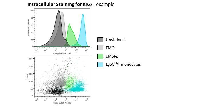
NOTES
    -   The number of leukocytes within bone marrow will differ according to the sex, age, housing conditions
        (i.e. germ-free or SPF), and weight of the mouse (especially with diet manipulation), so it is important to
        decide whether you will work with samples by taking a specific volume of resuspended bone marrow
        irrespective of sample source (i.e. per femur), or by counting and using a specific number of cells.

    -   Use FMO (flow-minus-one) and unstained controls prepared from pooled bone marrow from multiple
        samples, as well as an extra stained sample, if possible, to guide compensation and gating analysis.

EQUIPMENT
Bone Marrow Processing:
   • Dissection tools – tweezers, scissors
   • Kimwipes or gauze
   • 50 mL Falcon tube filled with 70% EtOH (for cleaning supplies)
   • 50 mL Falcon tube filled with sterile water (for cleaning supplies)
   • 50 mL Falcon tube filled with PBS (for short-term storage of samples)
   • 50 mL Falcon tube (empty)
   • 1x sterile PBS (prechilled at 4°C and maintained on ice)
   • 70% EtOH
   • Petri dishes (size is arbitrary)
   • 10 mL syringes
   • 18G and 26G needles

Bone Marrow Freezing (optional):
   • Fetal calf serum (FCS)
   • DMSO
   • 2 mL cryovials
   • Mr. Frosty or similar freezing container (prechilled to 4°C; isopropanol must be at indicated line and
       replaced after 5 ‘freeze/thaws’)
   • P200 and P1000 sterile tips and pipettors

Flow Cytometry – Staining
    • FACs Wash Buffer
           o 500 mL PBS
           o 2.5 g BSA (kept in the fridge)
           o 5 mL 0.5 M EDTA
           o dissolve and leave overnight in the fridge
           o filter through a 0.2 µM filter using vacuum pump
           o store at 4 °C
    • 1-step Fix/Lyse Solution (10X) (eBioscience, sold by ThermoFisher Scientific #00-5333)
           o prepare 1x solution with sterile H2O
           o store at 4 °C
    • Permeabilization Buffer (10x) (eBioscience, sold by ThermoFisher Scientific #88-8824-00)
           o Prepare fresh 1x solution with sterile H2O
    • 1x PBS (1.8 mM KH2PO4, 2.7 mM KCl, 10 mM Na2HPO4, 137 mM NaCl)
    • Fluorophore-conjugated antibodies (see protocol)
    • v-bottom 2 mL tubes (or v-bottom plates)

                                                                                                                 2
Flow Cytometry – Pre-analysis preparation
• Falcon Round-Bottom Polystyrene Test Tubes with Cell Strainer Snap Cap, 5mL (Falcon, sold by Fisher
Scientific #08-771-23)
• Falcon Round-Bottom Polystyrene Test Tubes, 5mL (Falcon, sold by Fisher Scientific # 14-959-6)
• CountBright Absolute counting beads (Invitrogen, sold by ThermoFisher Scientifc #C36950)
• OneComp eBeads Compensation Beads (Invitrogen, sold by ThermoFisher Scientifc #01-1111-41)
• Fluorophore-conjugated antibodies (see Tables 1-3); eBioscience antibodies are ordered through
ThermoFisher Scientific
PROTOCOL
FEMUR BONE MARROW COLLECTION
NB. Consider whether you should use femur or spine bone marrow. The protocols below are for femur bone
marrow extraction. A protocol for spine processing is available on the Bowdish website. Also consider whether
the bone marrow should remain sterile (i.e. if planning to freeze or culture). If processing bone marrow solely
for flow cytometry, it may not be necessary to work in a Biosafety Cabinet according to the mouse housing
conditions and experiment requirements.
Removal of leg bones
• Clean tools by immersing in 70% ethanol in 50 mL conical vial, and then wash tools by dipping in PBS in 50
mL conical vial before/after each sample.
• Euthanize mouse according to standard protocols and experimental procedures.
• Spray down fur on lower back/legs with 70% ethanol.
o Depending on available time, or if collecting muscles, you may choose to pin the body to a
dissection board.
• Using tweezers, lift skin from the middle of the back and cut with scissors to open skin parallel to the
spine. Peel skin from back/hip area by gently pulling to either side of cut. Remove skin from legs by
grasping top of femur at hip along spine and pulling skin away from the leg and over the foot. Use scissors
to cut additional skin if necessary. Be careful to not break/cut the leg bones (and spine if collecting).
o If working on a dissection board, remove fur and skin from legs by lifting skin at the base of each
leg with tweezers and cutting away skin across the thigh and down to the foot.
• Expose the hip joint of each leg and cut above the joint, making sure to not remove the top of the femur.
Be very careful not to cut bone as this will compromise the sterility of the bone marrow. Leave the foot
attached. If sterility is important, do not remove muscle until working in a Biosafety Cabinet. To remove
muscle, working parallel to the femur, carefully cut muscle layers away from the bone. Clean bones of
remaining muscle tissue using Kimwipes or gauze by gently rubbing the bone area.
o Alternatively, cut off legs first and then remove skin (and muscle if not concerned with sterility).
• Place leg(s) in a 50 mL tube containing PBS on ice (i.e. one tube per mouse).
• Discard remaining tissues (i.e. place in body bag and freeze to later discard according to facility
procedures). Clean and sterilize tools with 70% ethanol; if pins were used, discard into sharps container.
Bone marrow processing *work in the Biosafety Cabinet and use appropriate procedures if sterility is required*
• Prepare one 10 mL syringe of cold PBS with a 26G needle and 50 mL falcon tube for each leg.
• Pour out the PBS containing the leg(s) into a petri dish. If necessary, remove muscle as described above.
3o If desired to assure sterility, transfer the intact bones with sterile tweezers to a second petri dish
containing 70% EtOH for one minute and then to the third petri dish containing sterile PBS (this
step should be unnecessary if sterility has been maintained).
• Remove a leg from the PBS with tweezers and cut the ends as follows:
o Place the scissors just below the joint and adjust the tweezers so they are just above the scissors
to prevent the bone from shattering. Cut off the hip joint. Readjusting the scissors and tweezers,
cut off the knee joint. DO NOT let the ends of the bones touch anything as this will compromise
the sterility of the cells within.
o Ensure that tools are not coated in ethanol when cutting into bone. This can be avoided by
dipping tools into sterile water after cleaning in 70% ethanol.
• Flush out the bone marrow into an empty 50 mL Falcon tube by inserting the 26G needle attached to the
10 mL syringe filled with cold PBS at the knee side. Pass the PBS through the bone into the Falcon tube
until the colour of the bone turns from red to white, indicating that all marrow has been expelled. Discard
the bone into an empty petri dish. You should be able to see red bone marrow pieces within the PBS in
the 50 mL tube. Place the tube on ice.
o Perform this step for each bone IMMEDIATELY following the cutting of the bone.
o DO NOT put the bone down between cutting and flushing.
• Repeat steps above for each femur. Samples can be pooled into one Falcon tube if appropriate.
o Clean tools between mice and use separate ‘skin/muscle’ and ‘bone’ tool sets of scissors and
tweezers to reduce risk of contamination of bone marrow.
• For each Falcon tube of bone marrow, using a 10 mL syringe attached to an 18G needle, aspirate all liquid
and expel to break up clumps of bone marrow. Repeat once.
o Do not do this more than twice to avoid shearing cells.
• Centrifuge 50 mL Falcon tubes at 1500 rpm for 5 minutes at 4°C.
• Working one tube at a time, discard the PBS supernatant. Samples can be allocated for one purpose or
divided for freezing, flow cytometry, and/or cell culture according to experimental requirements.
o For freezing, resuspend pellet in 2 mL FCS and aliquot 1 mL each into 2 mL cryotubes. Add 0.1 mL
DMSO dropwise. Freeze in pre-chilled (in fridge) Mr. Frosty overnight at -80°C and then transfer
to liquid nitrogen for long term storage.
o For culturing, see bone marrow-derived macrophage protocol available on the Bowdish website.
o For flow cytometry, resuspend cells in up to 500 μL PBS and determine cell count by trypan blue
staining using a hemocytometer. Aliquot cells into a 2 mL v-shaped tube, and add FACs wash to
~500 μL. Keep samples on ice/in fridge.
▪ Plan to stain ~0.5 x 10⁶ cells for the monocyte surface stain, and at least ~1 x 10⁶ cells for
the progenitor stains, especially if you will also perform intracellular staining.
o Once all samples are prepared, centrifuge for at 4°C for 5 minutes at 2000 rpm.
BONE MARROW SURFACE STAINING FOR FLOW CYTOMETRY
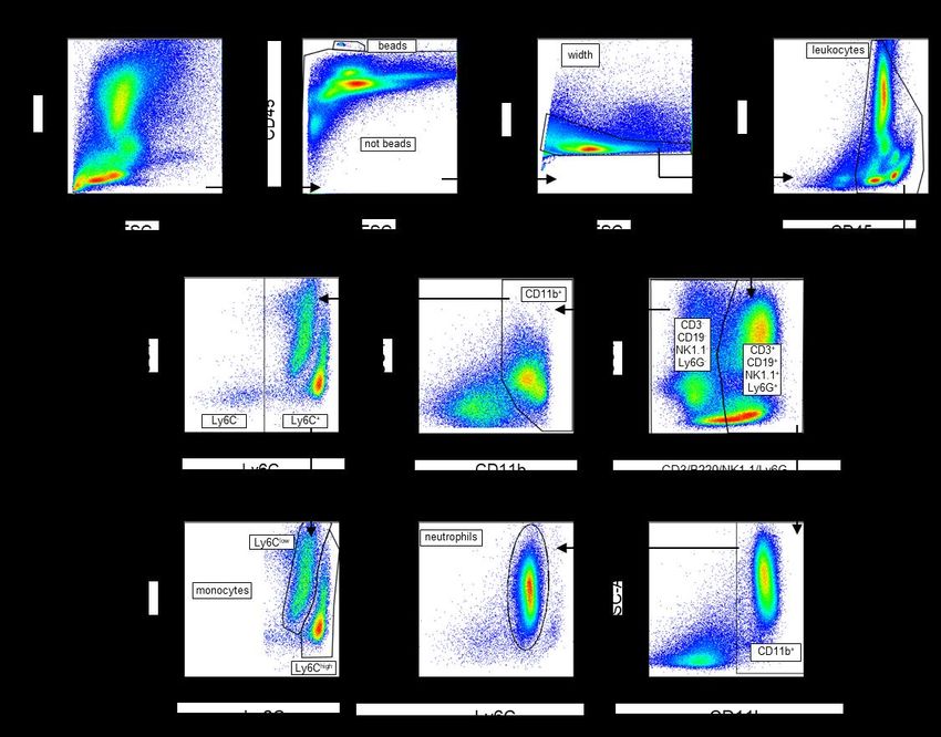
• As outlined in the introduction, the ‘monocyte’ stain (Table 1) is for identification of monocytes, the ‘basic
progenitor’ stain (Table 2) is for identification of HSPC, CLP, GMP, MEP, and CMP populations, while the
‘monocyte progenitor’ stain (Table 3) is for identification of those populations as well as a further
assessment of monocyte-myeloid progenitors.
• Antibodies should be titrated according to specific experimental requirements and cytometer use.
• For the ‘monocyte’ stain (Table 1), you should consider whether blocking with CD16/32 Fc block
(cat#101302, BioLegend) for 10 minutes, with a subsequent wash, and then staining samples, will reduce
any non-specific binding of antibodies. DO NOT block cells in the progenitor stains as CD16/32 is included
within the staining panels.
4Protocol:
• Resuspend cells in 100 μL FACs wash.
• Add 50 μL of bone marrow surface stain made in FACs wash as outlined in the tables on the next page.
• Mix by gently pipetting. Incubate for 30 minutes in the dark at 4°C.
• Add 1x Fix-Lyse up to 2 mL and incubator for 10 minutes at room temperature in the dark.
• Centrifuge at 4°C for 5 minutes at 2000 rpm. Remove supernatant.
• Resuspend cells in ~2 mL 1x PBS.
• Centrifuge at 4°C for 5 minutes at 2000 rpm. Remove supernatant.
o Intracellular staining protocol can start here.
• Resuspend cells in 200 μL FACs wash and store at 4°C in the dark until ready to run samples.
Table 1. Fluorophore-conjugated antibody mix for ‘monocyte’ surface stain
Conjugated Catalogue No.
Marker Company 1x Volume (μL)
Fluorophore and Clone
Ly6C AF 488 BioLegend 128022 (HK1.4) 0.125
CCR2 PE R&D Systems FAB5538P (475301) 0.4
CD11b PE-Cy7 eBioscience 25-0112 (M1/70) 0.125
CD45 eFluor 450 eBioscience 48‐0451 (30-F11) 0.34
CX3CR1 BV650 BioLegend 194033 (SA011F11) 0.1
F4/80 APC eBioscience 17-4801 (BM8) 0.1
CD3 AF 700 eBioscience 56-0032 (17A2) 0.2
CD19 AF 700 eBioscience 56‐0193 (eBio1D3) 0.2
NK1.1 AF 700 eBioscience 56-5941 (PK136) 0.1
Ly6G AF 700 BioLegend 127621 (1A8) 0.1
FACS Wash 48.21
Total volume 50
Table 2. Fluorophore-conjugated antibody mix for ‘basic progenitor’ surface stain
Conjugated Catalogue No.
Marker Company 1x Volume (μL)
Fluorophore and Clone
Sca-1 (Ly6A) PE-Dazzle 594 BioLegend 108137 (D7) 0.5
CD117 (cKit) BV421 BioLegend 105827 (2B8) 0.5
CD127 (IL7Rα) PerCP-Cy5.5 BioLegend 121114 (A7R34) 0.5
CD34 APC BioLegend 128606 (HM34) 0.5
CD16/32 BV711 BioLegend 101337 (93) 0.125
Gr-1 PE-Cy7 eBioscience 25-5931-83 (RB6-8C5) 0.25
CD3 PE-Cy7 eBioscience 25-0031-82 (145-2C11) 0.125
Ter119 PE-Cy7 eBioscience 25-5921-81 (TER-119) 0.125
B220/CD45R PE-Cy7 eBioscience 25-0452-81 (RA3-6B2) 0.125
CD11b PE-Cy7 eBioscience 25-0112-81 (M1/70) 0.25
FACS Wash 47.25
Total volume 50
5Table 3. Fluorophore-conjugated antibody mix for ‘monocyte-myeloid progenitor’ surface stain
Conjugated Catalogue No. and
Marker Company* 1x Volume (μL)
Fluorophore Clone
CD115 (M-CSF R) AF488 eBioscience 53-1152-80 (AFS98) 0.5
CD135 (Flt-3/Flk-2) PE BioLegend 135305 (A2F10) 0.5
Sca-1 (Ly6A) PE-Dazzle 594 BioLegend 108137 (D7) 0.5
CD127 (IL7Rα) PerCP-Cy5.5 BioLegend 121114 (A7R34) 0.5
Ter119 PE-Cy7 eBioscience 25-5921-81 (TER-119) 0.125
CD3 PE-Cy7 eBioscience 25-0031-82 (145-2C11) 0.125
B220/CD45R PE-Cy7 eBioscience 25-0452-81 (RA3-6B2) 0.125
CD117 (cKit) BV421 BioLegend 105827 (2B8) 0.5
Ly6C BV510 BioLegend 128033 (HK1.4) 0.125
CD16/32 BV711 BioLegend 101337 (93) 0.125
CD34 AF 647 BioLegend 128606 (HM34) 0.25
Ly6G AF 700 BioLegend 127622 (1A8) 0.125
CD11b APC-Cy7 BioLegend 101225 (M1/70) 0.125
FACS Wash 46.375
Total volume 50
NB. A live-dead stain is not used in these protocols as the viability of cells collected from fresh bone marrow is
generally high, but if there are concerns about viability, this can be determined (i.e. by Trypan blue staining and
hemocytometer) and the staining panels should be adjusted to incorporate a viability dye or live-dead marker
stain. In particular, if planning to use frozen bone marrow, a marker of viability should be incorporated, and it
should be carefully considered whether it is most appropriate to stain a specific number of ‘live’ cells (determined
by Trypan blue staining and hemocytometer) rather than a specific sample volume.
BONE MARROW INTRACELLULAR STAINING FOR KI67 STAINING (optional)
Intracellular staining can be performed without stimulation with permeabilization after surface staining.
If intracellular staining is performed, samples should be run on the cytometer on the same day.
• Starting point:
o If starting intracellular immediately after surface staining, remove the ~2 mL 1x PBS wash after
centrifugation.
o If samples were resuspended in 200 μL FACs wash and stored in fridge, add ~300 μL FACs wash
and centrifuge at 4°C for 5 minutes at 2000 rpm. Remove supernatant.
• Resuspend cells in 500 μL room temperature 1x Permeabilization Buffer.
• Incubate in the dark for 30 minutes at room temperature.
• Centrifuge at RT for 5 minutes at 2000 rpm. Remove supernatant.
• Prepare antibody mix in 1x Permeabilization Buffer (see table). Resuspend cell pellets in 50 μL of antibody
mix, and incubate in the dark for 30 minutes at room temperature.
• Add ~2 mL FACs wash and centrifuge for 5 minutes at 2000 rpm.
• Resuspend cells in 200 μL FACs wash and store at 4°C in the dark until ready to run samples.
Table 4. Fluorophore-conjugated antibody mix for intracellular Ki67 staining
Conjugated Catalogue No. and
Marker Company 1x Volume (μL)
Fluorophore Clone
Ki67 Brilliant Violet 605 BioLegend 652413 (16A8) 0.25
FACS Wash 47.75
Total volume 50
6PREPARATION OF SAMPLES FOR FLOW CYTOMETRY ANALYSIS
• After pipetting several times to resuspend cells, transfer total volume onto the mesh (40 µm) of 5 mL
polystyrene Falcon tubes (blue lids) (or filter through mesh into a U-bottom plate).
• Centrifuge tubes at 1500 rpm for 5 min at 4°C to remove cell clumps. Liquid should be at the bottom of
the tube, not on the mesh, after centrifugation.
• Protect from light by covering with aluminum foil.
• If using count beads for absolute quantification of cells in samples:
o Vortex count beads vial for 15 seconds and add 5 µL of beads per sample.
o Record concentration of count beads – this varies batch-to-batch.
PREPARATION OF COMPENSATIONS
• Prepare one 5 mL Falcon tube (without lid) for each fluorophore and the unstained control.
• Add 150-300 µL FACS Wash to each “fluorophore” tube.
• Add 1 mL FACS Wash to the “unstained” tube (100 µL x # of fluorophore tubes).
• Vortex OneCompBeads vial for 10 seconds.
• Add compensation beads into the “unstained” tube (1 drop for every 4-5 fluorophore tube).
• Vortex tube and distribute 100 µL from “unstained” tube to each “fluorophore” tube.
• Add 150-300 µL of FACS Wash (300 µL for flow beginners) to the “unstained” tube.
• Add 0.5 µL of appropriate antibody to each tube.
• Gently shake the rack and protect from light by covering with aluminum foil.
*See general flow cytometer protocol for further instructions*
ANALYSIS/SOFTWARE
- Machine: LSR Fortessa (or other suitable cytometer with appropriate lasers and configuration)
- Software: FlowJo (TreeStar)
- Gating: See attached diagram as a reference for surface staining. This varies depending on which markers
are included within the stain, as well as whether intracellular staining was included within the analysis.
LINKS AND REFERENCES
- Challen, G. A., Boles, N., Lin, K. K.-Y., & Goodell, M. A. (2009). Mouse hematopoietic stem cell identification and
analysis. Cytometry. Part A : The Journal of the International Society for Analytical Cytology, 75(1), 14–24.
http://doi.org/10.1002/cyto.a.20674
- Hettinger, J., Richards, D. M., Hansson, J., Barra, M. M., Joschko, A. C., Krijgsveld, J., & Feuerer, M. (2013). Origin
of monocytes and macrophages in a committed progenitor. Nature Immunology, 14(8), 821–830.
http://doi.org/10.1038/ni.2638
- Toxavidis, V., Tigges, J., & Alberich Jorda, M. Hematopoietic Bone Marrow Cell Sorting Without Compromise: 7
Lasers, Eight Colors, Six Way Sorting A Novel Approach. Beckman Coulter White Paper. Available from
https://www.beckman.com/gated-media?mediaId={38BF4CEA-379E-4B3A-8D98-F0932C9ABFA5}
- Loukov, D., Naidoo, A., Puchta, A., Marin, J.L.A.,Bowdish, D.M.E. (2016). Tumor necrosis factor drives increased
splenic monopoiesis in old mice. Journal of Leukocyte Biology, 100(1):121-9.
http://doi.org/10.1189/jlb.3MA0915-433RR
7Flow Cytometry Analysis - Bone Marrow ‘Monocyte’ Gating
8Flow Cytometry Analysis - Bone Marrow ‘Progenitor’ Gating
9Flow Cytometry Analysis - Bone Marrow Gating – Intracellular Staining
Intracellular staining of Ki67 should be assessed by measuring geometric mean fluorescence intensity.
10You can also read