Biomimetic Surfaces Coated with Covalently Immobilized Collagen Type I: An X-Ray Photoelectron Spectroscopy, Atomic Force Microscopy, Micro-CT and ...
←
→
Page content transcription
If your browser does not render page correctly, please read the page content below
International Journal of
           Molecular Sciences

Article
Biomimetic Surfaces Coated with Covalently
Immobilized Collagen Type I: An X-Ray
Photoelectron Spectroscopy, Atomic Force Microscopy,
Micro-CT and Histomorphometrical Study in Rabbits
Antonio Scarano 1,2, * , Felice Lorusso 1 , Tiziana Orsini 3 , Marco Morra 4 ,
Giorgio Iviglia 4,5 and Luca Valbonetti 6
 1    Department of Medical, Oral and Biotechnological Sciences, Center for Research on Aging and Translational
      Medicine (CeSI-MeT), University of Chieti-Pescara, 66100 Chieti, Italy; drlorussofelice@gmail.com
 2    Research staff at Zirconia Implant Research Group (Z.I.R.G), International Academy of Ceramic
      Implantology, USA
 3    CNR—National Research Council, Institute of Cell Biology and Neurobiology (IBCN), 00015 Roma, Italy;
      tiziana.orsini@cnr.it
 4    Nobil Bio Ricerche Srl, 14037 Portacomaro (AT), Italy; mmorra@nobilbio.it (M.M.); giviglia@nobilbio.it (G.I.)
 5    Department of Applied Science and Technology, Institute of Materials Physics and Engineering, Politecnico
      di Torino, 10121 Torino, Italy
 6    Faculty of Bioscience and Agro-Food and Environmental Technology, University of Teramo, 64100 Teramo,
      Italy; lvalbonetti@unite.it
 *    Correspondence: ascarano@unich.it; Tel.: +39-0871-3554099; Fax: +39-0871-3554173

 Received: 22 January 2019; Accepted: 6 February 2019; Published: 8 February 2019

 Abstract: Background: The process of osseointegration of dental implants is characterized by
 healing phenomena at the level of the interface between the surface and the bone. Implant surface
 modification has been introduced in order to increase the level of osseointegration. The purpose of
 this study is to evaluate the influence of biofunctional coatings for dental implants and the bone
 healing response in a rabbit model. The implant surface coated with collagen type I was analyzed
 through X-ray Photoelectron Spectroscopy (XPS), Atomic Force Microscopy (AFM), micro-CT and
 histologically. Methods: The sandblasted and double acid etched surface coated with collagen type I,
 and uncoated sandblasted and double acid etched surface were evaluated by X-ray Photoelectron
 Spectroscopy (XPS) and Atomic Force Microscopy (AFM) analysis in order evaluate the different
 morphology. In vivo, a total of 36 implants were positioned in rabbit articular femoral knee-joint,
 18 fixtures for each surface. Micro-CT scans, histological and histomorphometrical analysis were
 conducted at 15, 30 and 60 days. Results: A histological statistical differences were evident at 15,
 30 and 60 days (p < 0.001). Both implant surfaces showed a close interaction with newly formed
 bone. Mature bone appeared in close contact with the surface of the fixture. The AFM outcome
 showed a similar roughness for both surfaces. Conclusion: However, the final results showed that a
 coating of collagen type I on the implant surface represents a promising procedure able to improve
 osseointegration, especially in regions with a low bone quality.

 Keywords: collagen type I; implant surfaces; bone implant contact; hydrophilic surface; micro CT

1. Introduction
     Dental implants are successfully applied for partially or totally edentulous patients with missing
single or multiple teeth [1]. The osteointegration rate, bone quality and bone in contact with the dental
implant influence long-term success of oral implant rehabilitation [2,3]. One of the prerequisites for

Int. J. Mol. Sci. 2019, 20, 724; doi:10.3390/ijms20030724                              www.mdpi.com/journal/ijms
Int. J. Mol. Sci. 2019, 20, 724                                                                      2 of 16

success of an implant in oral rehabilitation is bone implant contact, bone density and quality around
titanium. Different implant surface treatments have been proposed by different authors to increase
bone implant contact and bone density. Micro- and macro-porous structures of titanium surfaces have
been evaluated to promote an enhanced peri-implant bone apposition during the early stages of bone
formation. Numerous animal studies have shown how the microstructure increases removal torque [4]
and macrostructure surface increases angiogenesis [5]. For roughening titanium implant surfaces
different methods are used and classified by subtraction and addition into mechanical, chemical and
physical methods. Subtractive techniques can be used such as sand-blasting with abrasives on TiO2 ,
AlO2 , hydroxyapatite (HA), Laser, electrochemical deposition, acid-etching or dual acid-etching, or
combinations of such treatments or organic biomaterials. The biomaterials used for implant surface
modification have been HA, CaP or micro/nano coating. All of these implant surface treatments
increased cell proliferation and growth, improved wettability and accelerated the osseointegration
process [6,7]. In vitro [8] and in vivo [9] studies have demonstrated that micro or macro implant
surfaces play a pivotal role, enhancing osteoblastic behavior and responses, such as increased cell
attachment, proliferation in bone implant contact and bone quality and density. New surfaces have
been proposed by different authors; for example a post-machining surface after treatment with
sandblasting and acid-etching or dual acid-etching was studied through a variety of techniques or the
incorporation of bioactive ceramic, HA and calcium phosphate (CaP), crystals polymer coatings [10]
or polyetheretherketone (PEEK) coating [11]. These coatings improved and increased the surface area
of the implants and increased dental implant stability and bone anchoring/biomechanics [12].
     Despite the excellent results obtained with the new implant surfaces in improving
osseointegration, the surface modification of Ti coatings is still an interesting viable. Recently, thermal
treatment has been proposed for improving the bioactivity of implant surfaces with increase the bone
implant contact [13].
     A new strategy to improve osseointegration is that of the immobilization on Ti of the main
components of the extracellular matrix, peptides or enzymes such as type I collagen for promoting the
adhesion of osteoblasts [14]. Collagen type I has been added onto implant surfaces when it is present
in the extracellular matrix of bone showing osteoconductivity, biocompatibility activity and it performs
the function of support by giving structural support to resident cells and ductility to the bone [15].
Collagen type I can be one of the prime candidate materials for realizing tissue-engineered grafts and
is one of the proteins that work critical roles in bone mineralization [16], bone healing [17], improving
blood compatibility and osteoblastic adhesion, differentiation, and extracellular-matrix secretion [18].
The present study was based on the hypothesis that collagen type I improves bone implant contact
(BIC) and osseointegration.
     The purpose of this study was to characterize two different surfaces by X-ray Photoelectron
Spectroscopy (XPS) and Atomic Force Microscopy (AFM) and compare, in a rabbit model, the BIC,
bone area inner threads (BAIT) and bone area outer threads (BAOT) by histologic analysis and
micro CT between collagen coated and uncoated implant surfaces using a split implant design.
The null hypothesis stated that there are no statistically significant differences in BIC, BAIT, BAOT and
osseointegration cell responses between collagen coated and uncoated implants.

2. Results

2.1. Surface Analysis by X-Ray Photoelectron Spectroscopy (XPS)

2.1.1. Surface (Uncoated Implant)
      The control group of samples were only sandblasted and acid etched. The XPS-detected surface
composition of uncoated and collagen coated samples is shown in Table 1. Briefly, the surface
of titanium is covered by a thin (about 4 nm thick) oxide layer, so that the maximum theoretical
concentration of Ti on pure titanium is 33%, the rest being oxygen (the most stable oxide is TiO2).
Surface contamination by adsorption of ubiquitous hydrocarbons from the atmosphere introduces a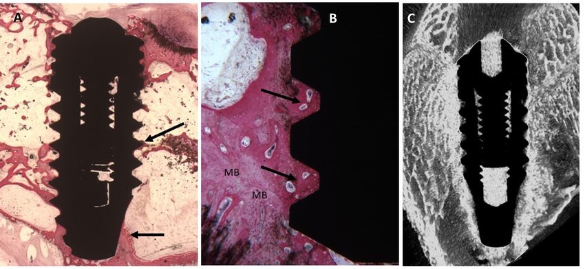
2.1.1. Surface (Uncoated Implant)
The control group of samples were only sandblasted and acid etched. The XPS-detected surface
composition of uncoated and collagen coated samples is shown in Table 1. Briefly, the surface of
Int. J. Mol. Sci. 2019, 20, 724 3 of 16
titanium is covered by a thin (about 4 nm thick) oxide layer, so that the maximum theoretical
concentration of Ti on pure titanium is 33%, the rest being oxygen (the most stable oxide is TiO2).
Surfaceoverlayer
surface contamination by adsorption
of carbon, of ubiquitous
readily captured hydrocarbonstechniques
by surface-sensitive from the atmosphere
such as XPS,introduces
decreasinga
surface
the overlayer of
concentration of Ti
carbon,
belowreadily capturedvalue
the theoretical by surface-sensitive
(Figure 1). techniques such as XPS, decreasing
the concentration of Ti below the theoretical value (Figure 1).
Table 1. Surface composition, as detected by X-ray Photoelectron Spectroscopy (XPS), of Ti and ColTi
samples
Table 1. (data arecomposition,
Surface expressed asas
% detected
at.) and suggests
by X-ray that the thickness
Photoelectron of the coating
Spectroscopy is very
(XPS), of Tithin.
and ColTi
samples (data are expressed as % at.) and suggests that the thickness of the coating is very thin.
Sample O C N Ti
Ti 49.2 Sample O
31.6 C N0.4 Ti 18.8
Ti 49.2 31.6 0.4 18.8
ColTi 23.8 ColTi 59.3 59.3 14.3
23.8 14.3 2.7 2.7
Figure1.1.High
Figure resolution
High C1s C1s
resolution peakpeak
obtained by XPSby
obtained of ColTi
XPS sample.
of ColTiThe figure shows
sample. the experimental
The figure shows the
curve
experimental curve (red) and the sum curve (brown) resulting from fitting with 1 (C–C, 2C–H)
(red) and the sum curve (brown) resulting from fitting with 1 (C–C, C–H) (green), (C–N, C–O)
(green),
(violet) and 3 (C=O, N–C=O functionalities) (azure).
2 (C–N, C–O) (violet) and 3 (C=O, N–C=O functionalities) (azure).
2.1.2. Surface (Coated Collagen Type I Implant)
2.1.2. Surface (Coated Collagen Type I Implant)
Observing the collagen coated sample, a completely different surface stoichiometry is detected.
Observing
The signal from the collagen
titanium coated
almost sample, awhile
disappears, completely
O/Ti anddifferent
C/Tisurface stoichiometry
ratio increase is detected.
and nitrogen rises
The signal from titanium almost disappears, while O/Ti and C/Ti ratio
up to over 14%. This is a clear indication that the coating process introduces on the surface increase and nitrogen rises an
up
to over 14%. This is a clear indication that the coating process introduces
organic molecule featuring multiple carbon-oxygen and carbon-nitrogen functionalities. Further on the surface an organic
molecule
clues featuringbymultiple
are supplied carbon-oxygen
the high-resolution and carbon-nitrogen
C1 peak of the collagen coatedfunctionalities. Further
sample shown clues are
in Figure 1,
supplied by the high-resolution C1 peak of the collagen coated
the experimental curve (red) is highly asymmetric and made up of different components. sample shown in Figure 1, the
experimental curve
Figure 1 also (red) the
shows is highly
resultsasymmetric and made up according
of peak deconvolution, of differenttocomponents.
literature procedures and
generalFigure
XPS 1approach
also shows the results
to extract of peakon
information deconvolution,
the carbon chemicalaccording to literature
environment. Theprocedures
peak is nicelyand
general XPS approach to extract information on the carbon chemical environment.
fitted by three components located at 285.00 eV (component 1, C–C, C–H), 286.3o eV (component 2, The peak is nicely
fittedC–O),
C–N, by three
andcomponents located at 285.00
288.10 eV (component 3, C=O,eV (component
N-C=O 1, C–C, C–H),
functionalities). So, the286.3o eV (component
combination of surface2,
C–N, C–O), and 288.10 eV (component 3, C=O, N-C=O functionalities). So, the
stoichiometry (Table 1) and curve fitting data indicates that the surface of the coated implant contains combination of surface
stoichiometry
an organic layer (Table
that,1) and curve
beside C–O fitting
and C–N data indicates
bonds, thatinthe
is rich surface of functionalities
amide-type the coated implant contains
(component
3anatorganic layerand
288.10 eV) that, beside C–O
confirms that and C–N molecules
collagen bonds, is rich coatinthe
amide-type
titanium functionalities (component
surface chemistry. (Table 1)3
at 288.10 eV) and confirms that collagen molecules coat the titanium surface
Incidentally, the small signal from titanium detected in the survey spectrum (Table 1), suggests that chemistry. (Table 1)
Incidentally, the small signal from titanium detected in the survey
the thickness of the coating is lower than the XPS-sampling depth (8 nm) (Figure 1). spectrum (Table 1), suggests that
the thickness of the coating is lower than the XPS-sampling depth (8 nm) (Figure 1).
2.2. Atomic Force Microscopy (AFM)
In both cases the topography of the disks shows the typical microroughness imparted by double
acid etching (the field of view is too small to also detect the longer-range roughness due to sandblasting).
No evidence of the organic layer detected by XPS is observed (Figure 2a). That is, even if the vertical
resolution of AFM is sub-nanometer, the microrough surface topography does not allow image
adsorbed collagen macromolecules (Figure 2b), which are instead plainly detected on flat surfaces [19].
For 10 × 10 micrometer areas, the Sdr roughness parameter (that indicates the percentage increase
of actual surface area with respect to the geometrical one) is 88 ± 7 for control sample and 85 ± 3
for the collagen coated one, without significant difference. On one hand, this result confirms that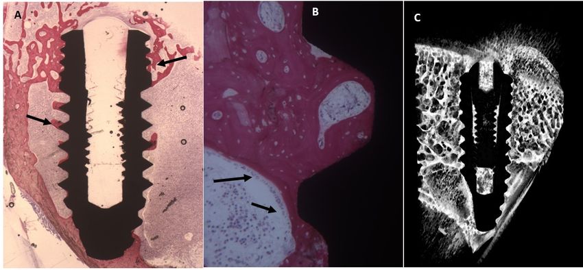
acid etching (the field of view is too small to also detect the longer-range roughness due to
sandblasting). No evidence of the organic layer detected by XPS is observed (Figure 2a). That is, even
if the vertical resolution of AFM is sub-nanometer, the microrough surface topography does not allow
image adsorbed collagen macromolecules (Figure 2b), which are instead plainly detected on flat
surfaces      [19].
 Int. J. Mol. Sci.     For
                   2019,     10 × 10 micrometer areas, the Sdr roughness parameter (that indicates4 of
                         20, 724                                                                                     the
                                                                                                                       16
percentage increase of actual surface area with respect to the geometrical one) is 88 ± 7 for control
sample and 85 ± 3 for the collagen coated one, without significant difference. On one hand, this result
 the process
confirms       thatdoes
                      the not   involve
                           process    does thenot
                                               deposition    of adeposition
                                                   involve the    thick coating,
                                                                             of abut   of acoating,
                                                                                   thick    conformal,   nanometer-thin
                                                                                                    but of  a conformal,
 surface    layer.    On   the   other  hand,   and  from   the point  of view of the   present
nanometer-thin surface layer. On the other hand, and from the point of view of the present       study, this observation
                                                                                                                  study,
 suggests     that   whatever      difference   is detected  in implantation  tests  it is not dictated
this observation suggests that whatever difference is detected in implantation tests it is not dictated by differences in
 surface    topography,       rather,  it genuinely   reflects the contribution  of the  different surface
by differences in surface topography, rather, it genuinely reflects the contribution of the different      chemistries to
peri-implant        new    bone   formation.
surface chemistries to peri-implant new bone formation.

                            (a)                                                         (b)
     Figure
     Figure 2.  10××1010micrometer
             2.10        micrometerimages
                                     imagesobtained
                                             obtainedby
                                                      bynon-contact
                                                         non-contactAFM
                                                                     AFManalysis
                                                                          analysisof
                                                                                   ofcontrol
                                                                                      control(a)
                                                                                              (a)and
                                                                                                  andcollagen
                                                                                                      collagen
     coated
     coated (b)
            (b) Ti
                 Ti disks subjected to the same treatment as the tested implants.

2.3. Micro-CT
2.3. Micro-CT Evaluation
              Evaluation
     Micrographs were
     Micrographs     were evaluated
                           evaluated for
                                       for bone
                                           bone BIC,
                                                 BIC, BAIT,
                                                      BAIT, BAOT
                                                             BAOT and
                                                                    and gaps
                                                                         gaps between
                                                                              betweenbone boneandandimplant.
                                                                                                      implant.
Radiographs   showed     new bone  in both  implants in intimate contact with the  implant
Radiographs showed new bone in both implants in intimate contact with the implant surface, nosurface,  no gaps
                                                                                                          gaps
were detected
were  detected at
                at 15
                   15 days.
                      days. At
                            At 30
                                30 days
                                   days the
                                         the BIC
                                             BIC and
                                                 andBAIT
                                                      BAITwere
                                                            were more
                                                                  more present
                                                                       present in
                                                                                in the
                                                                                    the coated
                                                                                        coated implant
                                                                                                implant with
                                                                                                          with
collagen I. No signs  of bone resorption  inflammation   and/or osteolysis were observed    in
collagen I. No signs of bone resorption inflammation and/or osteolysis were observed in either either surfaces.
surfaces.
2.4. Histological Evaluation
      Under light
2.4. Histological    microscopy, all 36 implants were surrounded by bone and showed good
                  Evaluation
osseointegration. The lower threads of the implants appeared to be in close contact with newly
     Under light microscopy, all 36 implants were surrounded by bone and showed good
formed bone or with marrow spaces, while along the upper threads there was contact with the cortical
osseointegration. The lower threads of the implants appeared to be in close contact with newly
bone. No evidence of soft tissue was present between bone and implant surfaces in all the specimens
formed bone or with marrow spaces, while along the upper threads there was contact with the
of either experimental groups.
cortical bone. No evidence of soft tissue was present between bone and implant surfaces in all the
specimens   of either
2.4.1. Fifteen Days experimental groups.

Uncoated Implant Surface
     The slide observed at low magnification showed a few trabeculae bone near or in contact with
the implant surface (Figure 3). Also, osteoblasts were present near and in contact with the implant.
Few inflammatory cells were observed at the interface implant bone.
     The mean BIC percentage was 22.42 ± 4.5%, bone area inner threads (BAIT) was 23 ± 0.8% and
bone area outer threads (BAOT) was 19 ± 0.8%.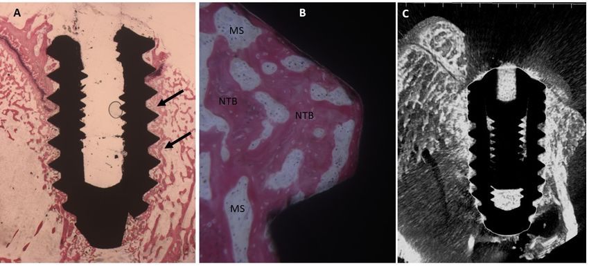
Uncoated Implant Surface
The slide
The slide observed
observed at at low
low magnification
magnification showed
showed aa few
few trabeculae
trabeculae bone
bone near
near or
or in
in contact
contact with
with
the implant surface (Figure 3). Also, osteoblasts were present near and in contact with
the implant surface (Figure 3). Also, osteoblasts were present near and in contact with the implant. the implant.
Few inflammatory
Few inflammatory cells cells were
were observed
observed atat the
the interface
interface implant
implant bone.
bone.
The mean
The mean BIC percentage
percentage waswas 22.42
22.42 ±± 4.5%,
4.5%, bone
bone area
area inner
inner threads
threads (BAIT)
(BAIT) was
was 2323 ±± 0.8%
0.8% and
and
Int. J. Mol. Sci. 2019,BIC
20, 724 5 of 16
bone area outer threads (BAOT) was
bone area outer threads (BAOT) was 19 ± 0.8%. 19 ± 0.8%.
Figure 3.
Figure 3. (A)
3. (A) Dental
Dental implants
implants surrounded
surrounded byby the
the trabeculae
the trabeculae of
trabeculae of newly-formed
of newly-formed bone.
newly-formed bone. Toluidine
Toluidine blue
blue
and acid
and acid fuchsin
fuchsin staining
fuchsin staining 22×.
staining 2×. (B)
×. (B) Higher
(B) Higher magnification.
Higher magnification.
magnification. A A few
A few bone
few bone trabeculae were
bone trabeculae
trabeculae were present
were present in
in the
the
implant
implant concavities
implant concavities (NTB).
concavities(NTB). Toluidine
(NTB).Toluidine blue
Toluidineblue and
blue and
and acid
acid fuchsin
fuchsin
acid 50×
fuchsin 50×. (C)
. (C)(C)
50×. Micro-CT
Micro-CT scans
scans
Micro-CT realized
realized
scans along
along
realized the
along
the transection
the transection
transection planes
planes of dental
of the
planes of the dental
the dental implant.
implant. Newly-formed
Newly-formed
implant. Newly-formed bone
bonebone was
waswas present.
present.
present.
Collagen-Coated
Collagen-Coated Surface
Surface
Collagen-Coated Surface
It
It was
was possible
possible toto observe
observe aaa trabecular
trabecular bone
bone in
in contact
contact with
with the
the fixture
fixture surface
surface (Figure
(Figure 4).
4). This
This
It was possible to observe trabecular bone in contact with the fixture surface (Figure 4). This
trabecular is
trabecular is present
is present
present inin the
in the concavity
the concavity
concavity ofof the
of the implant.
the implant. At
implant. At high
At high magnification
high magnification
magnification itit is
it is possible
is possible to
possible to observe
to observe
observe
trabecular
many
many osteoblasts
osteoblasts in
in direct
direct contact
contact with
with the
the titanium.
titanium. The
The mean
mean BIC
BIC percentage
percentage was
was 27.5
27.5±± 3.1%,
3.1%, bone
bone
many osteoblasts in direct contact with the titanium. The mean BIC percentage was 27.5 ± 3.1%, bone
area inner
area inner threads
inner threads (BAIT)
threads (BAIT) was
(BAIT) was 31
was 31 ± 0.8% and bone area outer threads (BAOT) was
31 ±± 0.8% and bone area outer threads (BAOT) was 21.8 ±± 1%. 21.8 ± 1%.
1%.
area
Figure
Figure 4. (A) New bone was present around the implants
4. (A) and in the implant
implants and concavities (arrows).
implant concavities
Figure 4. New bone was present around the implants in the implant (arrows).
Toluidine
Toluidine blue
blue and
and acid
acid fuchsin
fuchsinstaining
staining2 ×
2×.. (B) At higher magnification,
magnification, more
more trabeculae
trabeculae bone
bone was
was
Toluidine blue and acid fuchsin staining 2×. (B) At higher magnification, more trabeculae bone was
present
present in
in the
the implant
implant concavities
concavities (NTB)
(NTB) with
with a
a small
small medullary
medullary space
space (MS).
(MS). Toluidine
Toluidine blue
blue and
and acid
acid
present in the implant concavities (NTB) with a small medullary space (MS). Toluidine blue and acid
fuchsin
fuchsin staining
staining50 ×. (C)
50×. (C) Micro-CT
Micro-CT scans along transection planes of dental implant.
implant. AA newly-formed
newly-formed
fuchsin staining 50×. (C) Micro-CT scans along transection planes of dental implant. newly-formed
bone
bone was
was present.
present.
bone was present.
2.4.2. Thirty Days
Uncoated Implant Surface
The slide showed mature bone in contact with the implant, in only a few areas it is possible to
observe osteoblasts (Figure 5). No pathological infiltrate of inflammatory cells was observed.
The mean BIC percentage was 51.2 ± 3.9%, bone area inner threads (BAIT) was 28 ± 0.8% and
bone area outer threads (BAOT) was 36 ± 0.8%.
Uncoated
Uncoated Implant
Implant Surface
Surface
The
The slide
slide showed
showed mature
mature bone
bone in
in contact
contact with
with the
the implant,
implant, inin only
only aa few
few areas
areas it
it is
is possible
possible to
to
observe osteoblasts (Figure 5). No pathological infiltrate of inflammatory cells was
observe osteoblasts (Figure 5). No pathological infiltrate of inflammatory cells was observed. observed.
The
TheSci.mean
Int. J. Mol. mean BIC
BIC724percentage
2019, 20, percentage was
was 51.2
51.2 ±± 3.9%,
3.9%, bone
bone area
area inner
inner threads
threads (BAIT)
(BAIT) was
was 2828 ±± 0.8%
0.8% and
and
6 of 16
bone area outer threads (BAOT) was 36
bone area outer threads (BAOT) was 36 ± 0.8%. ± 0.8%.
Figure
Figure5.5.
Figure (A)
5.(A) The
(A)The Histological
TheHistological evaluation
evaluation showed
Histological evaluation showed organization
showed organizationand
organization andmineralization
and mineralizationof
mineralization ofofthe
thebone
the bonetissue
bone tissue
tissue
especially
especially near
especiallynear the
nearthe dental
thedental implant
dentalimplant (arrows).
implant(arrows). Toluidine
(arrows). Toluidine blue
Toluidine blue and acid fuchsin staining 2×. (B)
2×(B)
staining 2×.
blue and acid fuchsin staining At higher
. (B)AtAthigher
higher
magnification,
magnification,aaathin
magnification, thin trabecula
thintrabecula without
trabecula without osteoblastic
osteoblastic activity
without osteoblastic activitywas
activity waspresent
was presentin
present ininthe
theimplant
the implantconcavities
implant concavities
concavities
(arrows).
(arrows). Toluidine
(arrows).Toluidine blue
Toluidineblue and
blueand acid
andacid fuchsin
acidfuchsin staining
staining5050×.
fuchsinstaining (C)
×. (C)
50×. Micro-CT
(C)Micro-CT scans
Micro-CTscans along
scansalong transection
alongtransection planes
transectionplanes
planesof
of
the the
the dental
of dental
dental implant.
implant. Bone
Bone
implant. waswas
Bone present
present
was in
in contact
in contact
present with
with
contact thethe
with dental
dental
the implant.
implant.
dental implant.
Collagen-Coated
Collagen-Coated Surface
Collagen-Coated Surface
Surface
Mature
Mature bone
Mature bone was
wasfound
bone was found on
found on the
on the surface
the of titanium
surface of titanium with
titanium withcylindrical
with cylindricalstructures
cylindrical structuresthat
structures thatare
that arehaversian
are haversian
haversian
system.
system. No
system. No gaps
No gaps were
gaps were observed
were observed between
observed between implant
between implant and
implant and bone.
and bone.
bone. AA little
A little osteoblast
little osteoblast activity
osteoblast activity was
activity was present
was present
present
(Figure
(Figure6).
(Figure 6).
6).
Figure 6.(A)
Figure6.6.
Figure (A)Bone
(A) Bonewith
Bone withcomplete
with complete organization
complete and mineralization
organization and
and mineralizationespecially
mineralization especiallynear
especially nearthe
near thedental
the dentalimplant
dental implant
implant
(arrows).
(arrows). Toluidine
Toluidine blue
blue and
andacid
acid fuchsin
fuchsin staining
staining 2 ×
2×.. (B)
(B) At
At higher
higher magnification,
magnification, many
many osteoblasts
osteoblasts
(arrows). Toluidine blue and acid fuchsin staining 2×. (B) At higher magnification, many osteoblasts
(arrows)
(arrows)
(arrows)andand osteoid
andosteoid matric
osteoidmatric were
matric were present
were present
present in the fixture
in the fixture concavities.
fixture concavities.Toluidine
concavities. Toluidineblue
Toluidine blueand
blue andacid
and acidfuchsin
acid fuchsin
fuchsin
staining
staining
staining50 ×. (C) Micro-CT images
50×.
50×. (C) Micro-CT images along
images along transection
along transection planes
transectionplanes of the
planesofofthe dental
thedental implant.
dentalimplant. Mature
implant.Mature bone
bonewas
Maturebone was
was
present
presentinin
present contact
incontact with
contactwith the
withthe dental
thedental implant.
dentalimplant.
implant.
The mean BIC percentage was 55.3 ± 3.2%, bone area inner threads (BAIT) was 39 ± 2.2% and
bone area outer threads (BAOT) was 38 ± 2.2%.
2.4.3. Sixty Days
Uncoated Implant Surface
At lower magnification, evidence of a mature bone was observed in contact with the implant
surface. A few osteoblast activities were present (Figure 7). The mean BIC percentage was 56.32 ± 3.2%,
bone area inner threads (BAIT) was 35 ± 2.3% and bone area outer threads (BAOT) was 36 ± 2.3%.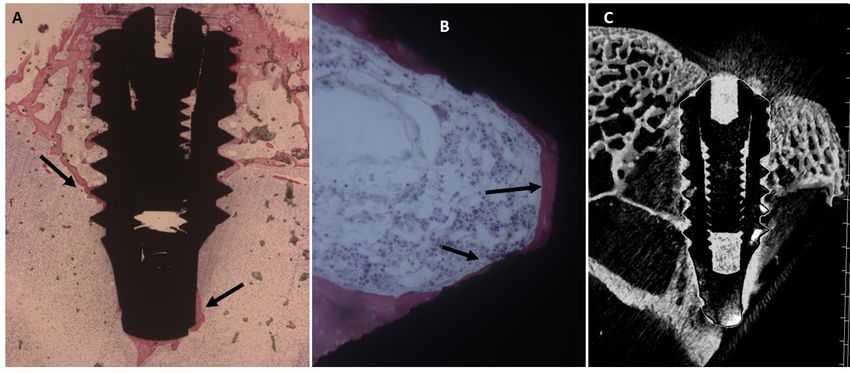
Uncoated Implant Surface
At lower magnification, evidence of a mature bone was observed in contact with the implant
surface. A few osteoblast activities were present (Figure 7). The mean BIC percentage was 56.32 ±
3.2%,
Int. bone
J. Mol. area 20,
Sci. 2019, inner
724 threads (BAIT) was 35 ± 2.3% and bone area outer threads (BAOT) was7 of
3616±
2.3%.
Figure 7.
Figure 7. (A)
(A) A
A thin
thin layer
layer of
of mature
mature bone
bone was
was present
present around
around the
the fixture.
fixture. Toluidine
Toluidine blue
blue and
and acid
acid
fuchsin
fuchsin staining ×. (B)
staining22×. (B) At
At higher
higher magnification,
magnification, a remodeling mature bone (MB) was evident
evident around
around
the
the implant surface
surface (Arrows).
(Arrows).Toluidine
Toluidineblue
blue and
and acid
acid fuchsin
fuchsin staining
staining 50×.50 ×.Micro-CT
(C) (C) Micro-CT
scans scans
along
along transection
transection planesplanes
of the of the dental
dental implant.
implant. Trabecular
Trabecular bone
bone was was present
present in contact
in contact with
with the the
dental
dental implant.
implant.
Collagen-Coated
Collagen-Coated Surface Surface
Low
Low magnification showed mature
magnification showed maturebone
bonetissues
tissuesalong
alongthethe periphery
periphery of of
thethe implant
implant andand in
in the
the thread of the implant. A few osteoblasts were also found around the
thread of the implant. A few osteoblasts were also found around the titanium. No gaps nor titanium. No gaps nor
inflammatory
inflammatory nor nor multinucleate
multinucleate cells
cells were
were detected
detected near
near the
the titanium
titanium surface
surface (Figure
(Figure 8).
8). The
The mean
mean
BIC was 63.6 ± 2.9%, bone area inner threads (BAIT) was 42 ±
BIC percentage was 63.6 ± 2.9%, bone area inner threads (BAIT) was 42 ± 2.3% and bone area outer
percentage 2.3% and bone area outer
threads
Int. J. Mol.(BAOT)
Sci. 2018,was FOR±PEER
19, x 44 2.3%.
REVIEW 8 of 16
threads (BAOT) was 44 ± 2.3%.
Figure8.8. (A)
Figure (A) Trabecular
Trabecularbone
bonewas wasmore
moreabundant
abundantaround
aroundthe
thedental
dentalimplant
implant(arrows).
(arrows). Toluidine
Toluidineblue
blue
and acid fuchsin staining 2
and acid fuchsin staining 2×. × . (B) At higher magnification, mature
mature bone (MB) was present aroundthe
bone (MB) was present around the
implant
implantsurface
surfaceand
andthe
theconcavities
concavitieswere
werecompletely
completelyfilled
filledby
bynew
newmature
maturebone
bone(Arrows).
(Arrows). Toluidine
Toluidine
blue
blueand
andacid
acidfuchsin
fuchsinstaining
staining50 ×. (C) Micro-CT scans along transection planes of the dental implant.
50×.
Trabecular
Trabecularbone
bonewas
waspresent
presentin incontact
contactwith
withthe
thedental
dentalimplant
implantand
andwas
wasalso
alsomore
morethan
thanthat
thatin
inthe
the
control implant.
control implant.
2.5. Statistical Evaluation
The means and SD values of BIC, BAIT and BAOT ratios are shown in Table 2. Significant
differences were found in the BIC, BAIT and BAOT ratios at 15, 30 and 60 days (p < 0.05).
The Pearson correlation test found significant correlation between the bone implant contact
(BIC), bone area inner threads (BAIT) and bone area outside threads (BAOT) ratios for either 15, 30
and 60 days (p < 0.05). (Tables 2 and 3; Figures 9–11).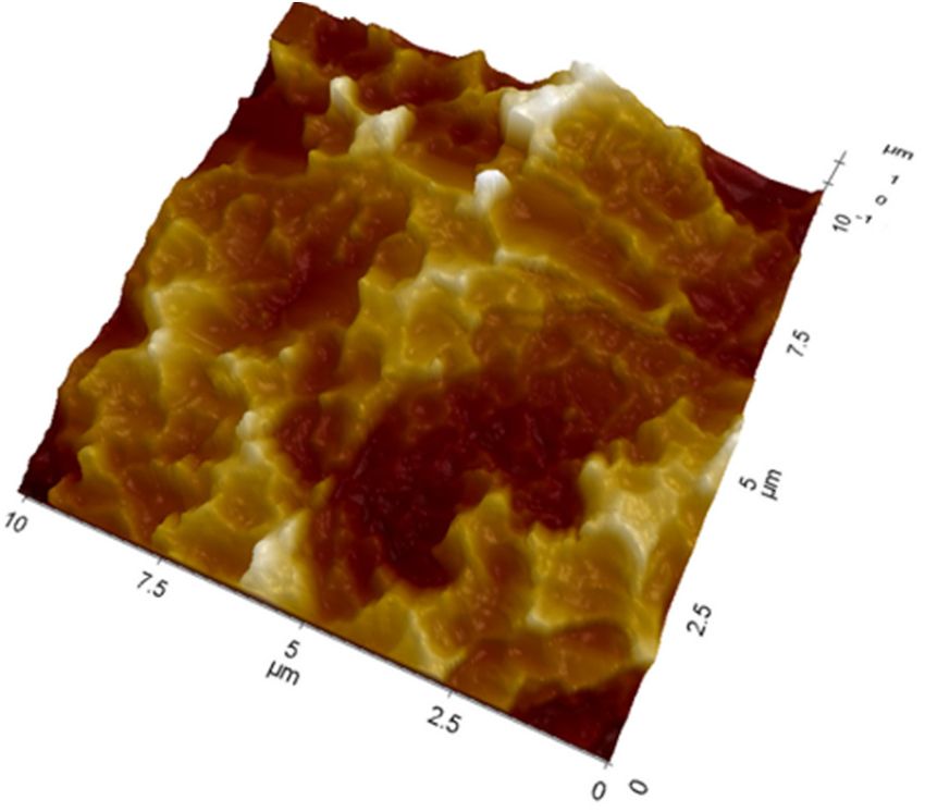
Int. J. Mol. Sci. 2019, 20, 724                                                                                           8 of 16

2.5. Statistical Evaluation
     The means and SD values of BIC, BAIT and BAOT ratios are shown in Table 2. Significant
differences were found in the BIC, BAIT and BAOT ratios at 15, 30 and 60 days (p < 0.05).

      Table 2. Means and SD of the measured Micro-CT BAIT and BOAT ratios and histomorphometric
      BIC ratios.

                                            BIC           p Value       BAIT       p Value         BAOT            p Value
                 Uncoated surface           22.4 ± 4.5                  23 ± 0.8                   19 ± 0.8
 15 Days                                                  p = 0.0005               p = 0.00004                     p = 0.00003
                 Collagen coated surface    27.5 ± 3.1                  31 ± 0.8                   21.8 ± 1
                 Uncoated surface           51.2 ± 3.9                  28 ± 0.8                   36 ± 0.8
  30 Days                                                 p = 0.0021               p = 0.00001                     p = 0.0008
                 Collagen coated surface    55.3 ± 3.2                  39 ± 2.2                   38 ± 2.2
                 Uncoated surface           56 ± 4.2                    35 ± 2.3                   36 ± 2.3
  60 Days                                                 p = 0.00001              p = 0.00006                     p = 0.0196
                 Collagen coated surface    63.6 ± 2.9                  42 ± 2.3                   44 ± 2.3

     The Pearson correlation test found significant correlation between the bone implant contact (BIC),
bone area inner threads (BAIT) and bone area outside threads (BAOT) ratios for either 15, 30 and
60 days (p < 0.05). (Tables 2 and 3; Figures 9–11).

      Table 3. The Pearson correlation results for the correlation between the Micro-CT BAIT and BAOT and
      histomorphometric BIC ratios. The Rho (ρ) values is the Pearson rank correlation coefficient. * p < 0.05;
      ** p < 0.01

                                                                        BIC            BAIT                   BOAT
                                                       BIC              -              0.620 *                −0.547 *
                      Uncoated surface                 BAIT             0.620 *        -                      −0.951 **
                                                       BOAT             −0.547 *       −0.951 **              -
 15 Days
                                                       BIC              -              0.576*                 0.578 *
                      Collagen coated surface          BAIT             0.576 *        -                      0.935 **
                                                       BOAT             0.578 *        0.935**                -
                                                       BIC              -              0.566 *                −0.536 *
                      Uncoated surface                 BAIT             0.566 *        -                      −0.766 **
                                                       BOAT             −0.536 *       −0.766 **              -
  30 Days
                                                       BIC              -              0.598 *                −0.603 *
                      Collagen coated surface          BAIT             0.598 *        -                      −0.894 **
                                                       BOAT             −0.603 *       -0.894 **              -
                                                       BIC              -              0.643 *                −0.512 *
                      Uncoated surface                 BAIT             0.643 *        -                      −0.857 **
                                                       BOAT             −0.582 *       -0.857 **              -
  60 Days
                                                       BIC              -              0.541 *                −0.611 *
                      Collagen coated surface          BAIT             0.541 *        -                      −0.863 **
                                                       BOAT             −0.611 *       −0.863 **              -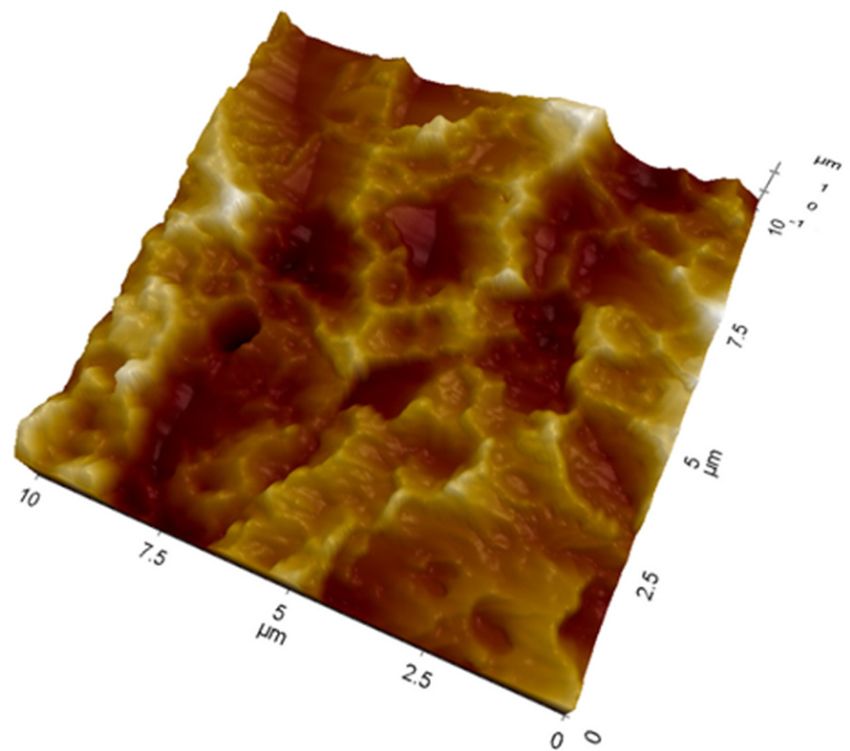
BOAT    −0.603 *   -0.894 **   -
                         60 Days Uncoated surface         BIC     -          0.643 *     −0.512 *
                                                          BAIT    0.643 *    -           −0.857 **
                                                          BOAT    −0.582 *   -0.857 **   -
                                  Collagen coated surface BIC     -          0.541 *     −0.611 *
Int. J. Mol. Sci. 2019, 20, 724                           BAIT    0.541 *    -           −0.863 **            9 of 16
                                                          BOAT    −0.611 *   −0.863 **   -

      Figure  9. Means
      Figure 9.  Meansand
                       andSD
                           SDofof
                                thethe measured
                                     measured BICBIC  ratios
                                                  ratios     at30
                                                         at 15, 15,and
                                                                    30 60
                                                                        and 60 days.
                                                                          days. Y axisYis axis
                                                                                          boneisimplant
                                                                                                 bone implant
                                                                                                        contact
      contact expressed
      expressed  in %. in %.

Figure 10. Means
     Figure      and SD
            10. Means   ofSD
                      and theof
                              measured  BAIT BAIT
                                the measured ratiosratios
                                                    at 15, at
                                                           3015,
                                                              and3060 days.
                                                                    and     Y axisYisaxis
                                                                        60 days.      BAIT   expressed
                                                                                          is BAIT      in %.
                                                                                                  expressed
      in %.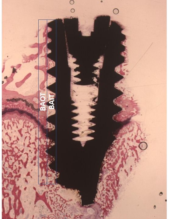
Int. J. Mol. Sci. 2019, 20, 724 10 of 16
Int. J. Mol. Sci. 2018, 19, x FOR PEER REVIEW 10 of 16
Figure
Figure 11.
11. Means
Means and
and SD
SD of
of the
the measured
measured BAOT
BAOT ratios
ratios at 15, 30
at 15, 30 and
and 60
60 days.
days. Y
Y axis
axis is
is BAOT
BAOT implant
implant
contact
contact expressed in %.
expressed in %.
3. Discussion
3. Discussion
The aimaim of ofthis
thisstudy
studywas wastoto investigate
investigate thethe effects
effects of collagen
of collagen typetypeI on Ibone
on bonehealinghealing and
and bone
bone formation
formation in a in a rabbit
rabbit model modelwhen when used usedas ascoating
coatingon on titanium
titanium implants.
implants. The The investigators
hypothesized that dental implant coated with collagen type I may
dental implant coated with collagen type I may not increase the amount not increase the amount of of
bone in
bone
contact
in contact with
withthetheimplant
implant surface.
surface.
Even if many in vivo and vitro studies have investigated collagen type I on titanium, there is a
lack of information
informationon onhowhowsuchsuchaacoated
coatedsurface
surfaceinfluences
influences BIC, BAIT
BIC, BAIT and andBAOT.
BAOT. TheTheoutcomes
outcomes of the
of
XPS evaluation
the XPS evaluationshowshow that titanium disk surfaces
that titanium evaluated
disk surfaces in the present
evaluated in thestudypresentare study
in good areagreement
in good
with the literature
agreement with the onliterature
titanium surfaces
on titanium in general
surfaces and,in ingeneral
particular,
and,on insurfaces
particular, of titanium
on surfaces dental
of
implants [20]. A large amount of literature exists on this subject, the value
titanium dental implants [20]. A large amount of literature exists on this subject, the value of about of about 18% Ti detected in
this case
18% is indicative
Ti detected in thisofcase
a comparatively
is indicative of clean surface [21,22].
a comparatively clean surface [21,22]
In the coated disks the XPS shows the presence of a proteinaceous layer on the implant surface
and it obviously confirms that collagen molecules coat the the titanium
titanium surface
surface chemistry.
chemistry. In In aa few
few areas,
areas,
a small
smallsignal
signalfrom from titanium
titanium in the survey
in the spectrum
survey is detected
spectrum (Table 1),
is detected suggesting
(Table that the thickness
1), suggesting that the
of the coating
thickness of theis lower
coating than the XPS-sampling
is lower depth (8 nm).
than the XPS-sampling Again,
depth this isAgain,
(8 nm). consistent
this iswith the nature
consistent of
with
the process,
nature ofwhich is basedwhich
the process, on interfacial
is basedadsorption
on interfacial of collagen
adsorption molecules
of collagenrather than the rather
molecules application
than
of aapplication
the thick coating. of aTitanium
thick coating.surfaces coatedsurfaces
Titanium with different
coated substances
with different as insubstances
dental implants have
as in dental
attracted have
implants increasing attention.
attracted increasing attention.
thisstudy
In this studysandblasted
sandblastedand and acid-etched
acid-etched implant
implant screws,
screws, uncoated
uncoated or coated
or coated withwith collagen
collagen type
type I were tested in a knee implantation model in rabbit. The outcomes
I were tested in a knee implantation model in rabbit. The outcomes of the present research showed a of the present research
showed a statistical
statistical differencedifference
in BIC, BAIT in BIC,
BAOTBAIT BAOT between
between titaniumtitanium
uncoated uncoated
or coated or with
coated with collagen
collagen type I.
typeresults
The I. The results of the present
of the present study support
study support the rejection
the rejection of the null of the null hypothesis.
hypothesis. The collagenThe collagen
coating
coating probably
probably influences influences cell migration,
cell migration, attachment, attachment,
proliferation, proliferation, and differentiation
and differentiation on the
on the titanium
titanium implants,
implants, but no bone butformation
no bone distant
formation fromdistant from surface.
the implant the implantThesesurface.
results areThesenot results
influencedare
notroughness,
by influencedinby roughness,
fact the AFM outcomein fact the AFMaoutcome
showed showed asurface
similar roughness similarforroughness
both implants. surface for
These
both implants. These findings were confirmed by Micro-CT.
findings were confirmed by Micro-CT. Type I collagen on the surface of titanium implants improvedType I collagen on the surface of
titanium
early implants improved
osteogenesis [23–26]. It early
wasosteogenesis
seen that [23–26].collagen Ittype was seen that collagen
I improved type I improved
osseointegration and
postcondition of titanium coated implants. Similar results were obtained in a rabbit model within
osseointegration and postcondition of titanium coated implants. Similar results were obtained a
femur condyle defect when the implants were coated in collagen type I [17]. It follows that coating
biomolecules positively influence mesenchymal cell proliferation, attachment, and differentiation
[24,27]. The positive influence of collagen coating was confirmed in a mini pig model. In theInt. J. Mol. Sci. 2019, 20, 724 11 of 16
a rabbit model with a femur condyle defect when the implants were coated in collagen type I [17].
It follows that coating biomolecules positively influence mesenchymal cell proliferation, attachment,
and differentiation [24,27]. The positive influence of collagen coating was confirmed in a mini pig
model. In the experimental model, the authors showed increased bone implant contact and bone
density [26]. In this study a rabbit model was chosen, where the knee served well for comparison of
osseointegration between different implant groups. Collagen type I improved bone inside the screw
threads and in contact with the surface when the implants were placed in the rabbit femur trabecular
bone [28]. Schliephake et al. studied collagen-coated implants and demonstrated an increase in the
percentage of BIC in a dog model [29]. Also, the study of Bae (2018) [30] in a rat tibia model showed
an enhancement in the osseointegration and bone healing with collagen type I crosslinked by gamma
irradiation. In this research we have not investigated what happens to the collagen during the phases
of osseointegration; it is probably resorbed during the phases of bone healing. The collagen type
I it could offer greater resistance to pre-implantitis and were used for treating the hard tissue loss
associated with peri-implantitis around SLA implants [31].
We used the rabbit model because it is a convenient model for skeletal research studies and
has been extensively used to test biomaterials [32] and osteoconductive/osteoinductive reactions in
implants [33]. Besides, this animal model provides an excellent cost-effective animal model, their
maintenance and housing are simple, and they recover very well postoperatively. In addition the
bone composition and morphology between rabbit and human are very similar [34], and there is a
sufficient correlation with maxilla bone [35]. This model was also chosen because the phenomenon
of osseointegration was discovered in the rabbit [36] and for this advantage they are often used in
skeletal research or for screening implant materials.
In this study, we chose a rabbit femur model to evaluate the potential ability of coating surfaces
to enhance bone formation when coated by collagen in the presence of large medullary spaces.
4. Materials and Methods
4.1. Surface Preparation by the Manufacturer
The surfaces evaluated in this investigation were prepared by the manufactures. Tests were
performed on grade 4 control and collagen coated dental implants. Both of them were sandblasted and
acid etched. The uncoated surface shows the typical topography of sandblasted-acid etched surfaces,
featuring a long-range roughness due to the plastic deformation of the surface induced by blasting,
and a short-range roughness due to acid etching. The Sa roughness value, as evaluated by Stereo
Scanning Microscopy analysis of 110 × 80 micrometers areas, is around 1.15 micrometers.
Collagen coated samples were further subjected to a proprietary treatment, based on the interfacial
adsorption and linking of collagen molecules to titanium, yielding a nanometer-thin, permanent
molecular layer on the implant surface. As discussed in the relevant section, no detectable modification
of surface topography and roughness, at the micrometer level, is induced by the collagen nanolayer.
Purified bovine collagen Type I, dry, obtained from calf hides through pepsin extraction, supplied
by Symatese (Chaponost, France) is used in the process. The product complies with the European
requirements: Regulation (UE) N◦ 722/2012 of 8 August 2012 with respect to active implantable
medical devices and medical devices manufactured utilizing tissues of animal origin; and it has
obtained a certificate of suitability with respect to Monograph N◦ 1483 of European Pharmacopieia:
“Products with risk of transmitting agents of animal spongiform encephalopathies”. Dry collagen is
dissolved in acidic (acetic acid) solution on coating process.
4.2. Surface Analysis by X-Ray Photoelectron Spectroscopy (XPS)
The roughness of the coated and uncoated implants was analyzed using atomic force microscopy.
The disks used in the present study were sandblasted and double acid etched.Int. J. Mol. Sci. 2019, 20, 724 12 of 16
Ten titanium sandblasted and double acid etched disks and ten titanium sandblasted and double
acid etched disks and coated with collagen type I were used for the evaluation of the surface
composition of uncoated and collagen coated disks by XPS analysis. All samples were titanium
grade 4 with 5 mm diameter subjected to the same treatments as implants.
XPS analysis was performed by a Perkin Elmer PHI 5600 ESCA system (PerkinElmer Inc., Waltham,
MA, USA). The instrument is provided with a monochromatized Al anode operating at 10 kV and 200 W.
The diameter of the analyzed spot is about 500 micrometers, the analyzed depth about 8 nanometers.
The base pressure was maintained at 10 Pa. The angle between the electron analyzer and the sample
surface was 45◦ . Analysis was performed by acquiring wide-range survey spectra (0–1000 eV binding
energy), using a pass energy of 117.4 eV, resolution of 1 eV/Step and acquisition time of 50 ms/Step.
After acquisition of the survey spectra, high resolution C1s peaks were acquired using a pass energy of
11.75 eV, resolution of 0.100 eV/Step and acquisition time of 50 ms/Step. Deconvolution of C1s peak
was performed using the instrument’s software, after correction of the peak position with reference to
the internal standard C–C component of the C1s peak positioned at 285.0 eV.
4.3. Atomic Force Microscopy (AFM)
Ten SLA titanium disks and ten titanium disks coated with collagen type I were studied for
the analysis of the surface topography by AFM. All of the disks were titanium grade 4 with 5 mm
diameter subjected to the same treatments as implants. Measurements were performed by a NX10
Park AFM instrument (Park System, Suwon, Korea), equipped with 20-bit closed-loop XY and Z
flexure scanners and a non-contact cantilever PPP-NCHR 5M. This instrument implements a True
Non-contact™ mode, allowing minimization of the tip-sample interaction, resulting in tip preservation,
negligible sample nanotopography modification and reduction of artefacts. On each sample, four
different areas were analyzed at a scan rate of 0.1 Hz. Surface topography parameters were obtained
using the instrument’s software.
4.4. In Vivo Experiment
A total of 18 white male mature New Zealand rabbits, about 2.5 kg of weight, were treated
in the investigation. The investigation was approved by the Ethical Committee of the University
of Chieti-Pescara, Chieti, Italy, N◦ 67/2017 del 20-01-2017. A total of 36 implants (4 × 13) were
used, 18 sandblasted and double acid etched coated with covalently immobilized collagen type I,
(ACTIGEN® , Ubigen Srl, Padova, Italy), and 18 uncoated sandblasted and double acid etched implants
(Ubigen, Srl, Padova, Italy) were used. A total of eighteen fixtures of each different surface were
positioned. The implants were randomly positioned into the articular femoral knee-joint of the animals.
Prior to the surgery, the animals were anesthetized with intramuscular infiltrations of fluanizone
(0.7 mg/kg b.wt.) and diazepam (1.5 mg/kg b.wt.), and local anesthesia was administered using 1
mL of 2% lidocain/adrenalin solution. In preparation, the skin at the surgical site was washed with
soap and water followed by disinfection with an aqueous solution of 10% povidone-iodine (Betadine®
Solution, McKesson, Canada). A skin incision with a periosteal flap was elevated to access the articular
surface. Dissection of the muscular plane was achieved with blunt scissors, and the knee-joint was
exposed using a periosteal elevator. The preparation of the bone implant bed was done with burs
under generous saline irrigation. The implant location followed manufacturer’s recommendation,
lanceolate drill, 2 mm, and a 3.8 mm under saline irrigation. For inserting the implants, a micromotor
was used for each implant. Each rabbit received two implants, one in each knee-joint. Four hours
postoperatively the animals were left fasting then food and water ad libidum were offered to the
rabbits. During the experimental program, two of the rabbits were substituted.
The animals were euthanized with an intravenous injection of Tanax at 15, 30 and 60 days. A total
of thirty-six samples were retrieved. The specimens and surrounding tissues retrieved were stored
in 10% buffered formalin and processed to obtain thin ground sections. Then, the samples were
dehydrated in a graded series of ethanol rinses and embedded in a glycolmethacrylate resin (TechnovitInt. J. Mol. Sci. 2019, 20, 724 13 of 16
7200 VLC, Kulzer, Wehrheim, Germany). At the end of the polymerization process, the specimens were
sectioned, along the longitudinal axis of the fixture, by a high-precision diamond disc at about 150 µm
at slow-speed precision to about 200 µm then ground down to about 40 µm with a specially designed
grinding machine Scan 1 Automated System (Pescara, Italy) [20]. A total of three slides were retrieved
from each fixture. These slides were stained with toluidine blue and acid fuchsin and observed in
Int. J. Mol. Sci. 2018, 19, x FOR PEER REVIEW 13 of 16
normal transmitted light under a Nikon microscope ECLIPSE (Nikon, Tokyo, Japan). New and old
bone
New couldand old bebone
distinguished according to color
could be distinguished (lightto
according red = old
color matrix,
(light red =dark
old red = new
matrix, matrix)
dark and
red = new
values
matrix)were and expressed
values were in expressed
percentagein(Mean ± SD).(Mean ± SD).
percentage
Bone
Bone implant contact (BIC) and bone area inner
implant contact (BIC) and bone area inner threads
threads (BAIT)
(BAIT) then
then bone
bone area
area outer
outer threads
threads
(BAOT)
(BAOT) were quantified to evaluate the osteogenic parameters around the implant surface and
were quantified to evaluate the osteogenic parameters around the implant surface and values
values
were expressed in percentage (Mean ± SD). The BAIT in close proximity to the
were expressed in percentage (Mean ± SD). The BAIT in close proximity to the fixture was evaluated fixture was evaluated
within
within the thread area,
the thread area, while
whilethe
theBAOT,
BAOT,distant
distanttotothe
theimplant,
implant, extended
extended forfor
thethesamesame
sizesize
intointo
the
the
adjacent new/old bone (Figure 12). The different BIC, BAIT and BAOT were measured by a by
adjacent new/old bone (Figure 12). The different BIC, BAIT and BAOT were measured a
light
light microscope
microscope connected
connected to atohigh-resolution
a high-resolution video
video camera(16.25-megapixel)
camera (16.25-megapixel)(Digital
(Digital Sight
Sight series
series
microscope
microscope cameras), a high definition monitor and a personal computer (Notebook Toshiba Satellite
cameras), a high definition monitor and a personal computer (Notebook Toshiba Satellite
pro
pro r50-c-15w).
r50-c-15w). The The optical
optical instrument
instrument waswas linked
linked toto aa dedicated
dedicated histometry
histometry software
software package
package ableable
to perform image capturing, recorded by a Sony α330 digital camera and subjected
to perform image capturing, recorded by a Sony α330 digital camera and subjected to morphometric to morphometric
analysis
analysis by by digital
digital image-analysis
image-analysis (NIS-Elements
(NIS-Elements AR AR 3.0
3.0 software,
software, Nikon,
Nikon, Minato,
Minato, Japan).
Japan). A A total
total of
of
18 fixtures for each experimental surface were evaluated in this
18 fixtures for each experimental surface were evaluated in this investigation. investigation.
Figure 12.
Figure 12.The
Thearea
areadirectly
directly adjacent
adjacent to the
to the dental
dental implant
implant was evaluated
was evaluated withinwithin the part
the thread thread part
(BAIT),
(BAIT), whereas the area far from the implant extended for the same size into the adjacent
whereas the area far from the implant extended for the same size into the adjacent new/old bone new/old
bone (BAOT).
(BAOT).
4.5.
4.5. Micro-CT
Micro-CT Analysis
Analysis
Computed
Computed tomography
tomography datasets
datasets were
were performed
performed byby Skyscan 1172G (Bruker,
Skyscan 1172G (Bruker, Kontich,
Kontich, Belgium),
Belgium),
aa high-resolution
high-resolution 3D
3D imaging
imaging system
system with
with aa L7901-20
L7901-20 Microfocus
Microfocus X-ray
X-ray Source
Source (Hamamatsu,
(Hamamatsu, Japan).
Japan).
The acquisition of volumes was performed with 0.5 mm Al filter, image pixel/size of 21.96
The acquisition of volumes was performed with 0.5 mm Al filter, image pixel/size of 21.96 µm, µm,
camera binning 4 × 4, source voltage of 70 kV, source current of 141 µA, exposure time of 500
camera binning 4 × 4, source voltage of 70 kV, source current of 141 µA, exposure time of 500 ms. The ms.
reconstructed tomographic volumes of the acquired scans were acquired by a built-in NRecon
Skyscan reconstruction software (Version:1.6.6.0; Skyscan Bruker).
The 3D images were generated using 3D Visualization Softwares CTvox v. 2.5 and DataViewer
v. 1.4.4 (Skyscan Bruker) to the volume rendering and virtual sectioning views. The analysis of the
samples was carried out using CT-Analyser software version 1.13.Int. J. Mol. Sci. 2019, 20, 724 14 of 16
The reconstructed tomographic volumes of the acquired scans were acquired by a built-in NRecon
Skyscan reconstruction software (Version:1.6.6.0; Skyscan Bruker).
The 3D images were generated using 3D Visualization Softwares CTvox v. 2.5 and DataViewer
v. 1.4.4 (Skyscan Bruker) to the volume rendering and virtual sectioning views. The analysis of the
samples was carried out using CT-Analyser software version 1.13.
4.6. Statistical Evaluation
The statistical evaluation was performed by GraphPad Prism 6 software (GraphPad Software,
Inc., San Diego, CA, USA). The outcome data about bone implant contact (BIC), bone area inner
threads (BAIT) and bone area outside threads (BAOT) were statistically evaluated between the two
experimental groups, and the Student t-Test was applied. The level of significance for the analysis was
set at p ≤ 0.05. The Pearson correlation test was performed to evaluate the correlation between the
bone implant contact (BIC), bone area inner threads (BAIT) and bone area outside threads (BAOT)
ratios for each group at 15, 30 and 60 days.
5. Conclusions
The outcome of the present research confirms that collagen type I on the implant surface is one
of the most efficient approaches for accelerating early osteogenesis and improving the bioactivity of
titanium implant surfaces. In the future, bone implant surfaces will be enriched with biomolecules
to increase the bone healing process [37–39]. In conclusion, these results show that surfaces coated
with collagen improve the bioactivity, BIC, and bone around the dental surface compared to control
implants, and could be clinically advantageous for shortening the implant healing period.
Author Contributions: Methodology, A.S., L.V.; software, A.S., F.L., T.O.; validation, A.S., L.V., M.M., G.I., T.O.;
formal analysis, A.S., L.V.; investigation, A.S., L.V.; resources, T.O.; data curation, A.S., F.L.; writing—original
draft preparation, A.S., F.L.; writing—review and editing, A.S., F.L.; visualization, L.V.; supervision, A.S.; project
administration, T.O., G.I.
Funding: The authors declare that no founding was received for this work.
Acknowledgments: The authors acknowledge the helpful technical assistance of Mauro di Berardino (X-Ray
Technician) in the elaboration of data.
Conflicts of Interest: The authors declaim no conflict of interest.
References
1. Rodriguez, G.M.; Bowen, J.; Grossin, D.; Ben-Nissan, B.; Stamboulis, A. Functionalisation of Ti6Al4V and
hydroxyapatite surfaces with combined peptides based on KKLPDA and EEEEEEEE peptides. Colloids Surf. B
2017, 160, 154–160. [CrossRef] [PubMed]
2. Albrektsson, T.; Brånemark, P.I.; Hansson, H.A.; Lindström, J. Osseointegrated titanium implants.
Requirements for ensuring a long-lasting, direct bone-to-implant anchorage in man. Acta Orthop. Scand.
1981, 52, 155–170. [CrossRef] [PubMed]
3. Scarano, A.; Piatelli, A.; Quaranta, A.; Lorusso, F. Bone response to two dental implants with different
sandblasted/acid-etched implant surfaces: A histological and histomorphometrical study in rabbits.
Biomed Res. Int. 2017, 2017, 8724951. [CrossRef] [PubMed]
4. Cordioli, G.; Majzoub, Z.; Piattelli, A.; Scarano, A. Removal torque and histomorphometric investigation of 4
different titanium surfaces: An experimental study in the rabbit tibia. Int. J. Oral Maxillofac. Implants 2000,
15, 668–674. [PubMed]
5. Scarano, A.; Perrotti, V.; Artese, L.; Degidi, M.; Degidi, D.; Piattelli, A.; Iezzi, G. Blood vessels are concentrated
within the implant surface concavities: A histologic study in rabbit tibia. Odontology 2014, 102, 259–266.
[CrossRef] [PubMed]
6. Mangano, F.; Maghaireh, H.; Calvo-Guirado, J. Engineering the Bone-Implant Interface. Biomed Res. Int.
2018, 2018, 4956491. [CrossRef]Int. J. Mol. Sci. 2019, 20, 724 15 of 16
7. Sinjari, B.; Traini, T.; Caputi, S.; Mortellaro, C.; Scarano, A. Evaluation of Fibrin Clot Attachment on Titanium
Laser-Conditioned Surface Using Scanning Electron Microscopy. J. Craniofac. Surg. 2018, 29, 2277–2281.
[CrossRef]
8. Coelho, P.G.; Granjeiro, J.M.; Romanos, G.E.; Suzuki, M.; Silva, N.R.F.; Cardaropoli, G.; Thompson, V.P.;
Lemons, J.E. Basic research methods and current trends of dental implant surfaces. J. Biomed. Mater. Res. B
2009, 88, 579–596. [CrossRef]
9. Ribeiro da Silva, J.; Castellano, A.; Malta Barbosa, J.P.; Gil, L.F.; Marin, C.; Granato, R.; Bonfante, E.A.;
Tovar, N.; Janal, M.N.; Coelho, P.G. Histomorphological and Histomorphometric Analyses of Grade IV
Commercially Pure Titanium and Grade V Ti-6Al-4V Titanium Alloy Implant Substrates: An In Vivo Study
in Dogs. Implant Dent. 2016, 25, 650–655. [CrossRef]
10. Orsini, G.; Piattelli, M.; Scarano, A.; Petrone, G.; Kenealy, J.; Piattelli, A.; Caputi, S. Randomized, controlled
histologic and histomorphometric evaluation of implants with nanometer-scale calcium phosphate added to
the dual acid-etched surface in the human posterior maxilla. J. Periodontol. 2007, 78, 209–218. [CrossRef]
11. Najeeb, S.; Zafar, M.S.; Khurshid, Z.; Siddiqui, F. Applications of polyetheretherketone (PEEK) in oral
implantology and prosthodontics. J. Prosthodont. Res. 2016, 60, 12–19. [CrossRef] [PubMed]
12. Mangano, F.G.; Iezzi, G.; Shibli, J.A.; Pires, J.T.; Luongo, G.; Piattelli, A.; Mangano, C. Early bone
formation around immediately loaded implants with nanostructured calcium-incorporated and machined
surface: A randomized, controlled histologic and histomorphometric study in the human posterior maxilla.
Clin. Oral Investig. 2017, 21, 2603–2611. [CrossRef] [PubMed]
13. Scarano, A.; Crocetta, E.; Quaranta, A.; Lorusso, F. Influence of the Thermal Treatment to Address a Better
Osseointegration of Ti6Al4V Dental Implants: Histological and Histomorphometrical Study in a Rabbit
Model. Biomed. Res. Int. 2018, 2018, 2349698. [PubMed]
14. Geissler, U.; Hempel, U.; Wolf, C.; Scharnweber, D.; Worch, H.; Wenzel, K. Collagen type I-coating of Ti6Al4V
promotes adhesion of osteoblasts. J. Biomed. Mater. Res. 2000, 51, 752–760. [CrossRef]
15. Takeuchi, Y.; Nakayama, K.; Matsumoto, T. Differentiation and cell surface expression of transforming
growth factor-beta receptors are regulated by interaction with matrix collagen in murine osteoblastic cells.
J. Biol. Chem. 1996, 271, 3938–3944. [CrossRef] [PubMed]
16. Scarano, A.; Lorusso, F.; Staiti, G.; Sinjari, B.; Tampieri, A.; Mortellaro, C. Sinus Augmentation with
Biomimetic Nanostructured Matrix: Tomographic, Radiological, Histological and Histomorphometrical
Results after 6 Months in Humans. Front. Physiol. 2017, 8, 565. [CrossRef] [PubMed]
17. Ao, H.-Y.; Xie, Y.-T.; Yang, S.-B.; Wu, X.-D.; Li, K.; Zheng, X.-B.; Tang, T.-T. Covalently immobilised type I
collagen facilitates osteoconduction and osseointegration of titanium coated implants. J. Orthop. Translat.
2016, 5, 16–25. [CrossRef]
18. Maghdouri-White, Y.; Bowlin, G.L.; Lemmon, C.A.; Dréau, D. Mammary epithelial cell adhesion, viability,
and infiltration on blended or coated silk fibroin-collagen type I electrospun scaffolds. Mater. Sci. Eng. C
2014, 43, 37–44. [CrossRef]
19. Morra, M.; Cassinelli, C.; Cascardo, G.; Bollati, D. Collagen I-coated titanium surfaces for bone implantation.
In Biological Interactions on Materials Surfaces; Springer: Heidelberg, Germany, 2009; pp. 373–396.
20. Kasemo, B.; Lausmaa, J. Biomaterial and implant surfaces: On the role of cleanliness, contamination, and
preparation procedures. J. Biomed. Mater. Res. 1988, 22, 145–158. [CrossRef]
21. Wieland, M. Measurement and evaluation of the chemical composition and topography of titanium implant
surfaces. Bone engineering 2000.
22. Morra, M.; Cassinelli, C.; Bruzzone, G.; Carpi, A.; Di Santi, G.; Giardino, R.; Fini, M. Surface chemistry
effects of topographic modification of titanium dental implant surfaces: 1. Surface analysis. Int. J. Oral
Maxillofac. Implants 2003, 18, 40–45. [PubMed]
23. Scharnweber, D.; Born, R.; Flade, K.; Roessler, S.; Stoelzel, M.; Worch, H. Mineralization behaviour of collagen
type I immobilized on different substrates. Biomaterials 2004, 25, 2371–2380. [CrossRef] [PubMed]
24. Morra, M.; Cassinelli, C.; Cascardo, G.; Bollati, D.; Baena, R.R.Y. Gene expression of markers of osteogenic
differentiation of human mesenchymal cells on collagen I-modified microrough titanium surfaces. J. Biomed.
Mater. Res. A 2011, 96, 449–455. [CrossRef] [PubMed]
25. Kung, S.; Devlin, H.; Fu, E.; Ho, K.-Y.; Liang, S.-Y.; Hsieh, Y.-D. The osteoinductive effect of chitosan-collagen
composites around pure titanium implant surfaces in rats. J. Periodont. Res. 2011, 46, 126–133. [CrossRef]
[PubMed]You can also read