Growth in fossil and extant deer and implications for body size and life history evolution
←
→
Page content transcription
If your browser does not render page correctly, please read the page content below
Kolb et al. BMC Evolutionary Biology (2015) 15:19
DOI 10.1186/s12862-015-0295-3

 RESEARCH ARTICLE                                                                                                                                Open Access

Growth in fossil and extant deer and implications
for body size and life history evolution
Christian Kolb1*, Torsten M Scheyer1, Adrian M Lister2, Concepcion Azorit3, John de Vos4, Margaretha AJ Schlingemann5,
Gertrud E Rössner6, Nigel T Monaghan7 and Marcelo R Sánchez-Villagra1

  Abstract
  Background: Body size variation within clades of mammals is widespread, but the developmental and life-history
  mechanisms by which this variation is achieved are poorly understood, especially in extinct forms. An illustrative
  case study is that of the dwarfed morphotypes of Candiacervus from the Pleistocene of Crete versus the giant deer
  Megaloceros giganteus, both in a clade together with Dama dama among extant species. Histological analyses of
  long bones and teeth in a phylogenetic context have been shown to provide reliable estimates of growth and life
  history patterns in extant and extinct mammals.
  Results: Similarity of bone tissue types across the eight species examined indicates a comparable mode of growth in
  deer, with long bones mainly possessing primary plexiform fibrolamellar bone. Low absolute growth rates characterize
  dwarf Candiacervus sp. II and C. ropalophorus compared to Megaloceros giganteus displaying high rates, whereas Dama
  dama is characterized by intermediate to low growth rates. The lowest recorded rates are those of the Miocene small
  stem cervid Procervulus praelucidus. Skeletal maturity estimates indicate late attainment in sampled Candiacervus and
  Procervulus praelucidus. Tooth cementum analysis of first molars of two senile Megaloceros giganteus specimens
  revealed ages of 16 and 19 years whereas two old dwarf Candiacervus specimens gave ages of 12 and 18 years.
  Conclusions: There is a rich histological record of growth across deer species recorded in long bones and teeth, which
  can be used to understand ontogenetic patterns within species and phylogenetic ones across species. Growth rates
  sensu Sander & Tückmantel plotted against the anteroposterior bone diameter as a proxy for body mass indicate three
  groups: one with high growth rates including Megaloceros, Cervus, Alces, and Dama; an intermediate group with
  Capreolus and Muntiacus; and a group showing low growth rates, including dwarf Candiacervus and Procervulus. Dwarf
  Candiacervus, in an allometric context, show an extended lifespan compared to other deer of similar body size such as
  Mazama which has a maximum longevity of 12 years in the wild. Comparison with other clades of mammals reveals
  that changes in size and life history in evolution have occurred in parallel, with various modes of skeletal tissue
  modification.
  Keywords: Island evolution, Pleistocene, Cervidae, Candiacervus, Megaloceros, Bone histology, Cementum analysis,
  Growth rates, Longevity, Skeletal maturity

Background                                                                             understand the mechanisms of life-history and size
Several lineages of mammals have evolved remarkable                                    evolution on islands but also in cases of significant
changes in body size following island isolation [1-3],                                 body size changes in mainland lineages, histology of
including among others dwarf hippopotamuses, elephants,                                hard tissues is a powerful tool, as has been demon-
and deer, and giant rabbits [4-6]. These patterns are the re-                          strated for ‘dwarf ’ and ‘giant’ sauropod [10-12] and
sult of complex interplay of multiple variables, including                             tyrannosaurid [13] dinosaurs, as well as early synapsids
resource limitation and ecological release [5,7-9]. To                                 [14,15] among fossil forms.
                                                                                         A remarkable example of island evolution is found in
* Correspondence: christian.kolb@pim.uzh.ch                                            the Pleistocene of Crete, where an endemic clade of deer,
1
 Paläontologisches Institut und Museum der Universität Zürich, Karl
Schmid-Strasse 4, CH-8006 Zürich, Switzerland
                                                                                       Candiacervus, including ‘dwarfed’ species, evolved from
Full list of author information is available at the end of the article                 the megacerine clade (Megacerini) of larger forms [16-19].
                                          © 2015 Kolb et al.; licensee BioMed Central. This is an Open Access article distributed under the terms of the Creative
                                          Commons Attribution License (http://creativecommons.org/licenses/by/4.0), which permits unrestricted use, distribution, and
                                          reproduction in any medium, provided the original work is properly credited. The Creative Commons Public Domain
                                          Dedication waiver (http://creativecommons.org/publicdomain/zero/1.0/) applies to the data made available in this article,
                                          unless otherwise stated.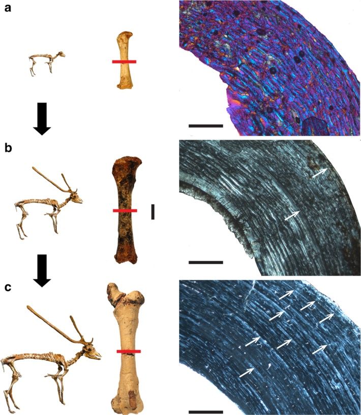
Kolb et al. BMC Evolutionary Biology (2015) 15:19                                                                 Page 2 of 15

Despite of the unresolved nature of megacerine phylogeny         absence of resorption [43] and more complete growth
[20], the small Candiacervus morphotypes must have               record, as shown by studies on growth marks in living
undergone size reduction since all their postulated main-        species, including deer of known age [43,44]. For example,
land sister-groups are significantly larger (e.g. Praemega-      using cementum analysis in molars, 99% of a sample of 51
ceros spp. with shoulder heights ranging from 0.9 m to           Spanish red deer could be aged within a one year confi-
1.50 m [18,19] or Cervus peloponnesiacus with a shoulder         dence interval [45].
height of just slightly less than one metre [21]. The kind          Palaeohistology previously led to the discovery in the
of dwarfism we observe in Candiacervus has been de-              island goat Myotragus balearicus from the Late Pleisto-
scribed as autapomorphic nanism by [22]. Candiacervus            cene of the Balearic Islands, of a ‘reptile’-like growth pat-
shows diversity in size, as six size classes of deer have been   tern consisting of lamellar-zonal bone throughout the
distinguished [16,17]. The smallest morphotype, C. ropalo-       cortex [46]. Myotragus was therefore hypothesized to
phorus, reached a shoulder height of about 40 cm, C. sp. II      have grown at low but variable rates and to have ceased
one of about 60 cm, and the largest one reached a height         its growth cyclically. Our investigation of Candiacervus
of about 1.65 m [23]. This phenomenon has been inter-            and of relevant mainland cervids, focusing on bone
preted as a case of adaptive radiation [24]. In the Middle       microstructure in growth series of various long bones,
to Late Pleistocene, Crete was characterized by dense for-       and dental histology in old adults, serves to examine
est as well as jagged rocks with several intermediate kinds      whether the pattern of growth of Myotragus is general
of environments, in which such a radiation could have oc-        among island artiodactyls. Longevity estimates, based on
curred [5]. Here we study Candiacervus ropalophorus and          the rest lines in the first molar of old individuals, were
C. sp. II, as these two size classes are small and are repre-    made. The first molar is the first permanent tooth to
sented by growth series we could sample. ‘Candiacervus           erupt [45], showing the most complete growth record in
sp. II’ may be a composite of three morphotypes of similar       deer. In order to further examine growth patterns across
size [17].                                                       cervids and to put life history data attained by histological
   Representing the other extreme of size with a shoulder        analyses into an allometric context, we investigated the re-
height of up to 2 m [20], Megaloceros giganteus has been         lation between body weight and growth rates [47-49].
a subject of extensive debates on evolutionary processes
[20,25,26]. It is best known from fossil occurrences in          Methods
Ireland from 11 to 12,000 BP [27] years ago and from             A total of 51 long bones, six phalanges, four lower first
possessing the largest antlers of any fossil or living spe-      molars and two upper first molars of Candiacervus sp. II
cies. Megaloceros was widespread in Europe and western           and Candiacervus ropalophorus, 14 long bones and five
Asia for 400,000 years and morphological and molecular           lower first molars of Megaloceros giganteus, and 13 long
analyses have supported a close relationship with fallow         bones and 2 lower first molars of Procervulus praeluci-
deer, Dama dama [20,28] (Figure 1a). The fossil record           dus were sampled (Table 1, see also Additional file 1:
of deer is long and complex, and Procervulus praeluci-           Methods). Sixteen long bones and two lower first molars
dus from the Early Miocene of Germany represents a               of Dama dama, and one femur each of Muntiacus
stem taxon that can help to reconstruct the evolution of         muntjak, Cervus elaphus and Alces alces, were sampled
life history features in deer [29] (Figure 1a).                  for comparison. Of Capreolus capreolus one femur and
   In order to enhance reproductive success life history         one metacarpal were sampled. Following standard pro-
traits can be selected by adjusting the developmental            cedures, the bones were coated and impregnated with
schedule to match environmental conditions [36]. Bone            epoxy resin (Araldite or Technovit) prior to sawing
microstructure can reveal such traits in mammals, which          and grinding. Long bones were transversely sectioned
generally exhibit bone matrices indicative of high rates         at mid-shaft where the growth record is most complete
of tissue deposition in juveniles, whereas after onset of        [e.g. 10]. For cementum analysis jaws were longitudin-
maturity a decrease in bone growth rate occurs resulting         ally cut through the cementum interroot pad of the
in deposition of highly organized bone tissue [37-39].           lower first molar and surfaces were impregnated with
Lines of arrested growth (LAGs) form from the first year         epoxy resin and finally ground and polished. Long
of an individual’s postnatal life as a result of annual ces-     bones of Megaloceros giganteus were also sampled by
sation of bone growth [40,41]. Counting these LAGs               using a diamond-studded core drill, with sampled
therefore provides the means to estimate minimum indi-           cores being subsequently processed [10,50]. Sections
vidual ages [40]. However, there can be decoupling of            were observed in normal transmitted and cross-
the number of LAGs in long bones and the actual age of           polarized light using a Leica DM 2500 M composite
old individuals leading to underestimation of individual         microscope equipped with Leica DFC 420 C digital
ages [42]. Dental cementum is a more accurate source             camera. Since there are no remarkable differences in
for estimating longevity in mammals, due to its usual            the bone tissue of the two Candiacervus morphotypes
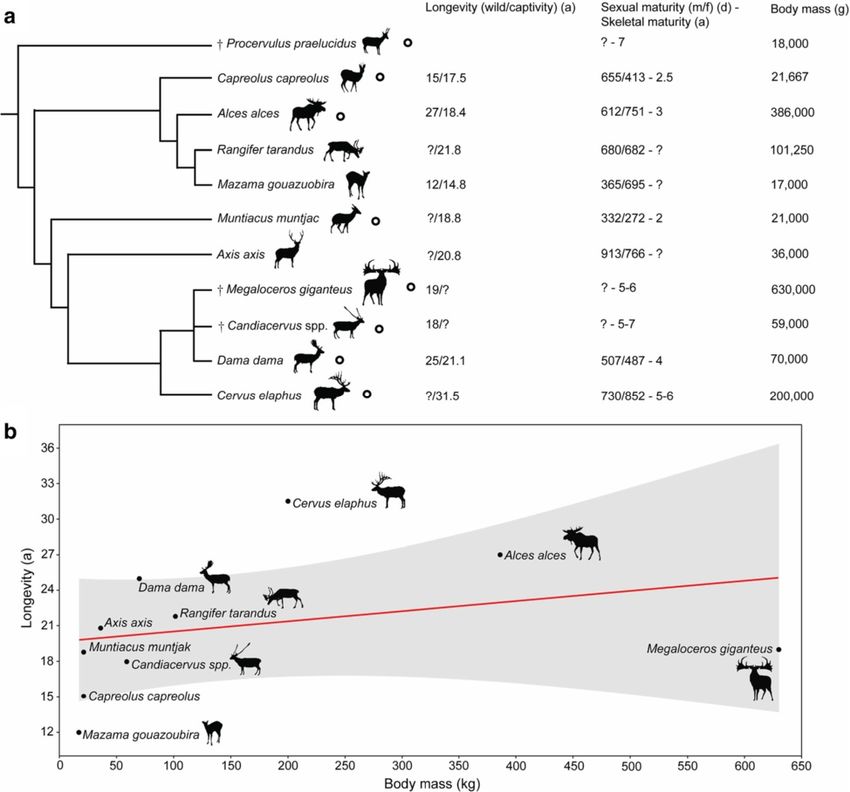
Kolb et al. BMC Evolutionary Biology (2015) 15:19                                                                                        Page 3 of 15

 Figure 1 Cervid phylogeny and life history. a) Phylogenetic relationships, maximal recorded life expectancy, onset of maturity, and average
 adult body mass of Candiacervus, Megaloceros, and relatives based on [17,20,23,28-35]. Cervid species studied are indicated by black circles. The
 trichotomy of the fallow deer (Dama) and its extinct relatives illustrates the unresolved nature of this well-supported clade. Body mass values are
 average. b) Linear regression relating maximum recorded longevity to body mass in cervids, and observed maxima within available samples of
 fossil species. Shaded region represents the 95% confidence interval. In cases in which unambiguous longevity data for wild animals were not
 available, data of captive specimens have been used.

sampled, they are treated together here. Polished tooth                      [41] and a 365 day growth period have been taken into
surfaces were observed using a Leica MZ 165 and MZ                           account as well (Additional file 2). Since growth zone
125 reflected-light microscope.                                              thickness may vary considerably within the cortex of one
   For quantification of growth rates, distances between                     bone, all measurements have been performed along the
LAGs, i.e. growth zones were measured with Leica IM                          anteroposterior axis in the anterior quadrant of each
50 Image Manager®, and annual growth rates per day                           section, whereas micrographs presented in this work
were calculated [51] by dividing growth zones by the                         have been taken from the best preserved and histologi-
number of days per growth period and year. The esti-                         cally most informative areas. Growth zone measure-
mate of number of days per growth period, i.e. 260 days,                     ments were performed for femora and tibiae since
is based on [41]. Growth period intervals (275–245 days)                     they are the most informative long bones in cervids
Kolb et al. BMC Evolutionary Biology (2015) 15:19                                                        Page 4 of 15

Table 1 Material used in this study
Species                         Object              Ontogenetic stage   Locality                   Specimen number
Candiacervus ropalophorus       Femur               adult               Gerani 4, Crete (Greece)   PIMUZ A/V 5195
"                               "                   adult               "                          PIMUZ A/V 5202
"                               "                   perinatal           "                          PIMUZ A/V 5207
"                               "                   perinatal           "                          PIMUZ A/V 5206
"                               Tibia               adult               "                          PIMUZ A/V 5188
"                               "                   adult               "                          PIMUZ A/V 5189
"                               "                   juvenile            "                          PIMUZ A/V 5208
"                               "                   juvenile            "                          PIMUZ A/V 5193
"                               "                   perinatal           "                          PIMUZ A/V 5191
"                               "                   perinatal           "                          PIMUZ A/V 5194
"                               Metatarsus          adult               "                          PIMUZ A/V 5192
"                               "                   juvenile            "                          PIMUZ A/V 5254
"                               "                   perinatal           "                          PIMUZ A/V 5205
"                               Humerus             adult               "                          PIMUZ A/V 5190
"                               "                   perinatal           "                          PIMUZ A/V 5187
"                               "                   perinatal           "                          PIMUZ A/V 5203
"                               Radius              adult               "                          PIMUZ A/V 5186
"                               "                   adult               "                          PIMUZ A/V 5199
"                               "                   perinatal           "                          PIMUZ A/V 5200
"                               Ulna                perinatal           "                          PIMUZ A/V 5255
"                               Metacarpus          adult               "                          PIMUZ A/V 5197
"                               "                   juvenile            "                          PIMUZ A/V 5198
"                               Lower M1            adult               "                          PIMUZ A/V 5196
Candiacervus sp. II             Femur               adult               Liko, Crete (Greece)       PIMUZ A/V 5218
"                               "                   juvenile            "                          PIMUZ A/V 5219
"                               "                   perinatal           "                          PIMUZ A/V 5244
"                               "                   perinatal           "                          PIMUZ A/V 5245
"                               Tibia               adult               "                          PIMUZ A/V 5222
"                               "                   juvenile            "                          PIMUZ A/V 5220
"                               "                   perinatal           "                          PIMUZ A/V 5221
"                               "                   perinatal           "                          PIMUZ A/V 5234
"                               Metatarsus          adult               "                          PIMUZ A/V 5240
"                               "                   adult               "                          PIMUZ A/V 5212
"                               "                   juvenile            "                          PIMUZ A/V 5213
"                               "                   juvenile            "                          PIMUZ A/V 5223
"                               "                   perinatal           "                          PIMUZ A/V 5224
"                               Humerus             adult               "                          PIMUZ A/V 5231
"                               "                   juvenile            "                          PIMUZ A/V 5236
"                               "                   perinatal           "                          PIMUZ A/V 5237
"                               Radius              adult               "                          PIMUZ A/V 5232
"                               "                   adult               "                          PIMUZ A/V 5233
"                               "                   juvenile            "                          PIMUZ A/V 5230
"                               "                   perinatal           "                          PIMUZ A/V 5211
"                               "                   perinatal           "                          PIMUZ A/V 5257
Kolb et al. BMC Evolutionary Biology (2015) 15:19                                                                      Page 5 of 15

Table 1 Material used in this study (Continued)
"                               "                   perinatal              "                                    PIMUZ A/V 5214
"                               Ulna                adult                  "                                    PIMUZ A/V 5215
"                               "                   juvenile               "                                    PIMUZ A/V 5225
"                               "                   perinatal              "                                    PIMUZ A/V 5226
"                               Metacarpus          adult                  "                                    PIMUZ A/V 5246
"                               "                   juvenile               "                                    PIMUZ A/V 5247
"                               "                   perinatal              "                                    PIMUZ A/V 5209
"                               "                   perinatal              "                                    PIMUZ A/V 5210
"                               1st Phalange        adult                  "                                    PIMUZ A/V 5238
"                               "                   juvenile               "                                    PIMUZ A/V 5239
"                               "                   perinatal              "                                    PIMUZ A/V 5216
"                               2nd Phalange        adult                  "                                    PIMUZ A/V 5217
"                               "                   juvenile               "                                    PIMUZ A/V 5235
"                               "                   perinatal              "                                    PIMUZ A/V 5227
"                               Rib                 adult                  "                                    PIMUZ A/V 5228
"                               Lower M1            adult                  "                                    PIMUZ A/V 5229
"                               "                   adult                  "                                    PIMUZ A/V 5243
"                               Upper M1            senescent (18 years)   "                                    PIMUZ A/V 5241
"                               "                   adult                  "                                    PIMUZ A/V 5242
Candiacervus sp.                Lower M1            senescent (12 years)   Bate cave, Crete (Greece)            PV M 82318 (NHML)
Procervulus praelucidus         Femur               adult                  Wintershof-West, Germany             BSPG 1937 II 23226
"                               "                   adult                  "                                    BSPG 1937 II 23227
"                               "                   juvenile               "                                    BSPG 1937 II 23228
"                               "                   juvenile               "                                    BSPG 1937 II 23229
"                               Tibia               adult                  "                                    BSPG 1937 II 23230
"                               "                   adult                  "                                    BSPG 1937 II 23231
"                               "                   juvenile               "                                    BSPG 1937 II 23232
"                               Humerus             adult                  "                                    BSPG 1937 II 23233
"                               "                   adult                  "                                    BSPG 1937 II 23234
"                               Radius              adult                  "                                    BSPG 1937 II 23235
"                               "                   adult                  "                                    BSPG 1937 II 23236
"                               "                   adult                  "                                    BSPG 1937 II 23237
"                               "                   adult                  "                                    BSPG 1937 II 23238
"                               Lower M1            adult                  "                                    BSPG 1937 II 12002
"                               "                   adult                  "                                    BSPG 1937 II 12040
Megaloceros giganteus           Femur               adult                  Craddanstown Rep. of Ireland         NMING:F7937/4
"                               "                   adult                  Baunmore Townland, Rep. of Ireland   NMING:F21306/13
"                               Tibia               adult                  Ballyragget, Rep. of Ireland         NMING:F22655/34
"                               "                   adult                  Buttevant, Rep. of Ireland           NMING:F22534/5
"                               "                   adult                  Baunmore Townland, Rep. of Ireland   NMING:F21306/14
"                               Metatarsus          adult                  North Sea sediments                  PIMUZ A/V 5256
"                               "                   adult                  Baunmore Townland, Rep. of Ireland   NMING:F21306/19
"                               "                   adult                  Buttevant, Rep. of Ireland           NMING:F22534/6
"                               Humerus             adult                  Ballyragget, Rep. of Ireland         NMING:F22655/37
"                               "                   adult                  Buttevant, Rep. of Ireland           NMING:F22534/2
Kolb et al. BMC Evolutionary Biology (2015) 15:19                                                                                              Page 6 of 15

Table 1 Material used in this study (Continued)
"                                  Radius-Ulna           adult                       Ballyragget, Rep. of Ireland                      NMING:F22655/36
"                                  "                     adult                       Buttevant, Rep. of Ireland                        NMING:F22534/3
"                                  Metacarpus            adult                       Ballyragget, Rep. of Ireland                      NMING:F22655/31
"                                  "                     adult                       Buttevant, Rep. of Ireland                        NMING:F22534/4
"                                  Lower M1              senescent (19 years)        Brühl (Schwetzingen), Deutschland                 PIMUZ A/V 2235
"                                  "                     senescent (16 years)        Kent'scavern, Torquay, UK                         PV OR 16800 (NHML)
"                                  "                     senescent (n.a.)            Wyhlen, Germany                                   BSPG 1957 I 398
"                                  "                     adult                       Rath, Rep. of Ireland                             NMING:F22654
"                                  "                     adult                       Craddanstown, Rep. of Ireland                     NMING:F7937/5
Dama dama                          Femur                 adult (wild)                Schrevenborn, Germany                             ZIUK 9630
"                                  "                     adult (captive)             Wildnispark Zürich, Switzerland                   PIMUZ A/V 5248
"                                  "                     adult (captive)             "                                                 PIMUZ A/V 5248
"                                  "                     juvenile (captive)          "                                                 PIMUZ A/V 5249
"                                  Tibia                 adult (wild)                Schrevenborn, Germany                             ZIUK 9630
"                                  "                     adult(captive)              WildnisparkZürich, Switzerland                    PIMUZ A/V 5248
"                                  "                     adult (captive)             "                                                 PIMUZ A/V 5248
"                                  "                     juvenile (captive)          "                                                 PIMUZ A/V 5249
"                                  Humerus               adult (wild)                Schrevenborn, Germany                             ZIUK 9630
"                                  "                     adult (captive)             Wildnispark Zürich, Switzerland                   PIMUZ A/V 5248
"                                  "                     adult (captive)             "                                                 PIMUZ A/V 5248
"                                  "                     juvenile (captive)          "                                                 PIMUZ A/V 5249
"                                  Radius-Ulna           adult (wild)                Schrevenborn, Germany                             ZIUK 9630
"                                  "                     adult (captive)             Wildnispark Zürich, Switzerland                   PIMUZ A/V 5248
"                                  "                     adult (captive)             "                                                 PIMUZ A/V 5248
"                                  "                     juvenile (captive)          "                                                 PIMUZ A/V 5249
"                                  Lower M1              adult (wild)                Schrevenborn, Germany                             ZIUK 9630
"                                  "                     adult (captive)             Wildnispark Zürich, Switzerland                   PIMUZ A/V 5248
Capreolus capreolus                Femur                 adult (wild)                Schrevenborn, Germany                             ZIUK 9872
"                                  Metatarsus            juvenile (wild)             Hittnau, Switzerland                              PIMUZ A/V 5251
Muntiacus muntjak                  Femur                 adult (captive)             Tierpark Hagenbeck, Hamburg, Germany              ZIUK 7994
Cervus elaphus                     "                     adult (wild)                Barmstedt, Germany                                ZIUK 23517
Alces alces                        "                     adult (wild)                Norway                                            ZMUZ 20242
Specimens used in this study with ontogenetic stage, locality of death/fossil site, specimen number and thin section number.
Institutional Abbreviations: BSPG Bayerische Staatssammlung für Paläontologie und Geologie, Munich, Germany; NBC Netherlands Centre for Biodiversity Leiden,
The Netherlands; NHML Natural History Museum London, UK; NMING National Museum of Ireland - Natural History; PIMUZ Paläontologisches Institut und Museum,
Universität Zürich, Switzerland; ZIUK Zoologisches Institut der Universität Kiel, Germany; ZMUZ Zoologisches Museum der Universität Zürich, Switzerland.

(see also Additional file 1: Methods). For growth rate                           woven-fibred bone as primary tissue (Figure 2a). In the
graphs Microsoft Office Excel 2010© has been used.                               inner cortex, vascularization tends to be reticular, whereas
Regression analyses (ordinary least squares) for aver-                           in the middle and outer cortex vascularization has a plexi-
age and growth rates sensu Sander & Tückmantel [51]                              form pattern. With increasing age, the amount of vascu-
were performed using Past3.0 [52]. All graphs have                               larisation and woven bone decreases, with the former
been redrawn using Adobe Illustrator CS5© .                                      changing from a plexiform to laminar organisation in the
                                                                                 middle and outer cortex, whereas the amount of lamellar
Results and discussion                                                           or parallel-fibred bone within the fibrolamellar matrix in-
Histological description of primary bone                                         creases (Figure 2a-c). The outermost layer of the outer
Newborn dwarf Candiacervus (C. ropalophorus and C.                               cortex in adult Candiacervus sampled is composed of a
sp. II) exhibit fibrolamellar bone with a high amount of                         narrow layer of avascular lamellar bone, called the outer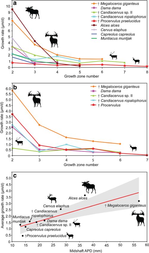
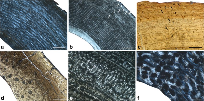
Kolb et al. BMC Evolutionary Biology (2015) 15:19                                                                                        Page 7 of 15

 Figure 2 Histological growth series of dwarf deer femora with skeletal reconstructions [55] and specimens sampled (anterior view).
 Red bars indicate plane of sectioning. Candiacervus sp. II under crossed polarised light (xpl). a) Bone cortex of a perinatal specimen (PIMUZ A/V
 5244) showing a mainly plexiform arrangement of vascular canals with a high amount of woven-fibred bone (violet areas; additional use of
 lambda compensator). Scale bar, 0.5 mm. b) Juvenile specimen (PIMUZ A/V 5219). Scale bars, 20 mm (femora all to scale), 1 mm (bone cortex).
 c) Adult specimen (PIMUZ A/V 5218). Note the increasing amount of parallel-fibred bone (bright areas) and the occurrence of lines of arrested
 growth (LAGs, white arrows) throughout ontogeny. Scale bar, 1 mm.

circumferential layer (OCL) [53] in this work and also                       Candiacervus, Procervulus, and Muntiacus it changes
referred to as external fundamental system (EFS, e.g.                        directly from plexiform/laminar to avascular in the outer
sensu [42], see also [54]). We prefer the term outer cir-                    circumferential layer. This is evidently a feature separating
cumferential layer for being more descriptive than the                       large and intermediate from small-sized deer, including
term external fundamental system. An inner circum-                           dwarf Candiacervus. Moreover, the density of vascu-
ferential layer [38] is well developed in all adult fem-                     lar canals is higher in intermediate-sized and larger
ora. Long bones of Candiacervus indicate, based on                           deer compared to smaller taxa (Figure 3a-c, see also
growth line counts, minimum ages of about two years                          Additional file 1: Table S2 and Figure S1). Because it
for the juveniles sampled.                                                   is a juvenile specimen and still shows less vascularisa-
   Adult Megaloceros, Dama, Cervus, and Alces show in                        tion than the adult Dama and Megaloceros, specimen
all sampled long bones a similar arrangement of bone                         BSPG 1937 II 23227 is especially illustrative concerning
tissue types to each other. Vascularisation in the outer                     the low amount of vascularization in the small sized
part of the cortex is partly longitudinal, whereas in dwarf                  cervid Procervulus (Figure 3a-c).
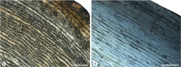
Kolb et al. BMC Evolutionary Biology (2015) 15:19                                                                                      Page 8 of 15

 Figure 3 Cervid bone tissue and growth marks. Increasing femoral vascularisation (a-c) accompanied by increasing body size in a) juvenile
 Procervulus specimen BSPG 1937 II 23227, medial cortex (xpl; scale bar, 0.5 mm), b) adult Dama specimen ZIUK 9630, medial cortex (xpl; scale bar,
 1 mm), and c) adult Megaloceros specimen NMING: F21306/13 under plane polarised light, anterior cortex (ppl, scale bar 1 mm). Note the low
 amount of vascularisation in the fibrolamellar bone of the juvenile Procervulus. Occurrence of LAGs indicated by black/white arrows and the outer
 circumferential layer by white brackets. Numbers indicate growth zones. Bone surfaces at the top, medullary cavities at bottom left. d) Tibia of
 adult Candiacervus sp. II (PIMUZ A/V 5222) showing Haversian bone in the inner part of the posterior cortex (bright area) and plexiform
 fibrolamellar bone in the middle part (lpl; scale bar, 1 mm). Occurrence of LAGs indicated by white arrows. Bone surface at top right, medullary
 cavity at bottom left. e) Radiating fibrolamellar bone in a metacarpal of adult Megaloceros giganteus (NMING:F22534/4, xpl; scale bar, 0.5 mm).
 f) First phalange of perinatal Candiacervus sp. II (PIMUZ A/V 5216) showing reticular vascularisation of mainly woven-fibred bone (xpl; scale
 bar, 0.2 mm).

   A longitudinal section of a Megaloceros femur confirms                  Dama and Megaloceros show areas of radiating fibrola-
the low amount of woven-fibred bone in plexiform bone                      mellar bone interdigitating with the otherwise plexi-
tissue [56] (Figure 4a,b). However, since woven-fibred                     form bone tissue. In general, adult radii of all sampled
bone is present, we follow [57] in using the term fibro-                   deer have a similar arrangement of bone tissue types, i.e.
lamellar bone. In general, the bone tissue found in the                    plexiform fibrolamellar bone with a varying amount of
femora and humeri gives a similar picture. The differ-                     Haversian bone. The amount of woven bone of perinatal
ences in the amount of vascularisation observed in                         ulnae of dwarf Candiacervus is high in the inner cortex.
dwarf Candiacervus and large/intermediate sized cer-                         Similarity of bone tissue types in Candiacervus,
vids are less obvious in the humeri than those seen in                     Megaloceros, and Capreolus shows a comparable mode
the femora. Unlike Candiacervus (Figure 3d), tibiae of                     of growth in the metapodials. A remarkable difference

 Figure 4 Bone cortex of Megaloceros giganteus. Femur (NMING: F21306/13) in transverse (a) and longitudinal (b) section under crossed
 polarised light (bone surface top right). Note the low amount of woven fibred bone (dark areas) in the longitudinal section.
Kolb et al. BMC Evolutionary Biology (2015) 15:19 Page 9 of 15
distinguishing adult Megaloceros from Candiacervus is the amount of remodelling is low enough to leave a
the occurrence of layers of radiating fibrolamellar bone sufficient growth record.
(Figure 3e) in the middle and outer cortex of Megaloceros. Tibiae of juvenile Candiacervus and Dama start being
The inner circumferential layer is relatively thicker in remodelled mainly in the medial and lateral parts of the
Megaloceros than in Candiacervus. middle and inner cortex, leading to the deposition of
Phalanges of newborn Candiacervus specimens show dense Haversian bone (Figure 3d). In rare cases, dense
fibrolamellar bone with reticular vascularisation (Figure 3f). Haversian bone is also found in the outermost part of
During ontogeny, the vascular organisation becomes plexi- the cortex in Megaloceros. Again, however, the amount
form, but with increasing age this is replaced by increasing of remodelling is low enough to leave a sufficient growth
amounts of poorly vascularised lamellar/parallel-fibred record.
bone in the sub-periosteal region. Haversian bone in juvenile radii of Candiacervus and
Dama indicates an early onset of secondary remodelling
Secondary bone and remodelling processes in the inner cortex. Strong remodelling in adult radii of
Perinatal specimens of Candiacervus show no signs of Candiacervus, Dama, and Megaloceros, especially in the
bone remodelling. In general, resorption of primary bone posterior area of the inner cortex, obscures the growth
and deposition of secondary osteons in cervid long bones record to a large degree in these bones.
starts in juveniles (Figure 5a). Large areas of Haversian Ulnae of all juvenile deer species sampled are already
bone in adults indicate strong bone remodelling during remodelled to a high degree, especially in the inner cor-
ontogeny. Apart from the femora, which have a mainly tex surrounding the medullary cavity, indicated by dense
circular outline in cross section, Haversian bone is most Haversian bone. Adult ulnae are strongly remodelled
dense where the curvature of the cortex is greatest, but in leaving only small areas of primary bone tissue in the
all long bones and specimens the area most affected by re- posterior part of the cortex. During ontogeny, the me-
modelling is the posterior area of the cortex. dullary cavity shifts to the anterior area of the cortex
In the femora, remodelling starts in the juvenile Can- being subsequently closed by the deposition of endosteal
diacervus specimens with scattered secondary osteons lamellar bone which is in turn subsequently replaced by
in the middle cortex, mainly in its posterior part. Adult dense Haversian bone (Figure 5b). Due to this strong re-
femora of all deer species sampled show strong remod- modelling of the ulnae in all deer species sampled, and
elling (i.e. Haversian bone) in the posterior part, ob- since only small areas of primary plexiform bone tissue
scuring the growth record in this area of the bone. are left in the bone cortex, skeletochronological inter-
Remodelling is strongest in the cortical area of the pretations are not feasible.
linea aspera. Remodelling in metapodials begins with the develop-
Similar to the femora, remodelling in the humeri of ment of Haversian bone in the inner cortex and is already
juvenile specimens of Candiacervus and Dama starts strongly developed in juvenile specimens. In all specimens,
in the middle zone of the medial part of the cortex the area most affected by remodelling is the posterior area
(Figure 5a). Adult humeri of all deer groups sampled of the cortex. Adult deer metapodials are strongly remod-
show more remodelling than the femora. Nevertheless, elled, occupying about half of the cortex and obliterating
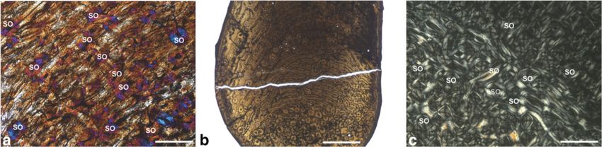
Figure 5 Cervid bone remodelling. a) Humerus of juvenile Candiacervus sp.II specimen PIMUZ A/V 5236 (xpl, lambda compensator,
scale bar 0.5 mm). Note the scattered secondary osteons (SO). b) Ulna of adult Candiacervus sp. II specimen PIMUZ A/V 5215 (lpl, scale
bar 1 mm) displaying plexiform fibrolamellar bone (centre) and dense Haversian bone (bottom). Note that the medullary cavity (bottom)
has been subsequently closed by the deposition of endosteal lamellar bone which was in turn replaced by secondary Haversian bone.
Anterior at the bottom. c) Dense Haversian bone in a metacarpal of adult Megaloceros giganteus specimen NMING: F22534/4 (xpl, scale
bar 0.5 mm).Kolb et al. BMC Evolutionary Biology (2015) 15:19 Page 10 of 15
the growth record by development of dense Haversian to and verification of known bone apposition rates in ex-
bone (Figure 5c). tant and fossil vertebrates [51,60].
In contrast to dwarfed forms of Candiacervus and to
Skeletochronology and growth mark analysis Dama, Megaloceros femora indicate up to five times
Cyclical growth patterns have been observed in many higher growth rates, with the second growth zone yielding
extant artiodactyls [41]. However, in mammals bone a rate of 7.69 μm/d, the third one 3.69 μm/d, and growth
resorption and remodelling may occur throughout zones four to six between 2.04 – 1.35 μm/d (Figure 6a).
ontogeny and LAG counts and age are apparently The femora of C. ropalophorus indicated a growth rate
decoupled in old individuals [40,42,43]. Therefore, of 2.19 μm/d to 1.81 μm/d in growth zones two to four
individual ages are often underestimated by bone histo- (Figure 6a). In the following year the growth rate slightly
logical studies, making cementum analysis a crucial decreased to 1.04 μm/d. The growth rate of Candiacer-
tool in order to study longevity in fossil cervids (see vus sp. II decreased from 3.34 μm/d in growth zone two
also Additional file 1: Discussion). to 1.19 μm/d in growth zone four. In growth zones five
LAGs are present in all deer taxa sampled. Femora, to seven the growth rate ranged from 0.69 μm/d to
tibiae, and humeri of adult specimens show, due to rela- 0.81 μm/d. The femoral growth rate of Dama is higher
tively low remodelling, the highest LAG counts. The max- than that of C. ropalophorus and equal to that of Can-
imum LAG counts seen in adult femora are eight in dwarf diacervus sp. II in the second growth zone (3.34 μm/d).
Candiacervus (n = 3, Figure 2c), six in Dama (n = 2), and After that, growth rate strongly decreased below the
10 in Megaloceros (n = 2). rates of C. ropalophorus and sp. II (0.73 and 0.84 μm/d).
In order to quantify growth rates in the sampled deer The growth rates recorded in the tibiae are similar to
taxa, we measured non-remodelled cortical growth the ones obtained for the femora. C. ropalophorus grew
zones until the “virtual end of circumferential bone in zone two at a rate of 2.47 μm/d, whereas Candiacer-
growth” [42]. Growth marks in the outermost part of the vus sp. II only grew at 0.69 μm/d (Figure 6b). D. dama,
bone cortex, not giving a signal because of similarity and at 2.54 μm/d, occupies an intermediate position between
diminutiveness of growth zone thickness, have been the dwarfed deer and Megaloceros (5.81 μm/d). Growth
omitted. It has recently been shown that in antelope rate strongly decreases from zone two to zone three in
(Addax nasomaculatus) femora the first LAG is resorbed most deer species sampled: 2.76 μm/d in Procervulus,
during ontogeny [39]. Ruminants such as antelopes and 3.07 μm/d in Megaloceros but only 0.34 μm/d in C. sp. II.
cervids show similar long bone morphology as well as There is discrepancy among taxa, and C. ropalophorus, al-
similar arrangement of bone tissue types, bone remodel- though the smallest species, shows about four times
ling, and resorption patterns [41,58]. Superimposition of higher growth rate in the second growth zone (similar
sections of femora and tibiae of perinatal, juvenile, and to Dama) compared to Candiacervus sp. II (Figure 6b).
adult dwarf Candiacervus, juvenile and adult specimens of This demonstrates the diversity of life history parameters
Procervulus praelucidus, and a juvenile as well as two across morphotypes of Candiacervus in Crete during the
adult specimens of known-age Dama dama however indi- Pleistocene [61,62].
cate that no LAG is lost during ontogeny in femora and Average growth rates of 0.46 μm/d (Figure 6c) in
tibiae of these cervids. On the grounds of phylogenetic femora of Procervulus were the lowest measured for all
parsimony we consider it as justified to assume that in the deer taxa sampled, lying below the lower limit of
general bone resorption patterns are identical throughout their 95% confidence interval. Muntiacus, Capreolus,
cervids. This approach made retrocalculation techniques Dama and dwarf Candiacervus show average to low
as performed for dinosaurs dispensable [12,59]. In order growth rates around 1.4 μm/d, whereas Cervus elaphus
to make growth rate measurements comparable, we (2.58 μm/d) had distinctly higher growth rates lying on
numbered the growth zones of adult specimens start- the upper limit of the 95% confidence interval. Alces
ing with two since the first growth zone is at least par- alces shows with 3.68 μm/d the highest average growth
tially resorbed and not available for skeletochronology rates. In contrast, the absolute high growth rates (based
(Figure 3b,c,d). on the growth zones preserved in the cortical bone tissue)
Additionally, and in order to verify our observations of Megaloceros are relatively low given the regression
made by growth zone counts and measurements, we (Figure 6c, average 3.23 μm/d), but still within the limits
followed [51] in determining how fast dwarf Candiacervus, of the 95% confidence interval.
Megaloceros, and Procervulus grew over a hypothetical Growth rates sensu Sander & Tückmantel plotted
365 days growth period by assessing growth rates sensu against the anteroposterior bone diameter as a proxy for
Sander & Tückmantel and comparing them to the values body mass indicate three groups (Figure 7): A group with
observed in extant cervids. Although bone growth rates high growth rates including Megaloceros (14.22 μm/d),
per day are always approximations, they allow comparison Cervus elaphus (ZIUK 23517; 12.66 μm/d), Alces (ZMUZKolb et al. BMC Evolutionary Biology (2015) 15:19 Page 11 of 15 Figure 6 Cervid growth rates. a) Graph of growth zone measurements of cervid femora sampled. Points indicate sample means or measurements of single specimens (see also Additional file 2). Note exceptionally high growth rates in the first two growth zones of Alces and Megaloceros as well as exceptionally low rates of Procervulus. Growth zones numbered, starting with two for the innermost complete zone of the cortex. b) Graph of cervid tibiae sampled. Note the eight times higher growth rate in growth zone two of Megaloceros compared to Candiacervus sp. II (and still twice as high as in C. ropalophorus). c) Regression of average growth rates in cervid femora (n = 12, r = 0.85111, p = 0.0036142.). Shaded region represents the 95% confidence interval. Anteroposterior diameter (APD) of femoral midshaft region is taken as proxy for body mass. 20242; 12.58 μm/d), and Dama (12.35 μm/d); an intermedi- Candiacervus ropalophorus to 2.6 μm/d in Procervulus ate group with Capreolus (ZIUK 9872; 6.79 μm/d) and (Figure 7). Dama and Cervus elaphus plot above the Muntiacus (ZIUK 7994; 5.75 μm/d); and a group showing upper limit of the 95% confidence interval whereas only low growth rates, including Candiacervus sp. II (PIMUZ Candiacervus sp. II lies well below the lower limit of the A/V 5218, 3.7 μm/d), and ranging from 4.16 μm/d in 95% confidence interval. All other cervids sampled show
Kolb et al. BMC Evolutionary Biology (2015) 15:19                                                                                    Page 12 of 15

 Figure 7 Regression of growth rates sensu Sander & Tückmantel in cervid femora (n = 12, r = 0.78168, p = 0.012835). Shaded region
 represents the 95% confidence interval. Anteroposterior diameter (APD) of femoral midshaft region is taken as proxy for body mass (see also
 Additional file 2).

growth rates within the 95% range given their body size                    context, show an extended lifespan compared to other deer
(see also Additional file 1: Discussion).                                  of similar body size such as Mazama with a maximum
                                                                           longevity of 12 years in the wild (Figure 1a). This is well in
Skeletal maturity estimates                                                accordance with observations of a recent study on popula-
Examination of femora of extant cervid taxa revealed the                   tion structure and dynamics in dwarf Candiacervus [62].
occurrence of OCLs not coeval with the timing of sexual                      A positive linear relationship between body mass and
maturity, as reported for femora of antelopes [39]. An                     longevity has been demonstrated in bats and mammals
adult specimen of Dama dama (ZIUK 9630; Figure 3b)                         in general [47,65], although this is difficult to assess in
shows three LAGs before the OCL, in contrast to the                        our cervid data set (Figure 1b) because of issues of com-
onset of sexual maturity which has been reported to                        parability: for example, the 32-year maximum age of a
occur during the second year of life in Dama dama [63]                     captive specimen of Cervus elaphus [63], a farmed and
(Figure 1a). These observations suggest that the transi-                   extensively studied species, is probably anomalously high.
tion of the fibrolamellar complex (FLC) to the OCL,                        Conversely, Megaloceros appears short-lived when body
which is not clearly definable in every specimen, is indi-                 mass is taken into account (Figure 1b), although the sam-
cating cervid skeletal maturity sensu [64] and not sexual                  ple size was small. Clearly, there is much diversity in life
maturity. This is well in accordance with known data of                    history across deer species, and examination of other pop-
skeletal maturity for Dama [30,31], and a recent study                     ulations of Megaloceros may reveal more diversity in the
on growth marks in the bone tissue of ruminants that                       giant deer than we have recorded in our study [18,19].
examined cervid bone histology in detail [41]. The bone
cortex of dwarf Candiacervus femora indicates skeletal                     Conclusions
maturity at five to seven years whereas Megaloceros                        Our histological observations indicate lower growth
reached skeletal maturity at five to six years. One Procer-                rates in dwarf Candiacervus than in Megaloceros. The
vulus specimen indicates attainment of skeletal maturity                   presence of laminar bone tissue in the middle and outer
at seven years whereas Cervus elaphus (four to six years)                  cortex of adults of small-sized deer (dwarf Candiacervus,
ranges with its timing of skeletal maturity between                        Procervulus and Muntiacus) suggests lower growth rates,
Megaloceros and Alces (three years) [31].                                  in contrast to the occurrence of plexiform bone in inter-
                                                                           mediate to large sized forms. Growth rates determined
Cementum analysis and longevity                                            by growth zone measurements in femora and tibiae indi-
Tooth cementum analysis of first molars of Candiacervus                    cate comparable growth rates of intermediate sized and
provided an age of four years for a juvenile Candiacervus                  small deer species, with slower growth in the stem group
sp. II and an age of at least nine years for an adult speci-               cervid Procervulus. Growth rates in the two small Can-
men of C. ropalophorus. Two senile Megaloceros giganteus                   diacervus morphotypes are different, underscoring the
specimens revealed ages of 16 and 19 years (Figure 8a,b).                  flexibility of growth strategies and the importance of a
Rest lines in two old Candiacervus specimens gave ages of                  resolved phylogenetic framework to study heterochrony.
12 (dwarf Candiacervus sp.) and 18 years (Candiacervus                     Skeletal maturity data suggest late maturation for dwarf
sp. II, Figure 8c). Dwarf Candiacervus thus, in an allometric              Candiacerus and Procervulus in comparison to a similarly
Kolb et al. BMC Evolutionary Biology (2015) 15:19 Page 13 of 15
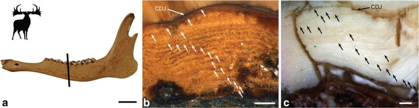
Figure 8 Cervid tooth histology. a) Senescent Megaloceros specimen PIMUZ A/V 2235. Left mandible in lateral view. Black bar indicates plane
of sectioning. Scale bar, 50 mm. b) Tooth cementum of same specimen showing 19 rest lines (white arrows). Scale bar, 0.5 mm. c) Tooth
cementum of the upper first molar of Candiacervus sp. II (PIMUZ A/V 5241) showing 18 rest lines (black arrows). CDJ = Cementum-dentine
junction. Scale bar, 0.5 mm. Direction of cementum apposition to the bottom.
small cervid such as Muntiacus attaining skeletal maturity shorter persistence time and perhaps to the larger size
in two years [32]. of Crete [3,8].
The landmasses of islands have been hypothesized of In life history theory, slow-developing long-lived species
being able to support only a limited number of primary are typically associated with low fecundity and rapidly-
producers affecting the energy flow at higher trophic developing short-lived species with high fecundity [36,70].
levels. As a consequence, energy-poor islands are ex- The condition found in Candiacervus has features in
pected to be impoverished in competitors and predators common with that of Myotragus [46], but is achieved with
making especially high growth rates and high reproduct- less far reaching modification of bone tissue, as indicated
ive rates dispensable to unnecessary [32,46,66-68]. A by the absence of lamellar-zonal bone throughout the cor-
delay in the attainment of maturity was recorded for the tex. Neither does the Myotragus pattern occur in the
dwarfed island bovid Myotragus balearicus [46], and was pygmy mammoth, Mammuthus exilis, from Santa Rosa
thought to be associated with synchronisation of meta- Island, California, whose bone cortex is characterized by
bolic requirements to fluctuating resource levels. The laminar fibrolamellar bone [71]. Therefore we suggest
delay of attainment of maturity in the island cervid variable modes of life history and size evolution among is-
Candiacervus and the continental Procervulus demon- land mammals in line with [65-68,72].
strates the variability of life history parameters in is-
land as well as continental cervids. This might point
towards fluctuating resource levels in the Late Pleisto- Availability of supporting data
cene Crete, selecting for a growth pattern recalling that The data sets supporting the results of this article are in-
of the stem-cervid Procervulus. cluded within the article (and its additional files).
The oldest individual seen in our cementum analysis High resolution versions of the histological figures
of Megaloceros was 19 years, comparable to maximum provided in this article are available on MorphoBank [73],
longevity in extant Dama. This find extends an age Project P2083 (http://www.morphobank.org/permalink/?
based on cementum analysis [25] by five years and lies P2083).
below another estimate [69] also based on cementum
analysis, by four years. However, [69] did not illustrate
cementum rest lines of the specimen studied. We there- Additional files
fore consider the result of our cementum analysis as the
highest rest line count in Megaloceros. The oldest indi- Additional file 1: Includes Table S1, additional discussion on
individual age estimates and growth rates, additional information
vidual seen in our cementum analysis of Candiacervus on methods used in this study, additional references, and
was 18 years, indicating prolonged longevity for a deer Figure S1.
of this body size. Additional file 2: Primary data for growth rate analysis: Growth zone
The exact persistence time of the Candiacervus radi- measurements, mean species growth rates, average species growth
rates, bone diameter, OCL thickness, and number of non-OCL lines of
ation on Crete is not known but was apparently much arrested growth (for growth rates sensu Sander & Tückmantel).
shorter, i.e., less than 0.5 myrs [5], compared to Myo-
tragus balearicus, which dwelt on Majorca for 5.2 myrs
[46]. The less extreme modification of bone tissue Competing interests
observed in dwarf Candiacervus could be related to The authors declare that they have no competing interests.Kolb et al. BMC Evolutionary Biology (2015) 15:19 Page 14 of 15
Authors’ contributions 13. Erickson GM, Makovicky PJ, Currie PJ, Norell MA, Yerby SA, Brochu CA.
CK and MRS-V designed the study and wrote the manuscript, CK, TMS, MAJS Gigantism and comparative life history parameters of tyrannosaurid
and CA collected and analysed histological data, JDV, AML, NTM, and GER dinosaurs. Nature. 2004;430:772–5.
provided materials and taxonomic/stratigraphical information, all authors 14. Chinsamy-Turan A (ed.): Forerunners of Mammals:Rradiation, Histology,
contributed to the final interpretation and editing of the manuscript. All Biology. Indiana: Indiana University Press;2012.
authors read and approved the final manuscript. 15. Huttenlocker AK, Botha-Brink J. Bone microstructure and the evolution of
growth patterns in Permo-Triassic therocephalians (Amniota, Therapsida) of
Acknowledgments South Africa. PeerJ. 2014;2:e325.
We thank Naturalis Biodiversity Center, National Museum of Ireland, Natural 16. de Vos J. The endemic Pleistocene deer of Crete. P K Ned Akad B.
History, Natural History Museum London (NHML), Bayerische Staatssammlung 1979;82(1):59–90.
für Paläontologie und Geologie, Wildnispark Zürich, Frank Zachos (now 17. de Vos J. The Endemic Pleistocene Deer of Crete, vol. 31. Amsterdam:
Naturhistorisches Museum Wien), Heiner Luttmann, Zoologisches Institut der North-Holland Publishing Company; 1984.
Universität Kiel, Marianne Haffner, Zoologisches Museum der Universität 18. Vislobokova IA. Giant deer: origin, evolution, role in the biosphere. Paleontol
Zürich, Emma Bernard (NHML) and Heinz Furrer, Paläontologisches Institut J. 2012;46(7):643–775.
und Museum der Universität Zürich (PIMUZ) for providing specimens for 19. Vislobokova IA. Morphology, taxonomy, and phylogeny of
histological study, Alexandra van der Geer (Naturalis Biodiversity Center, megacerines (Megacerini, Cervidae, Artiodactyla). Palaeontol J.
Leiden) and two anonymous reviewers for constructive and improving 2013;47(8):833–950.
criticism, as well as Vladimir Blagoderov, and Tony Wighton (NHML) for 20. Lister AM, Edwards CJ, Nock DAW, Bunce M, van Pijlen IA, Bradley DG, et al.
technical assistance. Vivien Jaquier, Fiona Straehl, Madeleine Geiger, James The phylogenetic position of the 'giant deer' Megaloceros giganteus. Nature.
Neenan, Juan Carrillo, Sarah Bolliger, Markus Hebeisen, Rosi Roth and Jérôme 2005;438(7069):850–3.
Gapany (all PIMUZ) are thanked for various support and discussion. This work 21. Sickenberg O. Eine Säugetierfauna des tieferen Biharium aus dem Becken
was funded by the SNSF (3100A0-133032/1 and 31003A-149605 to MRS-V; von Megalopolis (Peloponnes, Griechenland). Annales Géologiques des Pays
31003A-149506 to TMS) and the Forschungskredit of the University of Zurich Helléniques. 1975;27:25–73.
(No. 8264 to CK). 22. Gould GC, MacFadden BJ. Gigantism, dwarfism, and Cope’s rule: “Nothing in
evolution makes sense without a phylogeny”. B Am Mus Nat Hist.
Author details 2004;285:219–37.
1
Paläontologisches Institut und Museum der Universität Zürich, Karl 23. de Vos J. Pleistocene deer fauna in Crete: its adaptive radiation and
Schmid-Strasse 4, CH-8006 Zürich, Switzerland. 2Department of Earth extinction. Tropics. 2000;10(1):125–34.
Sciences, The Natural History Museum, Cromwell Road, London SW7 5BD, 24. de Vos J, Van der Geer A. Major patterns and processes in biodiversity:
UK. 3Department of Animal and Vegetal Biology and Ecology, Faculty of taxonomic diversity on islands explained in terms of sympatric speciation.
Experimental Sciences, University of Jaén, Jaén 23071, Spain. 4Naturalis In: WW H, EJ A, editors. World Islands in Prehistory, International Insular
Biodiversity Center, Postbus 9517, 2300 RA Leiden, The Netherlands. Investigations, V Deia International Conference of Prehistory. Oxford: British
5
Department of Integrative Zoology, IBL, Leiden University, Sylviusweg 72, Archaeological Reports International Series; 2002. p. 395–405.
Postbus 95052300 RA Leiden, The Netherlands. 6Bayerische Staatssammlung 25. Chritz KL, Dyke GJ, Zazzo A, Lister AM, Monaghan NT, Sigwart JD.
für Paläontologie und Geologie, Richard-Wagner-Strasse 10, D-80333 Palaeobiology of an extinct Ice Age mammal: Stable isotope and
München, Germany. 7National Museum of Ireland-Natural History, Merrion cementum analysis of giant deer teeth. Palaeogeogr Palaeocl.
Street, Dublin 2, Ireland. 2009;282(1–4):133–44.
26. Gould SJ. Positive allometry in antlers of the "Irish Elk", Megaloceros
Received: 22 July 2014 Accepted: 27 January 2015 giganteus. Nature. 1973;244:375–6.
27. Woodman PC, McCarthy M, Monaghan NT. The Irish Quaternary fauna
project. Quaternary Sci Rev. 1997;16:129–59.
References 28. Hughes S, Hayden TJ, Douady CJ, Tougard C, Germonpré M, Stuart A, et al.
1. Foster JB. Evolution of mammals on islands. Nature. 1964;202:234–5. Molecular phylogeny of the extinct giant deer, Megaloceros giganteus. Mol
2. Lomolino MV. Body size of mammals on islands: the island rule reexamined. Phylogenet Evol. 2006;40:285–91.
Am Nat. 1985;125:310–6. 29. Rössner GE. Odontologische und schädelanatomische Untersuchungen an
3. Lomolino MV, van der Geer AA, Lyras GA, Palombo MR, Sax DF, Rozzi R. Of Procervulus (Cervidae, Mammalia). Münchner Geowissenschaftliche
mice and mammoths: generality and antiquity of the island rule. J Biogeogr. Abhandlungen (A). 1995;29:1–128.
2013;40:1427–39. 30. McElligott AG, Mattiangeli V, Mattiello S, Verga M, Reynolds CA, Hayden TJ.
4. Lister AM. Rapid dwarfing of red deer on jersey in the last interglacial. Fighting tactics of fallow bucks (Dama dama, Cervidae): Reducing the risks
Nature. 1989;342:539–42. of serious conflict. Ethology. 1998;104:789–803.
5. van der Geer A, Lyras G, de Vos J, Dermitzakis M. Evolution of Island 31. Habermehl K-H. Altersbestimmung bei Wild- und Pelztieren - Möglichkeiten
Mammals. Adaptation and Extinction of Placental Mammals on Islands. und Methoden - Ein praktischer Leitfaden für Jäger, Biologen und Tierärzte.
Wiley-Blackwell: Sussex; 2010. Verlag Paul Parey: Hamburg, Berlin; 1985.
6. Quintana J, Köhler M, Moyà-Solà S. Nuragulus rex, gen. et sp. nov., an 32. Pei K. Post-natal growth of the Formosan Reeves' Muntjac Muntiacus reevesi
endemic insular giant rabbit from the Miocene of Minorca (Balearic Islands). micrurus. Zool Stud. 1996;35:111–7.
J Vertebr Paleontol. 2011;31(2):231–40. 33. Palombo MR, Köhler M, Moya-Sola S, Giovinazzo C. Brain versus body mass
7. Palkovacs EP. Explaining adaptive shifts in body size on islands: a life history in endemic ruminant artiodactyls: a case studied of Myotragus balearicus
approach. Oikos. 2003;103:37–44. and smallest Candiacervus species from Mediterranean Islands. Quatern Int.
8. Lomolino MV, Sax DF, Palombo MR, van der Geer A. Of mice and 2008;182:160–83.
mammoths: evaluations of causal explanations for body size evolution in 34. Gilbert C, Ropique A, Hassanin A. Mitochondrial and nuclear phylogenies of
insular mammals. J Biogeogr. 2012;39:842–54. Cervidae (Mammalia, Ruminantia): Systematics, morphology, and
9. Raia P, Meiri S. The island rule in large mammals: paleontology meets biogeography. Mol Phylogenet Evol. 2006;40:101–17.
ecology. Evolution. 2006;60:1731–42. 35. Hassanin A, Delsuc F, Ropiquet A, Hammer C, van Vuuren BJ, Matthee C,
10. Sander PM, Andrassy P. Lines of arrested growth and long bone histology et al. Pattern and timing of diversification of Cetartiodactyla (Mammalia,
in Pleistocene large mammals from Germany: what do they tell us about Laurasiatheria), as revealed by a comprehensive analysis of mitochondrial
dinosaur physiology? Palaeontogr Abt A. 2006;277:143–59. genomes. C R Biol. 2012;335:32–50.
11. Sander PM, Christian A, Clauss M, Fechner R, Gee CT, Griebeler E-M, et al. 36. Stearns SC. The Evolution of Life Histories. Oxford: Oxford University Press;
Biology of the sauropod dinosaurs: the evolution of gigantism. Biol Rev. 1992.
2011;86(1):117–55. 37. Garcia-Martinez R, Marin-Moratalla N, Jordana X, Köhler M. The ontogeny of
12. Erickson GM, Curry Rogers K, Yerby SA. Dinosaurian growth patterns and bone growth in two species of dormice: reconstructing life history traits.
rapid avian growth rates. Nature. 2001;412:429–33. C R Palevol. 2011;10(5–6):489–98.Kolb et al. BMC Evolutionary Biology (2015) 15:19 Page 15 of 15
38. Chinsamy-Turan A. Microstructure of bones and teeth of nonmammalian 63. Tacutu R, Craig T, Budovsky A, Wuttke D, Lehmann G, Taranukha D, et al.
therapsids. In Forerunners of Mammals: Radiation, Histology, Biology. Edited Human ageing genomic resources: integrated databases and tools for the
by Chinsamy-Turan A. Indiana: Indiana University Press; 2012;65–88. biology and genetics of ageing. Nucleic Acids Res. 2013;41(D1):D1027–33.
39. Marin-Moratalla N, Jordana X, Köhler M. Bone histology as an approach to 64. Huttenlocker AK, Woodward HN, Hall BK. The biology of bone. In Histology
providing data on certain key life history traits in mammals: implications for of Fossil Tetrapods - Advancing Methods, Analysis and Interpretation. Edited
conservation biology. Mamm Biol. 2013;78:422–9. by Padian K, Lamm E-T. Berkeley, Los Angeles, London: University of
40. Castanet J, Croci S, Aujard F, Perret M, Cubo J, de Margerie E. Lines of California Press; 2013: 13–34.
arrested growth in bone and age estimation in a small primate: Microcebus 65. Austad SN, Fischer KE. Mammalian aging, metabolism, and ecology:
murinus. J Zool. 2004;263:31–9. evidence from the bats and marsupials. J Gerontol. 1991;46(2):B47–53.
41. Köhler M, Marín-Moratalla N, Jordana X, Aanes R. Seasonal bone growth and 66. McNab B. Resource use and the survival of land and freshwater vertebrates
physiology in endotherms shed light on dinosaur physiology. Nature. on oceanic islands. Am Nat. 1994;144:643–60.
2012;487:358–61. 67. McNab BK. Minimizing energy expenditure facilitates vertebrate persistence
42. Woodward HN, Padian K, Lee AH. Skeletochronology. In Histology of Fossil on oceanic islands. Ecol Lett. 2002;5:693–704.
Tetrapods - Advancing Methods, Analysis and Interpretation. Edited by 68. McNab BK. Geographic and temporal correlations of mammalian size
Padian K, Lamm E-T. Berkeley, Los Angeles, London: University of California reconsidered: a resource rule. Oecologia. 2010;164:13–23.
Press; 2013: 195–215. 69. Aaris-Sorensen K, Liljegren R. Late Pleistocene remains of giant deer
43. Klevezal GA. Recording Structures of Mammals. Determination of Age and (Megaloceros giganteus Blumenbach) in Scandinavia: chronology and
Reconstruction of Life History. A.A.Balkema: Rotterdam/Brookfield; 1996. environment. Boreas. 2004;33:61–73.
44. Azorit C, Munoz-Cobo J, Hervas J, Analla M. Aging through growth marks in 70. Stearns SC. Life history evolution: successes, limitations, and prospects.
teeth of Spanish red deer. Wildl Soc Bull. 2004;32(3):702–10. Naturwissenschaften. 2000;87:476–86.
45. Azorit C, Analla M, Hervas J, Carrasco R, Munoz-Cobo J. Growth marks 71. Curtin AJ, Macdowell AA, Schaible EG, Roth L. Noninvasive histological
observation: preferential techniques and teeth for ageing of Spanish red comparison of bone growth patterns among fossil and extant neonatal
deer (Cervus elaphus hispanicus). Anat Histol Embryol-J Vet Med Ser C. elephantids using synchrotron radiation X-ray microtomography. J Vertebr
2002;31(5):303–7. Paleontol. 2012;32(4):939–55.
46. Köhler M, Moyà-Solà S. Physiological and life history strategies of a fossil 72. Raia P, Barbera C, Conte M. The fast life of a dwarfed giant. Evol Ecol.
large mammal in a resource-limited environment. P Natl Acad Sci USA. 2003;17(3):293–312.
2009;106(48):20354–8. 73. O'Leary, MA; Kaufman SG. MorphoBank 3.0: Web application for morphological
47. Calder WA. Size, Function, and Life History. Cambridge, Massachusetts, phylogenetics and taxonomy. 2012; http://www.morphobank.org.
London: Harvard University Press; 1984.
48. Case TJ. On the evolution and adaptive significance of postnatal growth
rates in the terrestrial vertebrates. Q Rev Biol. 1978;53(3):243–82.
49. Schmidt-Nielsen K. Scaling: Why is Animal Size so Important? Cambridge &
New York: Cambridge University Press; 1984.
50. Stein K, Sander M. Histological core drilling: a less destrcutive method for
studying bone histology. In: Methods in Fossil Preparation: Proceedings of
the First Annual Fossil Preparation and Collections Symposium. Petrified
Forest: Petrified Forest National Park; 2009. p. 69–80.
51. Sander PM, Tückmantel C. Bone lamina thickness, bone apposition rates, and age
estimates in sauropod humeri and femora. Palaeontol Z. 2003;77(1):161–72.
52. Hammer Ø, Harper DAT, Ryan PD. PAST: Paleontological statistics software
package for education and data analysis. Palaeontologia Electronica
2001;4(1): 9. http://palaeoelectronica.org/2001_1/past/issue1_01.htm
53. Ponton F, Elzanowski A, Castanet J, Chinsamy-Turan A, Margerie E, de
Ricqlès A, et al. Variation of the outer circumferential layer in the limb bones
of birds. Acta Ornithol. 2004;39(2):21–4.
54. Horner JR, Ricqlès AJD, Padian K. Variation in dinosaur skeletochronology
indicators: implications for age assessment and physiology. Paleobiology.
1999;25:295–304.
55. van der Geer A, de Vos J, Lyras G, Dermitzakis M. New data on the
Pleistocene Cretan deer Candiacervus sp. II (Cervinae, Mammalia). Cour
Forsch Senck. 2006;256:131–7.
56. Stein K, Prondvai E. Rethinking the nature of fibrolamellar bone: an
integrative biological revision of sauropod plexiform bone formation. Biol
Rev. 2013;1–24
57. Francillon-Vieillot H, de Buffrénil V, Castanet J, Géraudie J, Meunier FJ, Sire
JY, et al. Microstructure and mineralization of vertebrate skeletal tissues. In
Skeletal Biomineralization: Patterns, Processes and Evolutionary Trends.
Edited by Carter JG. New York: Van Nostrand Reinhold; 1990: 471–530. Submit your next manuscript to BioMed Central
58. Enlow DH, Brown SO. A comparative histological study of fossil and recent and take full advantage of:
bone tissues. Part III Tex J Sci. 1958;10:187–230.
59. Horner JR, Padian K. Age and growth dynamics of Tyrannosaurus rex. P Roy
• Convenient online submission
Soc Lond B Bio. 2004;271:1875–80.
60. de Ricqlès A, Meunier FJ, Castanet J, Francillon-Vieillot H. Comparative • Thorough peer review
microstructure of bone. In Bone Volume 3: Bone Matrix and Bone Specific • No space constraints or color figure charges
Products. Edited by Hall BK. Boca Raton: CRC Press; 1991;1–78.
• Immediate publication on acceptance
61. van der Geer A, Dermitzakis M, de Vos J. Relative growth of the metapodials
in a juvenile island deer: Candiacervus (Mammalia, Cervidae) from the • Inclusion in PubMed, CAS, Scopus and Google Scholar
Pleistocene of Crete. Hell J Geosc. 2006;41:119–25. • Research which is freely available for redistribution
62. van der Geer A, Lyras GA, MacPhee RDE, Lomolino MV, Drinia H. Mortality in
a predator-free insular environment: the dwarf deer of Crete. Am Mus Novit.
2014;3807:1–26. Submit your manuscript at
www.biomedcentral.com/submitYou can also read