Surface-treated 3D printed Ti-6Al-4V scaffolds with enhanced bone regeneration performance: an in vivo study - Annals of Translational Medicine
←
→
Page content transcription
If your browser does not render page correctly, please read the page content below
Original Article
                                                                                                                                    Page 1 of 15

Surface-treated 3D printed Ti-6Al-4V scaffolds with enhanced
bone regeneration performance: an in vivo study
Guangdao Zhang1#, Pengyu Zhao2,3#, Lin Lin4, Limei Qin1, Zhiguang Huan2,3, Sander Leeflang5, Amir A.
Zadpoor5, Jie Zhou5, Lin Wu1
1
Department of Prosthodontics, School of Stomatology, China Medical University, Shenyang, China; 2State Key Laboratory of High Performance
Ceramics and Superfine Microstructure, Shanghai Institute of Ceramics, Chinese Academy of Sciences, Shanghai, China; 3Center of Materials
Science and Optoelectronics Engineering, University of Chinese Academy of Sciences, Beijing, China; 4The First People’s Hospital of Shenyang,
Shenyang, China; 5Department of Biomechanical Engineering, Delft University of Technology, The Netherlands
Contributions: (I) Conception and design: G Zhang, Z Huan, L Wu; (II) Administrative support: Z Huan, L Wu; (III) Provision of study materials: G
Zhang, Pengyu Zhao, L Lin, J Zhou; (IV) Collection and assembly of data: G Zhang, L Lin, L Qin; (V) Data analysis and interpretation: G Zhang, P
Zhao, Z Huan, S Leeflang, AA Zadpoor, J Zhou, L Wu; (VI) Manuscript writing: All authors; (VII) Final approval of manuscript: All authors.
#
These authors contributed equally to this work.
Correspondence to: Lin Wu. Department of Prosthodontics, School of Stomatology, China Medical University, Shenyang, China.
Email: wulin13@163.com; Zhiguang Huan. State Key Laboratory of High Performance Ceramics and Superfine Microstructure, Shanghai Institute
of Ceramics, Chinese Academy of Sciences, Shanghai, China. Email: huanzhiguang@mail.sic.ac.cn.

                Background: Given their highly adjustable and predictable properties, three-dimensional(3D) printed
                geometrically ordered porous biomaterials offer unique opportunities as orthopedic implants. The
                performance of such biomaterials is, however, as much a result of the surface properties of the struts as
                it is of the 3D porous structure. In our previous study, we have investigated the in vitro performances of
                selective laser melted (SLM) Ti-6Al-4V scaffolds which are surface modified by the bioactive glass (BG) and
                mesoporous bioactive glass (MBG), respectively. The results demonstrated that such modification enhanced
                the attachment, proliferation, and differentiation of human bone marrow stromal cells (hBMSC). Here, we
                take the next step by assessing the therapeutic potential of 3D printed Ti-6Al-4V scaffolds with BG and
                MBG surface modifications for bone regeneration in a rabbit bone defect model.
                Methods: 3D printed Ti-6Al-4V scaffolds with BG and MBG surface modifications were implanted into
                the femoral condyle of the rabbits, the Ti-6Al-4V scaffolds without surface modification were used as the
                control. At week 3, 6, and 9 after the implantation, micro-computed tomography (micro-CT) imaging,
                fluorescence double-labeling to determine the mineral apposition rate (MAR), and histological analysis of
                non-decalcified sections were performed.
                Results: We found significantly higher volumes of regenerated bone, significantly higher values of the
                relevant bone morphometric parameters, clear signs of bone matrix apposition and maturation, and the
                evidence of progressed angiogenesis and blood vessel formation in the groups where the bioactive glass was
                added as a coating, particularly the MGB group.
                Conclusions: The MBG coating resulted in enhanced osteoconduction and vascularization in bone
                defect healing, which was attributed to the release of silicon and calcium ions and the presence of a nano-
                mesoporous structure on the surface of the MBG specimens.

                Keywords: Bone regeneration; 3D printing; bioactive glass; mesoporous bioactive glass; in vivo

                Submitted May 09, 2020. Accepted for publication Sep 25, 2020.
                doi: 10.21037/atm-20-3829
                View this article at: http://dx.doi.org/10.21037/atm-20-3829

© Annals of Translational Medicine. All rights reserved.                   Ann Transl Med 2021;9(1):39 | http://dx.doi.org/10.21037/atm-20-3829
Page 2 of 15                                                    Zhang et al. In vivo study of surface-treated Ti-6Al-4V scaffolds

Introduction                                                    were reported over 40 years ago (27). Since then, BG has
                                                                been extensively used in a variety of biomedical applications
The full regeneration of the bone defects caused by
                                                                including the repair of (mostly non-load-bearing)
trauma, infections, tumors, or the genetic disorders
                                                                bony defects. In recent years, BG has exhibited great
resulting in developmental skeletal anomalies, is a major
                                                                potential for bone tissue regeneration due to its excellent
clinical challenge (1,2). Bone grafting is one of the most
                                                                osteoconductive and osteostimulatory properties (28-32).
commonly used methods to treat bone defects. Autologous
                                                                Through the combination of the chemical sol-gel of the
bone, or autograft, is considered to be the gold standard
                                                                multi-component SiO2-CaO-P2O5 system of BG with the
in clinical practice and the most effective method for bone
                                                                supramolecular chemistry using surfactants as structure-
regeneration (3,4). However, autografting presents several
                                                                directing agents, mesoporous bioactive glass (MBG) with a
disadvantages, with limited bone supply and donor site
                                                                porous nanostructure, large specific surface area, and very
morbidity being the major drawbacks, especially if there
                                                                high bioactivity can be synthesized (33,34). MBG has been
is a massive bone loss due to trauma or disease (3,5-7).
                                                                applied to modify the surface of β-tricalcium phosphate
To overcome the limitations of the current treatment
                                                                (β-TCP) scaffolds (35). Due to the release of Si ions from
options, extensive research in the field of bone tissue
                                                                MBG and the mesoporous structure of MBG on the β-TCP
engineering (BTE) has been directed towards creating
                                                                strut surface, surface-modified β-TCP scaffolds show
novel alternatives to traditional bone grafts (8,9). Three-
                                                                significantly improved apatite mineralization, osteogenic
dimensional (3D) porous scaffolds prepared by using
                                                                and angiogenic activities, and enhanced bone regeneration
various fabrication methods and based on a wide range
                                                                performance as compared to bare β-TCP scaffolds (35). In
of biomaterials have been utilized to facilitate and guide
                                                                a previous study (23), BG and MBG coatings were applied
bone regeneration (10-12). An ideal porous scaffold
                                                                onto the strut surfaces of selective laser melted (SLM) Ti-
should be biocompatible and non-toxic, ensure normal cell
                                                                6Al-4V scaffolds. After spin-coating and heat treatment to
adhesion, and promote cell migration and differentiation
                                                                remove the organic compounds used in the preparation of
(13,14). Besides, the scaffold structure should be similar to
                                                                the coating, the characteristic mesoporous structure and
the natural bone tissue and have interconnected pores to
                                                                chemical composition of MBG were retained. The in vitro
facilitate the construction of a local vascular system during
                                                                study confirmed great improvements in the attachment,
bone regeneration (15,16). Furthermore, for a scaffold
                                                                proliferation, and differentiation of human bone marrow
to be used in a load-bearing area, it must possess suitable
                                                                stromal cells (hBMSCs) on the MBG-coated Ti-6Al-
mechanical properties to bear physiological loads on the
                                                                4V scaffolds (23). Here, we utilize the rabbit bone defect
one hand and to minimize the risk of stress shielding on the
                                                                model to further evaluate the bone tissue regeneration
other (17,18).
                                                                performance of BG and MBG modified 3D printed Ti-6Al-
   Titanium alloys have been widely used in orthopedic and
                                                                4V scaffolds.
dental treatments due to their excellent biocompatibility
                                                                   We present the following article in accordance with the
and favorable mechanical properties. In recent years, with
                                                                ARRIVE reporting checklist (available at http://dx.doi.
the ever-increasing application of additive manufacturing
                                                                org/10.21037/atm-20-3829).
(AM, or 3D printing) techniques in the medicine (19-22),
it has become feasible to process a chosen titanium alloy
into porous scaffolds with high structural complexity,          Methods
patient-specific geometry, and tunable mechanical
                                                                Preparation of the BG and MBG coated Ti-6Al-4Vporous
properties and permeability as a bone replacement
                                                                scaffolds
material (23,24). However, due to the inherent bio-
inertness of titanium, the performance of a titanium-based      Cylindrical porous titanium specimens (Φ5×10 mm2) were
biomaterial in osteoconduction and osteoinduction does          additively manufactured using an SLM machine (Realizer,
not meet the requirements of an ideal scaffold for bone         SLM-125, Borchen, Germany) equipped with a YLM-
tissue regeneration, as mentioned earlier, which limits         400-AC ytterbium fiber laser (IPG Photonics Corporation,
the applications of titanium alloy scaffolds in orthopedic      Oxford, MA, USA) under inert argon atmosphere (argon)
treatments (8,25,26).                                           with an oxygen content of less than 0.2%. A medical-grade
   The bone-bonding properties of bioactive glass (BG)          Ti-6Al-4V powder (grade 23) with a median particle size of

© Annals of Translational Medicine. All rights reserved.        Ann Transl Med 2021;9(1):39 | http://dx.doi.org/10.21037/atm-20-3829
Annals of Translational Medicine, Vol 9, No 1 January 2021                                                                   Page 3 of 15

 Table 1 The chemical composition of the Ti-6Al-4V powder (by weight percent)
 Element                             C              O         N             H           Fe             Al            V             Ti

 Content (wt.%)                     0.02           0.10     0.02         0.0017        0.19           6.4           4.0         Balance

36 μm (Advanced Powders and Coating, Boisbriand, QC,                    for comparison purposes. No P123 was added to the BG
Canada) was used in scaffold manufacturing. The chemical                precursor during preparation. The SiO2 precursor was the
composition of the powder is presented in Table 1. The                  same as that applied to the MBG-coated scaffolds (Figure 1).
scaffolds had a lattice structure of repeated diamond unit              The samples were ultrasonically cleaned and sterilized at
cells with a strut thickness of 300 μm, corresponding to a              134 ℃/0.21 MPa in an autoclave before implantation.
porosity value of 68% (design value). The as-built samples
were cleaned by wiping and ultrasonication in acetone to
                                                                        Animals and porous scaffolds implantation
remove powder particles loosely attached to the surfaces
of the samples. No further post-SLM processing was                      The general guidelines on the use of animals in research
performed.                                                              have complied with all these experiments. The study
   Then, a SiO 2 interlayer was applied to the sample                   was approved by the Animal Ethics Committee of China
surface before coating to avoid interactions between the                Medical University (No. K2016014). Eighteen mature
Ti-6Al-4V substrate and MBG coating, which might cause                  male New Zealand white rabbits (authorization number:
changes in the chemical composition and mesoporous                      SCXK LU 2015 0001), provided by the Xilingjiao Breeding
structure of MBG. Tetraethyl orthosilicate (TEOS) (20.1 g)              Center of China, Jinan, China with an average body weight
was hydrolyzed in 180 g ethanol solution, using 0.5 M                   of 2.5 kg (SD =0.3 kg) were used for the in vivo study.
HCl as the catalyzer. An MBG precursor solution (molar                  The rabbits were acclimated for 2 weeks before the study;
composition: 80Si-15Ca-5P) was prepared in the same                     they were housed in climate-controlled quarters with a
way as reported in the previous study (23). Briefly, 12 g of            12 h/12 h light/dark cycle, with free access to food and
nonionic block copolymer EO20PO70EO20 (P123, Sigma-                     water. These rabbits were randomly divided into three
Aldrich, St. Louis, MO, USA) was dissolved in 180 g ethanol             groups, namely Ti, Ti-BG, and Ti-MBG (n=6). General
and then stirred to clarification. Then, 20.1 g of TEOS,                anesthesia was induced by intramuscular injection with 3%
4.2 g Ca(NO 3) 2·4H 2O, 2.19 g triethyl phosphate (TEP,                 pentobarbital sodium (1 mg/kg of body weight, Sinopharm
99.8%), and 3 g of 0.5 M HCl were added. The solution                   Chemical Reagent, Shanghai, China). Local anesthesia
was stirred for 24 h. TEOS, Ca(NO3)2·4H2O, TEP, and                     was induced by subcutaneous injection with 1% lidocaine
HCl of the analytical reagent (AR) grade were purchased                 (0.5 mL, Sinopharm Chemical Reagent, Shanghai, China).
from Sinopharm Chemical Reagent, Shanghai, China.                       Under sterile conditions, a 2-cm skin incision was made
The obtained SiO2 precursor solution was used for coating               on the distal femoral condyle with split periosteum. An
on the surfaces of the samples by using the spin coating                implantation hole with a diameter of 5.5 mm and a depth
method four times successively, at a rotational speed of                of 10 mm was drilled using a W&H implanted motor SI-
500 rpm during the first 10 s and 2,000 rpm during the                  923 (W&H Group, Bürmoos, Austria) and a trephine drill
next 20 s. Between the two coating runs, the specimens                  (Ф =5.5 mm) on both femoral condyles (Figure 2A). During
were placed in a fume hood for 8 h to allow the organic                 drilling, the site was irrigated using a 0.9% 4 ℃ sterile
compounds to evaporate. Then, the MBG precursor                         saline solution to minimize friction and thermal necrosis.
solution was used for coating on the scaffold surfaces in               A cylindrical porous scaffold (with a diameter of 5 mm and
the same way. The evaporation-induced self-assembly                     a length of 10 mm) was then press-fitted into the defect
(EISA) method was used to obtain the dried gel. Finally,                (Figure 2B). The subcutaneous tissue and skin were sutured
the coated scaffold samples were heated at a rate of                    in different layers.
1 ℃/min to 650 ℃ and held at 650 ℃ for 5 h to extirpate the                Both sides of the femoral condyle were operated on,
organic compounds and form the mesoporous structure.                    and the same kinds of porous scaffolds were implanted into
Also, bioactive glass (BG) coating without mesopores was                the right and left defects in every group. After the surgery,
applied to the strut surfaces of the Ti-6Al-4V scaffolds                the rabbits were individually caged and fed. The animals

© Annals of Translational Medicine. All rights reserved.               Ann Transl Med 2021;9(1):39 | http://dx.doi.org/10.21037/atm-20-3829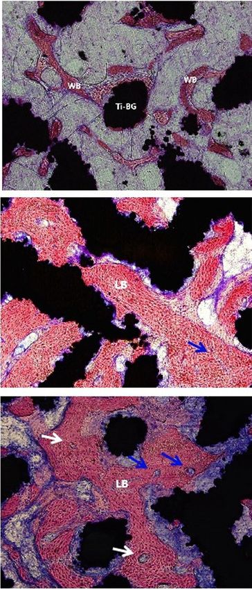
Page 4 of 15                                                          Zhang et al. In vivo study of surface-treated Ti-6Al-4V scaffolds

          A                                     B                                          C

  Ti-BG

                                       1 mm

          D                                      E                                         F

 Ti-MBG

                                       1 mm

Figure 1 The characteristics of the BG- and MBG-coated Ti-6Al-4Vporous scaffolds. Overview and SEM images of BG-coated Ti-6Al-
4Vporous scaffolds (upper row); Overview and SEM images of MBG-coated Ti-6Al-4V porous scaffolds (lower row). BG, bioactive glass;
MBG, mesoporous bioactive glass; SEM, scanning electron microscope.

were subcutaneously injected with 5 mg/kg calcein and                 scaffold in the center was selected as the region of interest
90 mg/kg xylenol orange (Sinopharm Chemical Reagent,                  (ROI), and 3D reconstruction was performed (Figure 3).
Shanghai, China), respectively, at 2 weeks and 1 week                    In this ROI, the bone volume/total volume (BV/TV) and
before being sacrificed for fluorescence double-labeling. At          structural bone parameters, including trabecular thickness
week 3, 6, and 9, two rabbits in every group were sacrificed          (Tb.Th), trabecular number (Tb.N), and trabecular
successively. Both the right and left femur condyles were             separation (Tb.Sp), were determined.
retrieved and preserved in 4% paraformaldehyde at 4 ℃ for
subsequent analyses.
                                                                      Preparation of hard tissue sections

                                                                      Following micro-CT imaging, the samples were processed
Micro-CT imaging
                                                                      for microscopy. They were rinsed with running water for
After being fixed in paraformaldehyde for 1 week, the                 24 h, dehydrated in a series of ascending ethanol
femoral condyles were scanned using micro-computed                    concentrations (50%, 75%, 95%, and 100%), and then
tomography (micro-CT) (YXLON Cheetah, Hamburg,                        embedded in methyl methacrylate (PMMA, Heraeus Kulzer,
Germany). Four samples were prepared for each group at                Hanau, Germany). The samples were trimmed and mounted
week 3, 6 and 9. The tube voltage, tube current, and voxel            for sectioning. Several sections (thickness =100–120 μm) were
sizes were 90 kV, 50 μA, and 20.0 μm × 20.0 μm × 20.0 μm,             cut in the central part of the samples using a Leica-SP 1600
respectively. An image analysis software suite (VGStudio              diamond-saw microtome (Leica Instruments, Nußloch,
Max 2.2.2, Volume Graphics, Heidelberg, Germany) was                  Germany) perpendicularly to the long axis of the porous
used to analyze and visualize the micro-CT data. To assess            scaffold. The sections were then reduced to a final thickness
the peripheral and central bone formation, respectively,              of approximately 50 μm by using silicon carbide grinding
around and inside the porous scaffolds, a cylindrical volume          cloths successively, followed by polishing with an alumina
(5.5 mm in diameter and 10 mm in height) with the porous              suspension (36). Two middle sections of each sample were

© Annals of Translational Medicine. All rights reserved.              Ann Transl Med 2021;9(1):39 | http://dx.doi.org/10.21037/atm-20-3829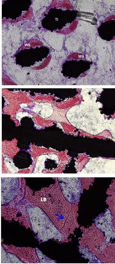
Annals of Translational Medicine, Vol 9, No 1 January 2021                                                                     Page 5 of 15

                    A                                                  B

Figure 2 Images of porous scaffolds implantation. The holes drilled in the femoral condyles of rabbits with a diameter of 5.5 mm and a
depth of 10 mm (A). The holes were subsequently filled with porous scaffolds with a diameter of 5 mm and a length of 10 mm (scale bars:
5 mm) (B).

Figure 3 Micro-CT images and 3D reconstruction. A cylindrical volume (with a diameter of 5.5 mm and a height of 10 mm) with the
titanium alloy porous scaffold in its center was selected as the region of interest (ROI). The volumes of the de novo bone formed in the
central part and around the periphery of the porous scaffold are shown in yellow and red, respectively. The porous titanium alloy scaffold is
depicted in white. Micro-CT, micro-computed tomography; 3D, three-dimensional.

selected for fluorescence microscopy and histological                      of the central newly-formed bone in the porous scaffolds
methylene blue acid fuchsin (BM-AF) staining.                              were detected on non-decalcified sections by a fluorescence
                                                                           microscope (Olympus BX51 + DP71, Japan). Four sections
                                                                           from each group of the scaffolds at each of the time
Fluorescence microscopy
                                                                           points were examined. Each section with a fluorescent
The fluorescent double labels (calcein and xylenol orange)                 dye was analyzed by using the ImageJ software (National

© Annals of Translational Medicine. All rights reserved.                 Ann Transl Med 2021;9(1):39 | http://dx.doi.org/10.21037/atm-20-3829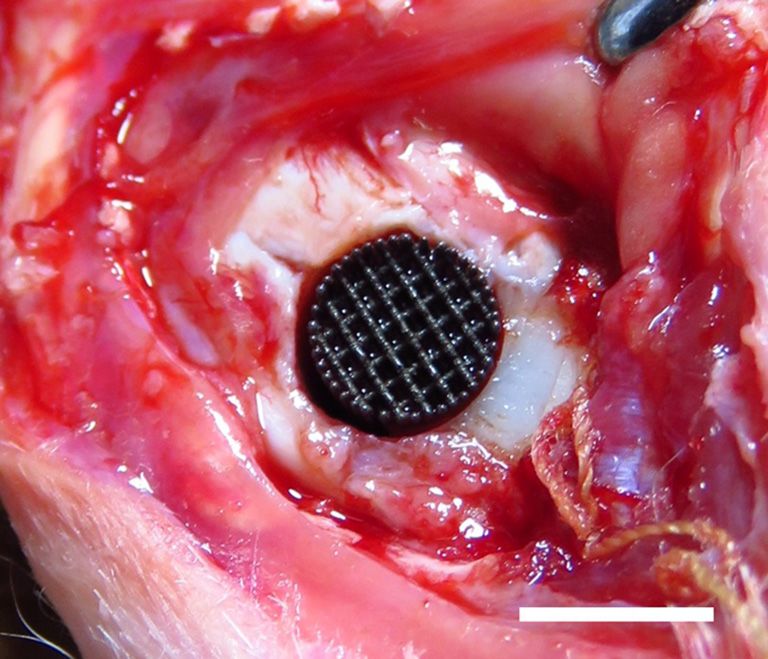
Page 6 of 15                                                     Zhang et al. In vivo study of surface-treated Ti-6Al-4V scaffolds

Institutes of Health, USA). As the different fluorescence        Ti group. At week 9, the Tb.Th values of the Ti-MBG and
emission spectra in the image represent the apposition of        Ti-BG groups were both significantly higher than the value
de novo bone at different time points, the area of interest      of the Ti group.
was selected to quantify the fluorescence intensity of the          As for the trabecular number (Tb.N), the value
labeled area, which reflected the formation of de novo bone      corresponding with the Ti-MBG group was significantly
during the corresponding period. One parallel fluorescence       higher than that of the Ti-BG group at week 6, while the
double labeling region was selected in each section, and the     Ti-BG group had a higher Tb.N value than that of the
distance between the red and green bands was measured at         Ti group at the same time point (Figure 5). At week 9,
both ends of the region. Then, the mineral apposition rate       however, the increase in the Tb.N value of the Ti-MBG
(MAR = the distance of fluorescence double labeling/time)        group slowed down, and there was no statistically significant
was determined. MAR represented the rate of de novo bone         difference between the Ti-BG and Ti-MBG groups. Both
mineralization in the three different groups of the scaffolds    values were still higher than the Tb.N value of the Ti
after implantation.                                              group.
                                                                    An analysis of the trabecular spacing (Tb.Sp) showed that
                                                                 the Tb.Sp values gradually decreasing with implantation
Statistical analyses                                             time and that Tb.Sp was inversely correlated with Tb.N
The experimental results are presented as mean ± SD              (the Pearson’s correlation coefficient: Ti =−0.9968, Ti-
(standard deviation). Statistical analyses were performed        BG =−0.8726, Ti-MBG =−0.9374). The obtained results
with the SPSS Statistics 21.0 software (IBM, Armonk, USA).       confirmed the regularity in the formation of de novo bone
Significant differences between the groups in the above-         and the maturation of the bone formed within the porous
mentioned measurements were compared using a one-way             scaffolds after implantation.
analysis of variation combined with the Student-Newman-             The same bone morphological parameters were also
Keuls (SNK) post hoc test. For all the tests, results with a P   calculated for the peripherally formed de novo bone
value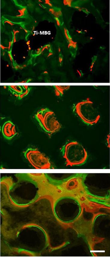
Annals of Translational Medicine, Vol 9, No 1 January 2021                                                                     Page 7 of 15

                                                                       3 Weeks

                                Ti

                            Ti-BG

                           Ti-MBG

                                                                       6 Weeks

                                Ti

                            Ti-BG

                           Ti-MBG

                                                                       9 Weeks

                                Ti

                            Ti-BG

                           Ti-MBG

Figure 4 3D reconstructions of the de novo bone at week 3, 6, and 9. The same color code as in Figure 3 is used here.

© Annals of Translational Medicine. All rights reserved.                 Ann Transl Med 2021;9(1):39 | http://dx.doi.org/10.21037/atm-20-3829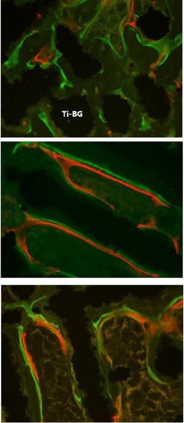
Page 8 of 15                                                                   Zhang et al. In vivo study of surface-treated Ti-6Al-4V scaffolds

                         0.3                     *            *                      0.20                                      *
                                                                  Ti                                                                   Ti
                                                         *        Ti-BG                                                                Ti-BG
                                                                  Ti-MBG             0.15                             *                Ti-MBG

                                                                           Tb. Th (mm)
                         0.2      *                                                                       *
                 BV/TV

                                                                                     0.10                         *

                         0.1
                                                                                     0.05

                         0.0                6W                                       0.00

                                                                                                                 6W
                                3W

                                                             9W

                                                                                                    3W

                                                                                                                             9W
                               Time after implantation (weeks)                                      Time after implantation (weeks)

                          4                                   *                                 5         *
                                                                  Ti                                  *                                Ti
                                                                  Ti-BG                         4                                      Ti-BG
                          3                          *            Ti-MBG                                                               Ti-MBG

                                                                                  Tb. sp (mm)
                                                                                                3                     *
                 Tb. N

                          2                  *                                                                    *
                                                                                                2
                                      *                                                                                            *
                          1                                                                                                   *
                                 *                                                              1

                          0                                                                     0
                                            6W

                                                                                                                 6W
                               3W

                                                             9W

                                                                                                     3W

                                                                                                                              9W
                               Time after implantation (weeks)                                      Time after implantation (weeks)

Figure 5 The different parameters describing the quantity and quality of centrally formed de novo bone in all experimental groups at week 3,
6, and 9. BV/TV, bone volume/total volume; Tb.Th, trabecular thickness; Tb.N, trabecular number; Tb.Sp, trabecular separation (*indicates
P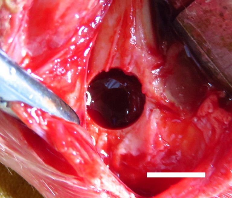
Annals of Translational Medicine, Vol 9, No 1 January 2021                                                                                  Page 9 of 15

                        0.5                                                          0.20                                      *
                                                 *            *   Ti                                                *              Ti
                                                                  Ti-BG                                                            Ti-BG
                        0.4                                                          0.15
                                                                  Ti-MBG                                                           Ti-MBG

                                                                           Tb. Th (mm)
                                             *                                                         *
                        0.3         *
                BV/TV

                                                                                     0.10
                        0.2
                                                                                     0.05
                        0.1

                        0.0                6W                                        0.00

                                                                                                             6W
                              3W

                                                         9W

                                                                                                 3W

                                                                                                                          9W
                              Time after implantation (weeks)                                    Time after implantation (weeks)

                         5                                                                 0.6
                                                                  Ti                               *                               Ti
                                                              *
                         4                           *            Ti-BG                                                        *   Ti-BG
                                                                                                                *
                                            *                     Ti-MBG                                                           Ti-MBG

                                                                             Tb. sp (mm)
                                                         *                                 0.4
                         3      *
                Tb. N

                         2
                                                                                           0.2
                         1

                         0                                                                 0.0
                                           6W

                                                                                                              6W
                              3W

                                                         9W

                                                                                                 3W

                                                                                                                          9W
                              Time after implantation (weeks)                                    Time after implantation (weeks)
Figure 6 The different parameters describing the quantity and quality of peripherally formed de novo bone in all experimental groups at
week 3, 6, and 9. BV/TV, bone volume/total volume; Tb.Th, trabecular thickness; Tb.N, trabecular number; Tb.Sp, trabecular separation
(*indicates P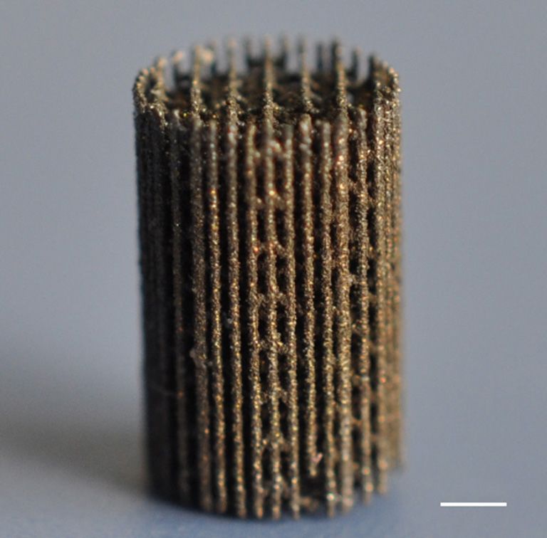
Page 10 of 15                                                             Zhang et al. In vivo study of surface-treated Ti-6Al-4V scaffolds

     3w

     6w

     9w

                              Ti                                       Ti-BG                                       Ti-MBG

Figure 7 The representative merged images of fluorescent double labeling of calcein (green color) and xylenol orange (red color), showing
bone formation in the three groups of the porous scaffolds at week 3, 6, and 9 (white scale bar =200 μm).

and osteoconductivity, the inherent fragility of MBG is                  regeneration capabilities in the central part of the scaffolds,
a major limitation for its clinical application, especially              as compared to the bare Ti-6Al-4V scaffolds. At week 6, the
at load-bearing sites. Coating additively manufactured                   quantitative analyses of BV/TV, Tb.Th, Tb.N, and Tb.Sp
lattice structures with MBG simultaneously resolves the                  showed that the bone regeneration capability of the Ti-
challenges associated with the fragility of MBG and the                  MBG group was significantly higher than that of the Ti-BG
limited osteoconductivity of bare metal scaffolds, leading               group (P
Annals of Translational Medicine, Vol 9, No 1 January 2021 Page 11 of 15
implantation site. The potential effects of the geometrical
4 *
*
*
* Ti design of the scaffolds as well as the implantation site should
Ti-BG be further investigated in future studies. In the design of
3
Ti-MBG
MAR (μm/day)
the coating protocol, different coatings could be applied to
2 the different parts of the porous scaffold to better guide the
process of bone tissue regeneration. For example, the MBG
1 coating may be applied to the struts in the central part of
the porous scaffolds, while the BG coating to the struts
0
on the outer surface. To increase the space for new bone
9W
3W
6W
formation and vascularization, one may consider a further
Time after implantation (weeks)
increase in the porosity of porous scaffolds, a reduction in
Figure 8 The mineral apposition rates (MAR = distance of
coating thickness, and an increase in coating roughness.
fluorescence double labeling/time) of the three groups at week 3,
Bone regeneration is a complex process. The mechanical
week 6, and week 9 (* indicates PPage 12 of 15 Zhang et al. In vivo study of surface-treated Ti-6Al-4V scaffolds
3w
6w
9w
Ti Ti-BG Ti-MBG
Figure 9 The representative histological images of non-decalcified sections obtained using methylene blue acid fuchsin staining. The blue
arrows indicate neovascularization, while the white arrows indicate the Haversian system. WB, woven bone; LB, lamellar bone (green scale
bar =200 μm).
improving their therapeutic efficacy in the treatment of capacity. The degree of restoration of the original anatomy
large bony defects such as human maxilla, mandible and is the other readout of such an experiment.
non-weight bearing parts of limbs. Further research needs
to be directed at understanding the coating-strut adhesion
Acknowledgments
under physiological loads, while the BG and MBG coatings
biodegrade overtime during the bone healing phase. Funding: We acknowledge the financial support from the
Besides, the osteogenesis capacity of porous scaffolds should National Key Research and Development Program of
be evaluated in critical size segmental defect models of load- China (No. 2017YFB1104101), Natural Science Foundation
bearing long bones (e.g., femur, tibia, and radius). Such of China (No. 81671830), and Science and Technology
types of in vivo models would also allow for biomechanical Commission of Shanghai Municipality (No. 19441902300).
testing, which could be used to evaluate the interface
strength of the bone-implant complex and to establish
Footnote
whether or not the process of bone tissue regeneration
has progressed far enough to have restored the functional Reporting Checklist: The authors have completed the
capacity of the long bone in terms of its load-bearing ARRIVE reporting checklist. Available at http://dx.doi.
© Annals of Translational Medicine. All rights reserved. Ann Transl Med 2021;9(1):39 | http://dx.doi.org/10.21037/atm-20-3829Annals of Translational Medicine, Vol 9, No 1 January 2021 Page 13 of 15
org/10.21037/atm-20-3829 chitosan/magnesium-calcium silicate composite for
orthopedic applications. Materials 2019;12:203.
Data Sharing Statement: Available at http://dx.doi. 6. Moore WR, Graves SE, Bain GI. Synthetic bone graft
org/10.21037/atm-20-3829 substitutes. ANZ J Surg 2001;71:354-61.
7. Tang W, Lin D, Yu Y, et al. Bioinspired trimodal macro/
Peer Review File: Available at http://dx.doi.org/10.21037/ micro/nano-porous scaffolds loading rhBMP-2 for
atm-20-3829 complete regeneration of critical size bone defect. Acta
Biomater 2016;32:309-23.
Conflicts of Interest: All authors have completed the ICMJE 8. Turnbull G, Clarke J, Picard F, et al. 3D bioactive
uniform disclosure form (available at http://dx.doi. composite scaffolds for bone tissue engineering. Bioact
org/10.21037/atm-20-3829). The authors have no conflicts Mater 2017;3:278-314.
of interest to declare. 9. Zadpoor AA, Bone tissue regeneration: the role of scaffold
geometry. Biomater Sci 2015;3:231-45.
Ethical Statement: The authors are accountable for all 10. Wu S, Liu X, Yeung KWK, et al. Biomimetic porous
aspects of the work in ensuring that questions related scaffolds for bone tissue engineering. Mater Sci Eng R Rep
to the accuracy or integrity of any part of the work are 2014;80:1-36.
appropriately investigated and resolved. The study was 11. Bose S, Roy M, Bandyopadhyay A. Recent advances in
approved by the Animal Ethics Committee of China bone tissue engineering scaffolds. Trends Biotechnol
Medical University (No. K2016014). The general guidelines 2012;30:546-54.
on the use of animals in research have complied with all 12. O'Keefe RJ, Mao J. Bone tissue engineering and
these experiments. regeneration: from discovery to the clinic-an overview.
Tissue Eng. Part B Rev 2011;17:389-92.
Open Access Statement: This is an Open Access article 13. Henkel J, Woodruff MA, Epari DR, et al. Bone
distributed in accordance with the Creative Commons regeneration based on tissue engineering conceptions - A
Attribution-NonCommercial-NoDerivs 4.0 International 21st century perspective. Bone Res 2013;1:216-48.
License (CC BY-NC-ND 4.0), which permits the non- 14. Bouet G, Marchat D, Cruel M. et al. In vitro three-
commercial replication and distribution of the article with dimensional bone tissue models: from cells to controlled
the strict proviso that no changes or edits are made and the and dynamic environment. Tissue Eng Part B Rev
original work is properly cited (including links to both the 2015;21:133-56.
formal publication through the relevant DOI and the license). 15. Rouwkema J, Rivron NC, van Blitterswijk C.
See: https://creativecommons.org/licenses/by-nc-nd/4.0/. Vascularization in tissue engineering. Trends Biotechnol
2008;26:434-41.
16. Barabaschi GDG, Manoharan V, Li Q, et al, Engineering
References
pre-vascularized scaffolds for bone regeneration. Adv Exp
1. García-Gareta E, Coathup MJ, Blunn GW. Med Biol 2015;881:79-94.
Osteoinduction of bone grafting materials for bone repair 17. Cheng XY, Li SJ, Murr LE, et al. Compression
and regeneration. Bone 2015;81:112-21. deformation behavior of Ti-6Al-4V alloy with cellular
2. Cuthbert RJ, Churchman SM, Tan HB, et al. Induced structures fabricated by electron beam melting. J Mech
periosteum a complex cellular scaffold for the treatment of Behav Biomed Mater 2012;l6:153-62.
large bone defects. Bone 2013;57:484-92. 18. Casalino G, Campanelli SL, Contuzzi N, et al.
3. Zhang J, Liu W, Schnitzler V, et al. Calcium phosphate Experimental investigation and statistical optimisation of
cements for bone substitution: Chemistry, handling and the selective laser melting process of a maraging steel. Opt
mechanical properties. Acta Biomater 2014;10:1035-49. Laser Technol 2015;65:151-8.
4. Oryan A, Alidadi S, Moshiri A, et al. Bone regenerative 19. Gibbs DMR, Vaezi M, Yang S, et al. Hope versus hype:
medicine: classic options, novel strategies, and future what can additive manufacturing realistically offer trauma
directions. J Orthop Surg Res 2014;9:18. and orthopedic surgery? Regen Med 2014;9:535-49.
5. Tsai CH, Hung CH, Kuo CN, et al. Improved bioactivity 20. Derby B, Printing and prototyping of tissues and scaffolds.
of 3D p rinted porous titanium alloy scaffold with Science 2012;338:921-6.
© Annals of Translational Medicine. All rights reserved. Ann Transl Med 2021;9(1):39 | http://dx.doi.org/10.21037/atm-20-3829Page 14 of 15 Zhang et al. In vivo study of surface-treated Ti-6Al-4V scaffolds
21. Melchels FPW, Domingos MAN, Klein TJ, et al. Additive study. Mater Sci Eng C Mater Biol Appl 2017;80:7-17.
manufacturing of tissues and organs. Prog Polym Sci 37. Lewis G, Properties of open-cell porous metals and
2012;37:1079-104. alloys for orthopedic applications. J Mater Sci Mater Med
22. Hutmacher DW, Cool S. Concepts of scaffold-based 2013;24:2293-325.
tissue engineering--the rationale to use solid free-form 38. Yazdimamaghani M, Razavi M, Vashaee D, et al. Porous
fabrication techniques. J Cell Mol Med 2007;11:654-69. magnesium-based scaffolds for tissue engineering, Mater
23. Ye X, Leeflang S, Wu C, et al. Mesoporous bioactive glass Sci Eng C Mater Biol Appl 2017;71:1253-66.
functionalized 3D Ti-6Al-4V scaffolds with improved 39. Almubarak S, Nethercott H, Freeberg M, et al. Tissue
surface bioactivity. Materials (Basel) 2017;10:1244. engineering strategies for promoting vascularized bone
24. Yin B, Xue B, Wu Z, et al. A novel hybrid 3D-printed regeneration. Bone 2016;83:197-209.
titanium scaffold for osteogenesis in a rabbit calvarial 40. Yin S, Zhang W, Zhang Z, et al. Recent advances in
defect model. Am J Transl Res 2018;10:474-82. scaffold design and material for vascularized tissue-
25. Roseti L, Parisi V, Petretta M, et al. Scaffolds for bone engineered bone regeneration. Adv Healthc Mater
tissue engineering: state of the art and new perspectives. 2019;8:e1801433.
Mater Sci Eng C Mater Biol Appl 2017;78:1246-62. 41. Javadi A, Solouk A, Nazarpak MH, et al. Surface
26. Tang D, Tare RS, Yang L-Y, et al. Biofabrication of bone engineering of titanium-based implants using
tissue: approaches, challenges and translation for bone electrospraying and dip coating methods. Mater Sci Eng C
regeneration. Biomaterials 2016;83:363-82. Mater Biol Appl 2019;99:620-30.
27. Hench LL, Splinter RJ, Allen WC, et al. Bonding 42. Huynh V, Ngo NK, Golden TD. Surface activation
mechanisms at the interface of ceramic prosthetic and pretreatments for biocompatible metals and
materials. J Biomed Mater Res 1971;5:117-41. alloys used in biomedical applications. Int J Biomater
28. Fernandes JS, Gentile P, Pires RA, et al. Multifunctional 2019;2019:3806504.
bioactive glass and glass-ceramic biomaterials with 43. Lee MN, Hwang H-S, Oh S-H, et al. Elevated
antibacterial properties for repair and regeneration of bone extracellular calcium ions promote proliferation and
tissue. Acta Biomater 2017;59:2-11. migration of mesenchymal stem cells via increasing
29. Brauer DS. Bioactive Glasses-Structure and Properties, osteopontin expression. Exp Mol Med 2018;50:1-16.
Angew Chem Int Ed Engl 2015;54:4160-81. 44. Shi M, Zhou Y, Shao J, et al. Stimulation of osteogenesis
30. Kaur G. Pandey OP, Singh K, et al. A review of bioactive and angiogenesis of hBMSCs by delivering Si ions and
glasses: Their structure, properties, fabrication and apatite functional drug from mesoporous silica nanospheres. Acta
formation. J Biomed Mater Res A 2014;102:254-74. Biomater 2015;21:178-89.
31. Jones JR. Review of bioactive glass: From Hench to 45. Lammens J, Laumen A, Delport H, et al. The
hybrids, Acta Biomater 2013;9:4457-86. Pentaconcept in skeletal tissue engineering. A combined
32. Miguez-Pacheco V, Hench LL, Boccaccini AR. Bioactive approach for the repair of bone defects. Acta Orthop Belg
glasses beyond bone and teeth: Emerging applications in 2012;78:569-73.
contact with soft tissues. Acta Biomater 2015;13:1-15. 46. Baino F, Fiorilli S, Vitale-Brovarone C. Bioactive glass-
33. Yan XX, Deng HX, Huang XH, et al. Mesoporous based materials with hierarchical porosity for medical
bioactive glasses. I. Synthesis and structural applications: Review of recent advances. Acta Biomater
characterization, J Non-Cryst Solids 2005;351:3209-17. 2016;42:18-32.
34. Yan X, Huang X, Yu C, et al. The in-vitro bioactivity 47. Kang MS, Lee N-H, Singh RK, et al. Nanocements
of mesoporous bioactive glasses. Biomaterials produced from mesoporous bioactive glass nanoparticles.
2006;27:3396-403. Biomaterials 2018;162:183-99.
35. Zhang Y, Xia L, Zhai D, et al. Mesoporous bioactive 48. Lavenus S, Poxson DJ, Ogievetsky N, et al. Stem cell
glass nanolayer-functionalized 3D-printed scaffolds for behavior on tailored porous oxide surface coatings.
accelerating osteogenesis and angiogenesis. Nanoscale Biomaterials 2015;55:96-109.
2015;7:19207-21. 49. Romero-Sánchez LB, Marí-Beffa M, Carrillo P, et al.
36. Cao H, Feng L, Wu Z, et al. Effect of low-intensity pulsed Copper-containing mesoporous bioactive glass promotes
ultrasound on the biological behavior of osteoblasts on angiogenesis in an in vivo zebrafish model. Acta Biomater
porous titanium alloy scaffolds: An in vitro and in vivo 2018;68:272-85.
© Annals of Translational Medicine. All rights reserved. Ann Transl Med 2021;9(1):39 | http://dx.doi.org/10.21037/atm-20-3829Annals of Translational Medicine, Vol 9, No 1 January 2021 Page 15 of 15
50. Baino F, Novajra G, Pacheco VM, et al. Bioactive glasses: 52. Santos MI, Unger RE, Sousa RA, et al. Crosstalk
Special applications outside the skeletal system. J Non- between osteoblasts and endothelial cells co-cultured
Cryst Solids 2016;432:15-30. on a polycaprolactone-starch scaffold and the in
51. Stegen S, Gastel N, Carmeliet G. Bringing new life to vitro development of vascularization. Biomaterials
damaged bone: The importance of angiogenesis in bone 2009;30:4407-15.
repair and regeneration. Bone 2015;70:19-27.
Cite this article as: Zhang G, Zhao P, Lin L, Qin L, Huan Z,
Leeflang S, Zadpoor AA, Zhou J, Wu L. Surface-treated 3D
printed Ti-6Al-4V scaffolds with enhanced bone regeneration
performance: an in vivo study. Ann Transl Med 2021;9(1):39. doi:
10.21037/atm-20-3829
© Annals of Translational Medicine. All rights reserved. Ann Transl Med 2021;9(1):39 | http://dx.doi.org/10.21037/atm-20-3829You can also read