Photoacoustic needle improves needle tip visibility during deep peripheral nerve block - Nature
←
→
Page content transcription
If your browser does not render page correctly, please read the page content below
www.nature.com/scientificreports
OPEN Photoacoustic needle improves
needle tip visibility during deep
peripheral nerve block
Kunitaro Watanabe1, Joho Tokumine1*, Alan Kawarai Lefor2, Harumasa Nakazawa1,
Katsuya Yamamoto3, Hiroyuki Karasawa3, Miki Nagase4 & Tomoko Yorozu1
We developed a novel technology using the photoacoustic effect that improve needle tip visibility. We
evaluated whether this technology improves needle tip visibility when performing a deep peripheral
nerve block in a cadaver model. A photoacoustic needle was developed using a conventional echogenic
needle with an intraluminal optical fiber. A pulsed laser sends light from a source through the fiber,
which is converted to ultrasound at the needle tip using the photoacoustic effect. A nerve block expert
performed deep nerve blocks using the photoacoustic needle and the ultrasound views recorded,
with or without photoacoustic ultrasound at the needle tip. Needle tip visibility was evaluated by
questionnaire (Likert scale 1: very poor, 5: very good) completed by anesthesiologists evaluating
recorded images. The score was presented as median [first quartile, third quartile]. Statistical analysis
was performed using the Wilcoxon matched-pairs signed rank test. The scores of needle tip visibility
with photoacoustic ultrasound from the needle tip (4.3 [4.0, 4.5]) was significantly higher than that
without photoacoustic ultrasound (3.5 [3.2, 3.8]) (p < 0.01). Ultrasound emitted at the needle tip using
the photoacoustic effect improves needle tip visibility during deep peripheral nerve blocks.
Clinical trial number University Hospital Medical Information Network Center Clinical Trials
Registration System (UMIN000036974).
Ultrasound-guided peripheral nerve block of deep targets remains a challenge in anesthesiology1. Needle trajec-
tory tends to be steep to approach deep neural targets. The needle tip is difficult to image because the reflection
of sound waves to the probe from the needle is reduced by the steep a ngle2,3. Therefore, deep nerve blocks are
technically difficult, and considered as high-risk p rocedures1.
The photoacoustic effect was discovered in 1880 by Alexander Graham B ell4. Clinical applications of this
technology have been t ried5. Piras et al. first reported ultrasound-emission from a biopsy needle using the
photoacoustic effect6. In their report, the optical fiber was inserted into a biopsy needle for guidance, and then
was then removed during the biopsy. Kang also reported a photoacoustic biopsy n eedle7. We developed a pho-
toacoustic needle with a technological innovations (2017)8, allowing use of a smaller caliber needle, a flexible
optical fiber, and less invasive (the light source generates two microjoules). A novel photoacoustic needle for
ultrasound-guided peripheral nerve block was developed for this study.
We evaluated whether the ultrasound-emitting needle tip improved needle tip visibility compared to a con-
ventional echogenic needle during ultrasound-guided deep nerve blocks in a cadaver model.
Materials and methods
This study was reviewed and approved by the local ethics committee (Kyorin University Ethical Review Board,
Reception No. 1408, approved date 2019.12.6) and registered in the University Hospital Medical Information
Network Center Clinical Trials Registration System (UMIN000036974). The study was conducted in accordance
with Consolidated Standards of Reporting Trials (CONSORT) guidelines (Supplemental information; “CON-
SORT Diagram”).
The cadaver used in this study was donated to Kyorin University School of Medicine for anatomical education,
research, and clinical skills training. The research protocol was prepared in strict accordance with the "Guide-
lines for the research involving cadavers" of the Japanese Association of Anatomists, and was approved by the
1
Department of Anesthesiology, Kyorin University School of Medicine, 6‑20‑2 Shinkawa, Mitaka City,
Tokyo 181‑8611, Japan. 2Department of Surgery, Jichi Medical University, Shimotsuke, Tochigi, Japan. 3Medical
System Research & Development Center, FUJIFILM Corporation, Kanagawa, Japan. 4Department of Anatomy,
Kyorin University Faculty of Medicine, Mitaka, Tokyo, Japan. *email: dg274825@cf6.so-net.ne.jp
Scientific Reports | (2021) 11:8432 | https://doi.org/10.1038/s41598-021-87777-9 1
Vol.:(0123456789)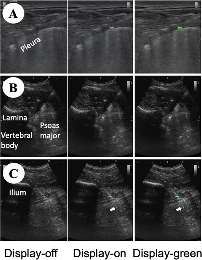
www.nature.com/scientificreports/
Figure 1. Configuration of the system for photoacoustic needle. Ultrasound machine (a) supplies power to
an output regulation port (b), which controls timing of electric power for generating laser light (c). The laser
is transmitted through the optical fiber integrated in the lumen of the needle (d). The black resin (e) absorbs
the light, and then converts it to ultrasound by a photoacoustic effect. The ultrasound probe detects the
photoacoustic ultrasound. (a) Ultrasound machine, (b) electrical output regulation port, (c) laser light source,
(d) photoacoustic needle, (e) black resin, (f) ultrasound probe.
Ethics Committee of Kyorin University Faculty of Medicine (Permit No. 986). A comprehensive consent form
was obtained from the donor prior to donation and from the family prior to and at the time of donation. The
research outline was published on the Kyorin University website to ensure that the families had the opportunity
to refuse (opt-out).
Ultrasound‑emitting from needle tip using photoacoustic effect. The principle of an ultrasound-
emitting system using a photoacoustic effect is described (Fig. 1)8. An optical fiber is inserted into the lumen
of a needle, and fixed in place maintaining the lumen open for administration of local anesthetic agents. The
fiber tip is covered with black resin containing carbon-black pigment, and fixed to the inside wall at the needle
bevel. Pulsed laser from an external laser light source is transmitted through the optical fiber. The black resin at
the needle tip absorbs the pulsed laser light, which causes adiabatic thermal expansion, and is translated to high
frequency vibrations. As a result, ultrasound waves are generated by the photoacoustic effect. The ultrasound
wave is received by the ultrasound transducer in the ultrasound probe, which is converted into electrical signals
and transferred to the ultrasound unit for imaging. Typical ultrasound imaging for scanning tissue involves a
“round trip” for the ultrasound waves, emitted from the transducer in the probe and reflected by the needle back
to the probe. However, the ultrasound wave generated by the photoacoustic effect is a one-way trip, emitted at
the needle tip to the probe. The ultrasound frame to create a view for the ultrasound wave generated by the pho-
toacoustic effect needs 11% of all frames. This reduction in the frame rate did not inhibit smooth dynamic ultra-
sound views in the preliminary study. Safety evaluation of the system was proven in a previous study according
to the “Safety of laser products—Part 1: Equipment classification and requirements: IEC60825-1:2014”8. The
ultrasound waves generated by the photoacoustic effect is colored green or white.
In the study, we used an ultrasound machine FC1 (FUJIFILM Medical Co., Ltd., Tokyo, Japan) and ultrasound
probe L38xp/13-6 and C35xp/8-3 (FUJIFILM SonoSite, Inc., Bothell, WA, USA). The nerve block needle was
a Stimuplex Ultra 360 (insulated echogenic needle; size 22 G, length 80 mm, bevel angle 30°, B. Braun Medical
Inc., Melsungen, Germany), in which an optical fiber was incorporated as described above). Figure 2 shows
ultrasound views at several trajectory needle angles of the echogenic needle or ultrasound-emitting needle
using the simulator.
Ultrasound‑guided deep nerve block. An expert (K.W.) in ultrasound-guided nerve blocks performed
these nerve blocks on a cadaver under ultrasound guidance using the novel ultrasound-emitting needle. The
cadaver (an 85-year-old man fixed with N-vinyl-2-pyrrolidone) had soft and pliable tissue9. The deep nerve
blocks performed included a paravertebral block (transversal technique with lateral to medial direction, in-plane
approach), lumbar plexus block (transverse technique with lateral to medial direction, in-plane approach) and
sciatic nerve block (parasacral approach).
Scientific Reports | (2021) 11:8432 | https://doi.org/10.1038/s41598-021-87777-9 2
Vol:.(1234567890)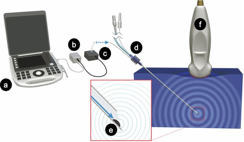
www.nature.com/scientificreports/

                                  Figure 2.  Needle tip visibility at several trajectories. Stimuplex Ultra 360 (B. Braun Medical Inc., Melsungen,
                                  Germany) and a photoacoustic needle (an optical fiber is incorporated into Stimuplex Ultra 360) are inserted
                                  into UGP GEL (main ingredient; 20% Agar, Alfabio Co., Gunma, Japan) at trajectories from 30° to 80°.
                                  Although needle shafts of both needles are always clearly visible at any trajectory, needle tip visibility of the
                                  conventional needle tip gradually decreases with an increased trajectory angle. However, the needle tip of the
                                  photoacoustic needle can be identified clearly even at 80°. (A) Stimuplex Ultra 360, (B) Photoacoustic needle.

                                      On the ultrasound display, ultrasound views with and without the ultrasound generated by the photoacoustic
                                  effect were displayed during the procedure. In the study, the ultrasound generated by the photoacoustic effect
                                  was shown in white to prevent information bias.
                                      The side showing ultrasound generated by the photoacoustic effect was blinded to the expert with an opacity
                                  board. Then, the expert performed the nerve block watching with a typical ultrasound view, then both of the
                                  movies were recorded. To confirm whether the nerve blocks were successful, 10 ml of acrylic paint solution was
                                  injected instead of a local anesthetic agent, and dissection performed after the experiment.

                                  Survey to evaluate needle tip visibility.             Needle tip visibility was evaluated by a survey of participating
                                  anesthesiologists acting as volunteers. Recruitment of participants was performed through the local community
                                  of anesthesiologists, including department colleagues and anesthesiologists in associated hospitals. Exclusion
                                  criteria was rejection to participate in the survey. The authors and collaborators of the study were excluded from
                                  participating in the survey.
                                      Ultrasound movies were randomly selected using a random number table. Participating anesthesiologists
                                  watched the ultrasound images and evaluated needle tip visibility in each movie using a Likert scale (score 1:
                                  very poor, 2: poor, 3: fair, 4: good, 5: very good). Demographic data of participants were also collected, including
                                  overall clinical experience and experience with ultrasound-guided nerve block. The survey was conducted on the
                                  Internet using questions posted with an invitation limit. Informed consent was obtained from all participants
                                  prior to the Internet questions. The primary outcome of the study was evaluation of needle tip visibility with
                                  or without the photoacoustic ultrasound. The secondary outcome was a relationship between experience with
                                  ultrasound-guided nerve block and needle tip visibility with or without the photoacoustic ultrasound.

                                  Statistical analysis. Likert scale scores from participant responses are expressed as median [first quartile,
                                  third quartile]. Clinical experience of participants is expressed in four rank groups (1–5, 6–10, 11–15, > 15 years)
                                  and the number of ultrasound-guided nerve block procedures expressed in four rank groups (0–50, 51–100,
                                  101–200, > 200). Wilcoxon matched-pairs signed rank test was used to evaluate the scores. Spearman’s correla-
                                  tion coefficients (rS) were used to evaluate strength of associations among the variables. A preliminary study for
                                  estimating power analysis was performed using the authors and collaborators. The median score and standard
                                  deviation of photoacoustic ultrasound were expected to be superior in one score than without photoacoustic
                                  ultrasound. The sample size required for 80% power at ɑ = 0.05 was estimated to be sixteen participants. A
                                  p-value less than 0.05 was considered statistically significant. Statistical analyses were performed with GraphPad
                                  Prism, version 7.02 (GraphPad Software Inc., San Diego, USA).

Scientific Reports |   (2021) 11:8432 |                https://doi.org/10.1038/s41598-021-87777-9                                                     3

                                                                                                                                                 Vol.:(0123456789)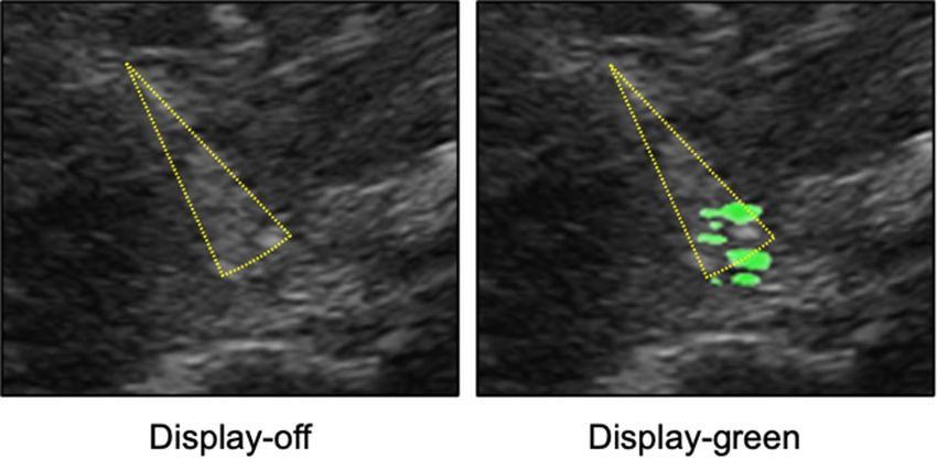
www.nature.com/scientificreports/

                                            Figure 3.  Ultrasound views of nerve blocks. The pictures show nerve blocks (at right side of cadaver).
                                            These pictures are all obtained using the photoacoustic needle. The picture of display-off does not show the
                                            photoacoustic ultrasound, the pictures of display-on and display-green show the photoacoustic ultrasound.
                                            Mirror image artifact of the needle tip is observed in the display-on and display-green of the (C) (white arrows
                                            indicate the mirror image artifact). (A) paravertebral block (A), (B) lumbar plexus block, (C) sciatic nerve block,
                                            display-off: no photoacoustic ultrasound, display-on: photoacoustic ultrasound using white color, display-green:
                                            photoacoustic ultrasound using green color (See Supplemental Digital Contents nos. 13, 15 and 17).

          Scientific Reports |   (2021) 11:8432 |                https://doi.org/10.1038/s41598-021-87777-9                                                  4

Vol:.(1234567890)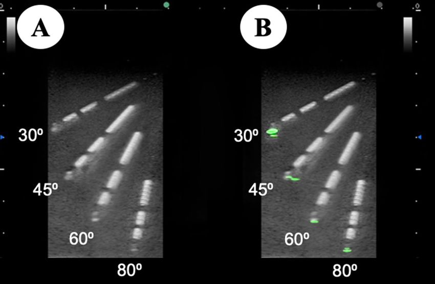
www.nature.com/scientificreports/

                                  Figure 4.  Dissected region in the cadaver. Paint injected during the deep nerve block at right- or left-side
                                  of a cadaver are green or pink, respectively. Pink stain at the left-side lumbar plexus block (B. Lt) and sciatic
                                  nerve block (C, Lt) are undetermined due to weak and blurred stain in connective tissue over the nerves. (A)
                                  Paravertebral block, (B) lumbar plexus block, (C) sciatic nerve block. Rt right-side of the cadaver, Lt left-side of
                                  the cadaver.

Scientific Reports |   (2021) 11:8432 |                https://doi.org/10.1038/s41598-021-87777-9                                                        5

                                                                                                                                                Vol.:(0123456789)
www.nature.com/scientificreports/
Figure 5. Needle tip visibility. The asterisk indicates a statistically significant difference between the display-off
and display-on groups.
Figure 6. Artifacts cause multiple needle tips. Sciatic nerve block on the left side of the cadaver (See
Supplemental Digital Content nos. 11 and 18). Combination of reverberation artifact and bayonet artifact
(dotted yellow triangle) may cause an illusion of multiple needle tips (display-green; Supplemental Digital
Content no. 18). The needle tip without displaying photoacoustic ultrasound cannot be identified (display-off;
Supplemental Digital Content no. 11). Display-off: no photoacoustic ultrasound, display-green: photoacoustic
ultrasound using green color.
Results
Twelve ultrasound movies were recorded (See Supplemental Digital Contents nos. 1–12, Fig. 3: sample scenes
of the movies). Each nerve block was recorded using two types of movies, with or without the photoacoustic
ultrasound. One of the movies was displayed on the photoacoustic ultrasound (display-on), but the other movie
was displayed off of the photoacoustic ultrasound (display-off).
We invited forty anesthesiologists to participate in the study. Thirty anesthesiologists participated in this
study with no exclusions. The clinical experience of participants in each group; 1–5, 6–10, 11–15, and > 15 years
of experience were 11 (37%), 6 (20%), 7 (23%), and 6 (20%), respectively. The numbers of ultrasound-guided
nerve block procedures performed by participants in each group; 0–50, 51–100, 101–200, and > 200 procedures
were 12 (40%), 7 (23%), 2 (7%), and 9 (30%), respectively.
Needle trajectories of the deep nerve blocks were 35–40° in the paravertebral block (transverse technique,
in-plane approach), and 60° in the lumbar plexus block (transverse technique, in-plane approach) or sciatic
nerve block (parasacral approach). Dissection of the cadaver was performed to evaluate the quality of the nerve
blocks. Most of the nerve blocks were confirmed as being successful. However, success of the left lumbar plexus
block and the left sciatic nerve block could not be confirmed due to loss of paint (Fig. 4).
Scientific Reports | (2021) 11:8432 | https://doi.org/10.1038/s41598-021-87777-9 6
Vol:.(1234567890)www.nature.com/scientificreports/
The Likert scale score for needle tip visibility in the display-on movie (4.3 [4.0, 4.5]) was significantly higher
than that in the display-off movie (3.5 [3.2, 3.8]) (p < 0.01) (Fig. 5). Subgroup analysis of each nerve block had
the same tendency (the paravertebral nerve block; display-on movie 4.5 [4.0, 4.5], display-off movie 3.5 [3.0, 4.0]
(p < 0.01), the lumbar plexus block; display-on movie 4.5 [4.0, 5.0], display-off movie 4.0 [3.5, 4.5] (p = 0.01), the
sciatic nerve block; display-on movie 4.0 [3.9, 4.5], display-off movie 3.3 [3.0, 3.6] (p < 0.01).
Correlation was shown between experience with ultrasound-guided nerve block and the score of needle tip
visibility in the display-off movie (rS 0.43, p = 0.02). There was no correlation between other outcomes or the
demographics of the participants.
Discussion
This study demonstrates that a novel needle with an ultrasound-emitting needle-tip using the photoacoustic effect
resulted in significantly improved visibility of the needle tip while performing simulated deep nerve blocks on
a cadaver model. However, the difference in needle tip visibility scores between the display-on and display-off
movies was only a median of 0.8, lower than that expected. The needle-tip was colored white on the ultrasound
to prevent information bias. If we colored the needle-tip green instead of white during the study, it may greatly
improve needle tip visibility. The visibility of the ultrasound-emitting needle tip is not dependant on sound waves
reflected from the needle tip so a steeper angle of trajectory should not reduce tip visibility because ultrasound
is emitted from the needle tip itself.
Commonly used needles for nerve blocks have a tendency to have decreased needle tip visibility at steeper
trajectories. Several types of echogenic needles have been developed to improve needle tip v isibility10, which
may increase safety and efficacy11. Studies have shown that the visibility of an echogenic needle tip at a trajec-
tory of 60° in a simulated environment is effective2,3. However, these ex-vivo results are difficult to translate
directly to clinical p ractice10. The echogenic needles were not easily visible during ultrasound-guided injection
in cadaver models with a needle trajectory greater than 60°12. Uultrasound beam steering increases needle
visibility3. However, the beam steering dose not improve needle visiblity at a 70° trajectory angle3. The visibility
of the photoacoustic needle tip is effective at a needle angle of 80° (Fig. 2 and “Supplemental Digital Contents”).
We suggest that the clinical efficacy of a photoacoustic needle may facilitate performing ultrasound-guided deep
nerve blocks, especially using the out-of-plane t echnique13.
Visualization of the needle during insertion is a basic skill during ultrasound-guided peripheral nerve b lock14.
The training needed to achieve skill in needle tip visualization varies among trainees15. In the study, correlation
was found between experience in ultrasound-guided nerve block and the scores of needle tip visibility in the
display-off movie. In another words, participants with more extensive experience in nerve blocks tended to
appreciate conventional echogenic needles. In the display-on movie, high-power ultrasound from the needle tip
may cause a reverberation artifact16, which induces an illusion of multiple needle tips (Fig. 6). That artifact may
induce discomfort in participants with greater experience in nerve blocks. We think that anesthesiologists may
overcome pitfalls induced by these artifacts by understanding details of adjusting the appropriate ultrasound
gain and physical characteristics of ultrasound.
In the system, the ultrasound frame rate is reduced due to sharing frames for detecting photoacoustic ultra-
sound. The frame rate constructing the ultrasound view keeps 89% of the total frame rate, which may not reduce
image quality and add no stress to the operator during the nerve block without sensing time lag. Hence, we
believe that the reduced frame rate of 11% may not influence in the study result.
Needle tip visibility in tissues in a live human was reported to be less than that in cadaveric t issue17. Hence,
there is still a question whether good visibility of the photoacoustic needle tip can be achieved a live human. This
is a limitation of this study. Lediju Bell and Shubert have demonstrated good visibility of photoacoustic needles
in fat and other t issues18,19. These results suggest the efficacy of photoacoustic needles in obese patients. Photoa-
coustic ultrasound in clinical use has been a subject of very considerable interest during the past few years. We
believe that this innovation will resolve technical problems associated with deep nerve blocks.
Conclusion
Ultrasound-emitted from a needle tip using the photoacoustic effect improved needle tip visibility during deep
nerve blocks in a cadaver model.
Received: 16 January 2021; Accepted: 5 April 2021
References
1. Wadhwa, A., Kandadai, S. K., Tongpresert, S., Obal, D. & Gebhard, R. E. Ultrasound guidance for deep peripheral nerve blocks:
A brief review. Anesthesiol. Res. Pract. 2011, 262070 (2011).
2. Prabhakar, C., Uppal, V. & Sondekoppam, R. V. Effect of beam steering on echogenic and nonechogenic needle visibility at 40°,
50°, and 60° needle insertion angles. Anesth. Analg. 126(6), 1926–1929 (2018).
3. Uppal, V., Sondekoppam, R. V. & Ganapathy, S. Effect of beam steering on the visibility of echogenic and non-echogenic needles:
A laboratory study. Can. J. Anaesth. 61(10), 909–915 (2014).
4. Bell, A. G. The Photophone. Science 1(11), 130–134 (1880).
5. Biagi, E., Brenci, M., Fontani, S., Masotti, L. & Pieraccini, M. Photoacoustic generation: Optical fiber ultrasonic sources for non-
destructive evaluation and clinical diagnosis. Opt. Rev. 4(4), 481–483 (1997).
6. Piras, D., Grijsen, C., Schütte, P., Steenbergen, W. & Manohar, S. Photoacoustic needle: Minimally invasive guidance to biopsy. J.
Biomed. Opt. 18(7), 070502 (2013).
7. Kang, H.J., Guo, X., Cheng, A., Choti, M., Boctor, E.M. Needle visualization using photoacoustic effect. In Photons Plus Ultrasound:
Imaging and Sensing 2015 (eds. Oraevsky, A.A., Wang, L.V.) 93232Y-1-7. (San Francisco, CA, USA, SPIE 2015).
Scientific Reports | (2021) 11:8432 | https://doi.org/10.1038/s41598-021-87777-9 7
Vol.:(0123456789)www.nature.com/scientificreports/
8. Irisawa, K., Murakoshi, D., Hashimoto, A., Yamamoto, K., Hayakawa, T. Needle tip visualization by bevel-point ultrasound gen-
erator and prototype photoacoustic imaging system. In Photons Plus Ultrasound: Imaging and Sensing 2017 (eds. Oraevsky, A.A.,
Wang, L.V.) vol. 10064, 1006448. (San Francisco, CA, USA: SPIE 2017).
9. Nagase, M., Kimoto, Y., Sunami, E., Matsumura, G. A new human cadaver model for laparoscopic training using N-vinyl-2-pyr-
rolidone: a feasibility study. Anat. Sci. Int. 95(1), 156–164 (2020). https://www.scientificamerican.com/article/the-photophone/
(Accessed 19 December 2020).
10. Sviggum, H. P., Ahn, K., Dilger, J. A. & Smith, H. M. Needle echogenicity in sonographically guided regional anesthesia: Blinded
comparison of 4 enhanced needles and validation of visual criteria for evaluation. J. Ultrasound Med. 32(1), 143–148 (2013).
11. Hocking, G. & Mitchell, C. H. Optimizing the safety and practice of ultrasound-guided regional anesthesia: The role of echogenic
technology. Curr. Opin. Anaesthesiol. 25(5), 603–609 (2012).
12. Guo, S. et al. Echogenic regional anaesthesia needles: A comparison study in Thiel cadavers. Ultrasound. Med. Biol. 38(4), 702–707
(2012).
13. Scholten, H. J., Pourtaherian, A., Mihajlovic, N., Korsten, H. H. M. & Bouwman, R. A. Improving needle tip identification during
ultrasound-guided procedures in anaesthetic practice. Anaesthesia 72(7), 889–904 (2017).
14. Sites, B. D. et al. The American Society of Regional Anesthesia and Pain Medicine and the European Society of Regional Anaesthesia
and Pain Therapy joint committee recommendations for education and training in ultrasound-guided regional anesthesia. Reg.
Anesth. Pain Med. 35(2 Suppl), S74-80 (2010).
15. Barrington, M. J., Wong, D. M., Slater, B., Ivanusic, J. J. & Ovens, M. Ultrasound-guided regional anesthesia: How much practice
do novices require before achieving competency in ultrasound needle visualization using a cadaver model. Reg. Anesth. Pain Med.
37(3), 334–339 (2012).
16. Reusz, G., Sarkany, P., Gal, J. & Csomos, A. Needle-related ultrasound artifacts and their importance in anaesthetic practice. Br.
J. Anaesth. 112(5), 794–802 (2014).
17. Wiesmann, T., Bornträger, A., Neff, M., Wulf, H. & Steinfeldt, T. Needle visibility in different tissue models for ultrasound-guided
regional anaesthesia. Acta Anaesthesiol. Scand. 56(9), 1152–1155 (2012).
18. Lediju Bell, M. A. & Shubert, J. Photoacoustic-based visual servoing of a needle tip. Sci. Rep. 8(1), 15519 (2018).
19. Shubert, J., Lediju Bell, M.A. Photoacoustic based visual servoing of needle tips to improve biopsy on obese patients. In Ultrasonics
Symposium (IUS) 2017 IEEE International. IEEE, 1–4 (2017).
Acknowledgements
The authors thank generous support from Dr. George Matsumura, M.D., Ph.D., Specially Appointed Professor
of Kyorin University School of Medicine.
Author contributions
K.W., J.T., K.Y., H.K. and M.N. conceived the study. H.N. performed data collection. A.K.L. and T.Y. analysed the
results. K.W. and J.T. wrote the original draft. A.K.L. edited the manuscript. All authors reviewed the manuscript.
Funding
This study was supported by a research Grant from the FUJIFILM Co. (Tokyo, Japan) (A Study on the Usefulness
of Ultrasound-Guided Needle Tip Enhancement).
Competing interests
Katsuya Yamamoto (the fifth coauthor) and Hiroyuki Karasawa (the sixth coauthor) are employee of the FUJI-
FILM Co. The other authors declare no competing interests.
Additional information
Supplementary Information The online version contains supplementary material available at https://doi.org/
10.1038/s41598-021-87777-9.
Correspondence and requests for materials should be addressed to J.T.
Reprints and permissions information is available at www.nature.com/reprints.
Publisher’s note Springer Nature remains neutral with regard to jurisdictional claims in published maps and
institutional affiliations.
Open Access This article is licensed under a Creative Commons Attribution 4.0 International
License, which permits use, sharing, adaptation, distribution and reproduction in any medium or
format, as long as you give appropriate credit to the original author(s) and the source, provide a link to the
Creative Commons licence, and indicate if changes were made. The images or other third party material in this
article are included in the article’s Creative Commons licence, unless indicated otherwise in a credit line to the
material. If material is not included in the article’s Creative Commons licence and your intended use is not
permitted by statutory regulation or exceeds the permitted use, you will need to obtain permission directly from
the copyright holder. To view a copy of this licence, visit http://creativecommons.org/licenses/by/4.0/.
© The Author(s) 2021
Scientific Reports | (2021) 11:8432 | https://doi.org/10.1038/s41598-021-87777-9 8
Vol:.(1234567890)You can also read