BOXER DOG CARDIOMYOPATHY: AN UPDATE - KATHRYN M. MEURS, DVM, PHD
←
→
Page content transcription
If your browser does not render page correctly, please read the page content below
Vet Clin Small Anim
                                    34 (2004) 1235–1244

     Boxer dog cardiomyopathy: an update
                    Kathryn M. Meurs, DVM, PhD
           Department of Veterinary Clinical Sciences, The Ohio State University,
       College of Veterinary Medicine, 601 Vernon Tharp, Columbus, OH 43210, USA

   Dr. Neil Harpster first described myocardial disease in the Boxer dog in
the early 1980s. It was characterized as a degenerative myocardial disease
with unique right ventricular histologic findings that include myocyte
atrophy and fatty infiltration [1,2]. Affected dogs could be asymptomatic
or syncopal with ventricular arrhythmias, and they sometimes developed
congestive heart failure. The disease seemed to have a greater prevalence in
certain families of dogs. In the early 1990s, Dr. Bruce Keene described
a family of Boxers with myocardial dysfunction, tachyarrhythmias and
congestive heart failure, and decreased myocardial carnitine levels [3].

Arrhythmogenic right ventricular cardiomyopathy
   Careful evaluation of the disease by these investigators as well as others
has demonstrated that boxer dog cardiomyopathy has striking similarities
to a human myocardial disease called arrhythmogenic right ventricular
cardiomyopathy (ARVC) [4,5]. ARVC is an inherited disorder that is
characterized by fatty or fibrofatty replacement of right and, sometimes, left
ventricular myocardium. Ventricular tachycardia, which often exhibits an
upright or left bundle branch configuration, is a common clinical manifes-
tation. Affected individuals are at risk for sudden cardiac death. Similarities
in clinical presentation, pathologic findings, and presumed etiologic basis
have supported interest in reclassification of the disease as boxer arrhyth-
mogenic right ventricular cardiomyopathy.

  This work was generously supported by the American Kennel Club–Canine Health
Foundation and the American Boxer Trust.
  E-mail address: meurs.1@osu.edu

0195-5616/04/$ - see front matter Ó 2004 Elsevier Inc. All rights reserved.
doi:10.1016/j.cvsm.2004.05.003
1236 K.M. Meurs / Vet Clin Small Anim 34 (2004) 1235–1244 Cause ARVC in the Boxer dog is a familial disease apparently inherited as an autosomal dominant trait [5]. The presentation of the disease in affected offspring is quite variable, however, suggesting that incomplete penetrance of the disease may be involved. Affected dogs may experience lethal ventricular arrhythmias and sudden cardiac death, may develop systolic dysfunction and congestive heart failure, or may live an asymptomatic life with frequent ventricular ectopy. Clinical presentation Boxer ARVC is an adult-onset myocardial disease and, as originally proposed by Harpster, there seems to be three forms of the disease, now referred to as concealed, overt, and myocardial dysfunction [1]. The concealed form is characterized by an asymptomatic dog with occasional ventricular premature complexes (VPCs). The overt form is characterized by a dog with tachyarrhythmias and syncope or exercise intolerance. The third group, diagnosed least frequently, is characterized by a dog that has developed myocardial systolic dysfunction, sometimes with evidence of congestive heart failure. Although it is likely that these three forms represent a continuum of the disease (particularly the concealed and overt forms), this has not been well documented. Diagnosis The diagnosis of canine ARVC can be challenging. Unfortunately, a single diagnostic test for ARVC does not exist. The diagnosis is best based on the presence of a combination of factors, including a family history of disease, the presence of a ventricular tachyarrhythmia, a history of syncope or exercise intolerance, and the postmortem finding of fibrofatty infiltration into the myocardium. Physical examination Many affected dogs have a normal physical examination. In some cases, an occasional ventricular premature beat may be detected. Heart murmurs are infrequently heard, although the presence of a left apical systolic murmur may suggest the myocardial dysfunction form of the disease. Caution when evaluating heart murmurs in the Boxer is suggested, however, because many adult Boxers have a left basilar systolic heart murmur that may be physiologic or, in some cases, may be associated with subvalvular or valvular aortic stenosis [6,7]. These murmurs should not be confused with the left apical systolic murmur associated with the myocardial dysfunction form of ARVC.
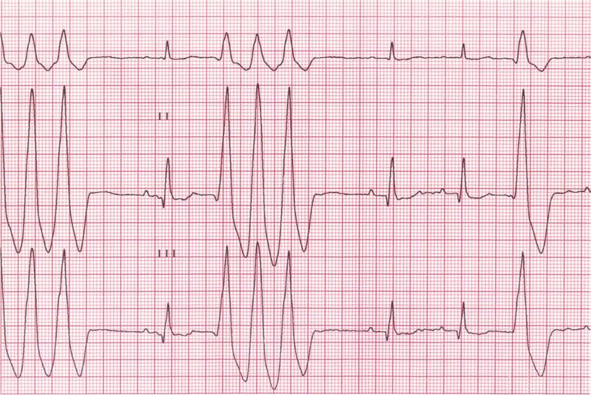
K.M. Meurs / Vet Clin Small Anim 34 (2004) 1235–1244            1237

Electrocardiography
   Affected dogs should have increased ventricular ectopy, but it may be
intermittent. The presence of an upright VPC on a lead II (left bundle
branch block morphology) electrocardiogram is suggestive of the disease
(Fig. 1). Some affected dogs have a different morphology to their VPCs or
may not have any VPCs detected on an electrocardiogram, however. A
normal electrocardiogram does not exclude a diagnosis of ARVC because
of the intermittent nature of the arrhythmia. If suspicion exists because of
auscultation of an arrhythmia, suggestive clinical signs (eg, syncope, exercise
intolerance), or a family history of disease, a 24-hour Holter monitoring
study is strongly suggested.

Holter monitoring
   Holter monitoring is an important part of the diagnosis, screening, and
management of canine ARVC. Even if the diagnosis is suspected based on
the identification of occasional VPCs on an in-house electrocardiogram,
a Holter study can provide the best assessment of overall frequency and
complexity of the arrhythmia and serve as an important guide for
monitoring treatment. It is uncommon for normal healthy adult dogs to
have any VPCs. In one study, healthy adult large-breed dogs had a median
of 2 VPCs in 24 hours [8]. An evaluation of more than 300 asymptomatic

Fig. 1. Upright ventricular premature complexes (left bundle branch block morphology)
observed in electrocardiogram leads I, II, and III.
1238           K.M. Meurs / Vet Clin Small Anim 34 (2004) 1235–1244

adult Boxers in a study performed in our practice found that 75% of the
population had less than 75 VPCs in 24 hours. Therefore, the identification
of frequent ventricular ectopy (>100 VPCs over 24 hours) in an adult Boxer
is strongly suggestive of a diagnosis of ARVC, particularly if there is signi-
ficant complexity (couplets, triplets, bigeminy, or ventricular tachycardia) to
the arrhythmia.
    Alternatively, a strong suspicion of ARVC sometimes exists but the
Holter monitor reading is not clearly abnormal. This may be because of the
significant day-to-day variability of VPC number in affected dogs. Consider-
able day-to-day variability in VPC number exists, and affected untreated
Boxers have been shown to have up to an 83% change in VPC number from
one day to the next [9]. Therefore, if a strong suspicion of ARVC exists but
the Holter reading was not diagnostic, a second Holter monitoring study
should be performed. If the dog is syncopal, an event monitor may be
considered. Additional testing may identify other possible causes of
syncope.

Echocardiography
    Although ARVC is a myocardial disease, most of the myocardial changes
are abnormalities noted at histologic examination as opposed to abnormal-
ities obvious on gross examination of the ventricle. Therefore, most affected
dogs have normal echocardiograms, particularly with regard to evaluation
of the size and function of the left ventricle. In some cases, careful
echocardiographic evaluation of the right ventricle may detect right
ventricular enlargement and, possibly, right ventricular dysfunction. Thor-
ough evaluation of the right ventricle by echocardiography is difficult,
however, because of the complex anatomy of the right ventricle, and subtle
changes may be frequently overlooked. A small percentage of adult Boxers
with tachyarrhythmias are observed to have left ventricular dilation with
systolic dysfunction [10]. Because these cases are observed infrequently, it
should be remembered that most affected dogs do not have echocardio-
graphically apparent abnormalities.

Pathology
   Postmortem findings can be helpful in the evaluation of sudden cardiac
death in the Boxer. Many affected dogs have a grossly normal appearance to
their heart at the time of death; however, some cases may show evidence of
right ventricular enlargement and, in some cases, left ventricular enlarge-
ment. Careful histologic evaluation should identify fatty, and sometimes
fibrofatty, segmental or diffuse replacement of the right ventricular free wall
from the epicardium toward the endocardium (Fig. 2). Occasionally, the
interventricular septum and left ventricular free wall are also involved [1,4].
K.M. Meurs / Vet Clin Small Anim 34 (2004) 1235–1244 1239 Fig. 2. Right ventricular myocardial sample from a Boxer with arrhythmogenic right ventricular cardiomyopathy. Note the fibrofatty myocardial infiltration observed histologically. The epicardium is located on the left aspect of the image. Screening The familial nature of ARVC has led to increased interest by Boxer breeders in the screening of breeding dogs for the disease. Unfortunately, because of the absence of a perfect diagnostic test for ARVC, screening the asymptomatic dog is challenging, and substantial efforts are being directed toward the development of a genetic test. Careful consideration of multiple criteria should be used to help determine the likelihood that an individual asymptomatic dog is affected. Important factors might include a familial history of ARVC in association with repeatable abnormal Holter monitor readings. Holter monitoring results should be evaluated for the number of VPCs as well as the complexity of arrhythmia (eg, single, couplets, triplets, ventricular tachycardia). As mentioned previously, the identification of frequent ventricular ectopy (>100 VPCs over 24 hours) in an adult Boxer is strongly suggestive of a diagnosis of ARVC, particularly if there is sig- nificant complexity (couplets, triplets, bigeminy, or ventricular tachycardia) to the arrhythmia. Long-term studies that evaluate the predictive value of these findings for identifying dogs at risk of dying from ARVC have not been completed, however. Some affected dogs have an abnormal degree of ectopy but never develop clinical signs, whereas some affected dogs with the same degree of ectopy gradually progress and develop more severe arrhythmias as they mature. Finally, some Boxers have thousands of VPCs
1240           K.M. Meurs / Vet Clin Small Anim 34 (2004) 1235–1244

over 24 hours and a high grade of complexity and remain asymptomatic
[11]. The factors that determine which dogs eventually progress to the
clinical form of the disease are not known and increase the frustration of
screening for this disease. Although breeders are concerned about passing
on this trait, they should be advised about the significant complexities of
performing screening and counseled not to remove dogs completely from
a breeding program because of a single abnormal Holter reading. Annual
Holter monitoring is strongly recommended, and significant emphasis on
a single Holter monitor reading is strongly discouraged. ARVC is an adult-
onset disease, and the degree of ventricular ectopy seems to increase with
age; thus, a single Holter monitoring study performed in a young adult dog
may not detect an affected dog that is not yet demonstrating the trait.
Additionally, in some cases, systemic disease or other forms of cardiac
disease may lead to the development of VPCs on a Holter monitoring study
that may not be apparent on subsequent ones. Inadvertently removing
a significant number of unaffected dogs from breeding programs may have
a detrimental effect on the overall gene pool of a purebred dog.
   A possible system for screening asymptomatic dogs might include the
following Holter monitoring criteria:

 1. None to 20 single VPCs over 24 hours: interpreted as within normal limits
 2. Twenty to 100 VPCs over 24 hours: interpreted as indeterminate, suggest
    repeating in 6 to 12 months
 3. One hundred to 300 single VPCs over 24 hours: interpreted as suspicious,
    consider keeping out of the breeding program for 1 year and repeating
    the Holter study
 4. One hundred to 300 VPCs over 24 hours with increased complexity (fre-
    quent couplets, triplets, and ventricular tachycardia) or 300 to 1000 single
    VPCs over 24 hours: interpreted as likely affected
 5. More than 1000 VPCs over 24 hours: interpreted as affected, may con-
    sider treatment as discussed below

   The criteria listed are based on the appearance of the particular
prevalence of the arrhythmias in the asymptomatic population as opposed
to long-term outcome studies of dogs with the arrhythmias. They are given
as one possible screening method. It is strongly advised to consider all issues
for each dog, including family history, evidence of ongoing systemic disease,
and repeated Holter studies, before making strict recommendations.

Treatment
   Treatment considerations for affected dogs are generally directed toward
the use of ventricular antiarrhythmics, because most affected dogs do not
have systolic dysfunction and do not seem to progress to heart failure. When
K.M. Meurs / Vet Clin Small Anim 34 (2004) 1235–1244 1241 prescribing antiarrhythmics for the affected dog, several considerations should be taken into account. There are known benefits to the use of certain antiarrhythmics in affected dogs, including the ability to decrease the number of VPCs and the complexity of the arrhythmia. Additionally, syncopal episodes have been shown to decrease with antiarrhythmic therapy [12]. There is no evidence that the use of antiarrhythmics alters the risk of sudden cardiac death in affected dogs, however. Additionally, there is a risk of a proarrhythmic effect associated with the use of many antiarrhythmics, and the arrhythmia could potentially be exacerbated. Finally, antiarrhythmic therapy adds increased expense to the management of the case as well as increased labor for the owner. Therefore, the risks and benefits of treatment should be carefully considered and discussed with owners before starting therapy. Ideally, therapy should be managed by assessing a pretreatment and posttreatment (2–3 weeks later) Holter monitoring study. Comparing the pretreatment and posttreatment readings should help to detect a proarrhythmic effect as well as help to evaluate the efficacy of treatment. Considerable day-to-day variability in VPC number exists, and affected untreated Boxers have been shown to have up to an 83% change in VPC number from one day to the next [9]. Therefore, a positive treatment response is best attributed to treatment when at least an 80% reduction in VPC number as well as a reduction in the complexity of the arrhythmia on the posttreatment Holter reading is observed. Additionally, an increase in symptoms after starting treatment or a significant increase in the number of VPCs may suggest a proarrhythmic effect. The significant day-to-day variability in VPC number even in untreated affected Boxers underscores the inability to make an accurate assessment of treatment on a brief in-house electrocardiogram. Asymptomatic Boxers The best indication to start therapy in an asymptomatic affected Boxer is not well understood, and the risks and benefits of therapy should be carefully evaluated. Certainly, some dogs die from their arrhythmia before ever developing clinical signs; thus, the absence of clinical signs does not mean that there is no risk of sudden death. If an arrhythmia is detected on routine examination in an asymptomatic dog, a Holter monitoring study should be performed to evaluate for the frequency and complexity of the arrhythmia. Although a strict relation between the development of symptoms and the number of VPCs does not exist, the development of syncope is associated with higher numbers of VPCs and a greater degree of complexity [11]. Sustained ventricular tachycardia (ie, ventricular tachycardia run that lasts longer than 30 seconds) might be a risk factor for sudden death in people with ARVC [12]. A VPC that occurs early in diastole, the R on T phenomenon, is believed to be dangerous, because the nadir of the ventricular fibrillation threshold occurs at about the time that
1242           K.M. Meurs / Vet Clin Small Anim 34 (2004) 1235–1244

the T wave is inscribed on the electrocardiogram. Based on this, treatment
may be reasonable if there are at least 1000 VPCs over 24 hours or if runs of
ventricular tachycardia or evidence of the R on T phenomenon exists.
Nevertheless, it should be recognized that risk factors for sudden death in
Boxer dogs with ARVC have been incompletely described; furthermore,
studies that evaluate the impact of antiarrhythmic therapy on long-term
survival have not been done.

Boxers with syncope/exercise intolerance
   Boxers with syncope and ventricular arrhythmias are generally started on
treatment, ideally after a 24-hour Holter monitoring study. In some cases, if
the syncope is frequent or ventricular tachycardia is observed, a Holter
study may not be performed so as to start therapy as soon as possible. In
some cases, however, the absence of a pretreatment study may make it
difficult to assess the response to therapy fully. Therefore, when possible, a
Holter monitoring study is performed and the owner is advised to start
therapy immediately after removing the Holter monitor (before the results
are available) if great concern exists.

Treatment options
   A reduction in VPC number and complexity of the arrhythmia in affected
Boxers with frequent ectopy has been demonstrated with two treatment
protocols [13]. The first is sotalol (1.5–2.0 mg/kg administered orally every
12 hours), and the second is a combination of mexiletine (5–8 mg/kg
administered orally every 8 hours) and atenolol (12.5 mg per dog
administered orally every 12 hours). In some cases, a loss of appetite or
mild gastrointestinal upset may be observed with mexiletine; however,
theoretically, the addition of atenolol allows the low end of the mexiletine
dose to be used to help reduce this side effect. Additionally, when mexiletine
is given with food, side effects may be less frequently observed. It is likely
that variations in drug response exist in individual dogs. Therefore, if a poor
response is observed with one drug, a different one may prove to be more
effective.

Boxers with left ventricular dilation and systolic dysfunction
   A small percentage of Boxers with tachyarrhythmias present with systolic
dysfunction and, in many cases, left or biventricular heart failure [10]. If
echocardiography demonstrates systolic dysfunction and ventricular di-
lation, treatment for canine dilated cardiomyopathy is warranted. Addi-
tionally, supplementation with L-carnitine might be considered at a rate of
50 mg/kg administered orally every 8 to 12 hours, because a small number of
affected dogs have demonstrated improvement in systolic function and
prognosis after supplementation [3,14].
K.M. Meurs / Vet Clin Small Anim 34 (2004) 1235–1244                  1243

Prognosis
   Dogs with ARVC are always at risk of sudden death. Many affected dogs
live for years even without treatment, however. Many dogs may be
managed, symptom free, for years on antiarrhythmics. A small percentage
of these dogs may eventually develop ventricular dilation and systolic
dysfunction.

L-carnitine   cardiomyopathy in Boxers
   A deficiency in myocardial L-carnitine levels was observed in a family of
Boxers with left ventricular dilation and systolic dysfunction. The tachyar-
rhythmias included atrial fibrillation and VPCs. The dogs had normal to
high levels of plasma L-carnitine but low levels of myocardial carnitine.
Supplementation with L-carnitine as well as standard heart failure therapy
resulted in significant improvement in myocardial function and clinical
condition but did not result in total regression of the disease [3]. In general,
myocardial carnitine deficiency does not seem to be a common cause of
myocardial disease in the Boxer and is not likely to be directly associated
with boxer ARVC. Because it is difficult to assess myocardial carnitine levels
accurately without an endomyocardial biopsy and because plasma levels are
not an accurate assessment of myocardial levels, the true role of carnitine in
the boxer with systolic dysfunction and dilation is not known. Supplemen-
tation of these cases with L-carnitine, 50 mg/kg every 8 hours, may be
considered [14].

References
[1] Harpster N. Boxer cardiomyopathy. In: Kirk R, editor. Current veterinary therapy VIII.
    Philadelphia: WB Saunders; 1983. p. 329–37.
[2] Harpster N. Boxer cardiomyopathy. Vet Clin N Am Small Anim Pract 1991;21(5):
    989–1004.
[3] Keene BW, Panciera DP, Atkins CE, Regizt V, Schmidt MJ, Shug AL. Myocardial
    L-carnitine deficiency in a family of dogs with dilated cardiomyopathy. J Am Vet Med
    Assoc 1991;198:647–50.
[4] Basso C, Fox PR, Meurs KM, Towbin JA, Spier AW, Calabrese F, et al. Arrhythmogenic
    right ventricular cardiomyopathy causing sudden cardiac death in Boxer dogs: a new
    model of human disease. Circulation 2004;109:1180–5.
[5] Meurs KM, Spier AW, Miller MW, Lehmkuhl LB, Towbin JA. Familial ventricular
    arrhythmias in Boxers. J Vet Intern Med 1999;13:437–9.
[6] Koplitz S, Meurs K, Spier A, Bonagura JB, Luis Fuentes V, Wight N. Aortic ejection
    velocity in healthy boxers with soft murmurs and boxers without cardiac murmurs: 210
    cases (1997–2001). J Am Vet Med Assoc 2003;222:770–4.
[7] Kvart C, French AT, Luis Fuentes V, Haggstrom J, McEwan J, Schober K. Analysis of
    murmur intensity duration and frequency components in dogs with aortic stenosis. J Small
    Anim Pract 1998;39:318–24.
1244 K.M. Meurs / Vet Clin Small Anim 34 (2004) 1235–1244
[8] Meurs KM, Spier AW, Hamlin RL, Wright NA. Use of ambulatory electrocardiography
for detection of ventricular premature complexes in healthy dogs. J Am Vet Med Assoc
2001;218:1291–2.
[9] Spier AW, Meurs KM, Lehmkuhl LB, Wright NA. Spontaneous variability in the
frequency of ventricular arrhythmias in Boxers with arrhythmogenic cardiomyopathy. J
Am Vet Med Assoc 2004;224:538–41.
[10] Meurs KM, Baumwart R, Atkins CE, Bonagura JB, DeFrancesco T, Keene BW, et al.
Myocardial dysfunction in boxer dogs with tachyarrhythmias [abstract]. J Vet Intern Med
2003;17:439.
[11] Meurs KM, Spier AW, Wright NA. Evaluation of the ambulatory electrocardiogram of
boxer dogs with ventricular tachyarrhythmias and syncope [abstract]. J Vet Intern Med
2002;16:338.
[12] Priori SG, Aliot E, Blomstrom-Lundqvist C, Bossaert L, Breithardt G, et al. Task Force
on Sudden Cardiac Death of the European Society of Cardiology. Eur Heart J 2001;
22:1374–450.
[13] Meurs KM, Spier AW, Wright NA, Atkins CE, DeFrancesco T, Gordon S, et al.
Comparison of the effects of four antiarrhythmic treatments for familial ventricular
arrhythmias in Boxers. J Am Vet Med Assoc 2002;221:522–7.
[14] Keene B. L-carnitine supplementation in the therapy of dilated cardiomyopathy. Vet Clin
N Am Small Anim Pract 1991;21(5):1005–9.You can also read