HHV-8 and EBV Positive Lymphoproliferative Disease: A Challenging Case
←
→
Page content transcription
If your browser does not render page correctly, please read the page content below
Case Report                                                                                                                    doi: 10.5146/tjpath.2021.01540

        HHV-8 and EBV Positive Lymphoproliferative Disease:
                       A Challenging Case
                       Göksenil BÜLBÜL1                 , Gülen GÜL2           , Mehmet Ali ÖZCAN3                  , Sermin ÖZKAL1
Department of Pathology, 1Dokuz Eylül University, School of Medicine, IZMIR, TURKEY, 2University of Health Sciences, Tepecik Training and Research Hospital, IZMIR, TURKEY
                                       3
                                         Department of Hematology, Dokuz Eylul University, School of Medicine, IZMIR, TURKEY

   This case report has been submitted as an e-poster to the 30th National Pathology Congress, which has organized by the Federation of Pathology Societies and the
   Turkish Pathology Society and was held online on 20-23 May, 2021.

                                                                            ABSTRACT
   Human herpes virus-8 (HHV-8) is linked to four lymphoproliferative diseases: primary effusion lymphoma, HHV-8 positive multicentric
   Castleman disease (MCD), HHV-8 positive diffuse large B cell lymphoma and HHV-8 positive germinotropic lymphoproliferative disorder
   (GLPD). The diagnosis of HHV-8 associated lymphoproliferative diseases is quite challenging because each entity is rare and has a wide
   morphological spectrum. Our aim is to emphasize the overlapping histopathological features of MCD and GLPD as well as to underline the
   importance of clinicopathological correlation in case these two entities cannot be distinguished by pathological examination.
   We present here a case of an 82-year-old male patient who was examined for weight loss and multiple lymphadenopathy. Histopathological
   examination of the axillary lymph node revealed lymphoid follicle structures of varying shapes and sizes, including some atrophic germinal
   centers. Plasmablast-like cells were notable in some of these areas. HHV-8 and Epstein Barr Virus (EBV) positivity were noted in some of these
   cells and in a small number of cells in the mantle zone. Based on these findings; a diagnosis of “HHV-8 and EBV positive lymphoproliferative
   disease” was established.
   Since HHV-8 positive MCD and GLPD have similar histopathological features, it may not be possible to distinguish these two entities by
   histopathological examination only. At this point, the importance of clinicopathological correlation becomes more evident, especially in the
   determination of the treatment protocol to be applied to the patient.
   Keywords: Castleman disease, Germinotropic lymphoproliferative disorder, EBV, HHV-8

                             INTRODUCTION                                                                           CASE REPORT
   Human herpesvirus-8 (HHV8) is a herpes virus that                                    Here we report a case of an 82-year-old male from İzmir/
   infects the endothelium, lymphocytes, keratinocytes                                  Turkey diagnosed with schizophrenia, Parkinson’s disease
   and bone marrow stromal cells. It is associated with four                            and diabetes mellitus and who had been taking medications
   lymphoproliferative diseases: primary effusion lymphoma,                             for many years. He presented to a physician with increasing
   HHV-8 positive multicentric Castleman disease (MCD),                                 weight loss for the last one year in addition to fatigue.
   HHV-8 positive diffuse large B cell lymphoma and HHV-                                It was also learned that his brother had a diagnosis of
   8 positive germinotropic lymphoproliferative disorder                                lymphoma. Physical examination revealed conglomerated
   (GLPD) (1,2).                                                                        and fixed multiple lymphadenopathies, the largest of which
                                                                                        was 4 cm in the right inguinal region and 1 cm in the left
   The Epstein Barr Virus (EBV) is also a lymphotropic virus
                                                                                        supraclavicular region. Peripheral blood test revealed the
   from the herpesvirus family like HHV8 (3). Although both
                                                                                        following: hemoglobin 9.9 g/dL, white blood cells 13,200/
   viruses are associated with various lymphoid diseases,
                                                                                        mm3, and platelets 525,000/ mm3. There were abnormal
   HHV8 + / EBV + lymphoproliferation is a rare entity (4).
                                                                                        findings in routine blood tests: serum electrolytes were
   Because of its rarity, we present a case co-infected with                            generally low, BUN was 33 mg/dL, and CRP 58.6 mg/L;
   HHV8 and EBV resulting in a differential diagnosis                                   the IgG level was 3059.8 mg/dL (N: 700-1600) and the IgA
   difficulty due to the similar histopathological features of                          level 502.9 mg/dL (N: 70-400). On serological examination,
   HHV-8 associated lymphoproliferative diseases.                                       there was no evidence of HIV infection.

                                                                                        Correspondence: Göksenil BÜLBÜL
                                                                                        Department of Pathology,
   Received : 02.05.2021 Accepted : 23.05.2021                                          Dokuz Eylül University, School of Medicine, İZMIR, TURKEY
                                                                                        E-mail: goksenilbulbul@gmail.com Phone: +90 554 470 75 37

                                                                                                                                                                     1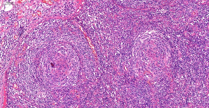
Turkish Journal of Pathology BÜLBÜL G et al: HHV8 & EBV Positive Lymphoproliferative Disease
Abdominopelvic ultrasound showed hepatosplenomegaly interfollicular areas were enlarged. In these areas, mostly
while PET revealed cervical, supraclavicular, axillary, CD3 positive T lymphocytes as well as CD38 positive
mediastinal-hilar, intraabdominal, bilateral inguinal plasma cells, some of which formed large aggregates, and
and femoral multifocal lymphadenopathy in addition to marked vascular proliferation in the endothelium were
bilateral pleural effusion. The largest lymph node was in the seen (Figure 6). Although plasma cells and plasmablast-like
right axillary with a size of 30x18 mm and SUVmax of 3.3. cells were predominantly lambda positive, some of them
were positive with lambda and some with kappa.
On evaluation of the resected right axillary lymph node
specimen measuring 23x13x7 mm, serial sections were gray- Finally the case was reported as “HHV-8 and EBV Positive
white colored and a nodular appearance was remarkable. Lymphoproliferative Disease” instead of giving a definite
In the sections of the total processed lymph node, the diagnosis. Two cycles of Rituximab one month apart were
normal structure was partially preserved and lymphoid administered to the patient.
follicle structures (CD20 and PAX5 positive) of varying DISCUSSION
shapes and sizes, including some atrophic germinal centers
(CD21 and CD23 positive, Bcl-2 negative) were observed Kaposi sarcoma associated herpesvirus (KSHV), also
(Figure 1). In some of the germinal central structures, it was known as HHV-8, is a lymphotropic virus and associated
seen that lymphoid cells were decreased and hyalinized. with 4 lymphoproliferative diseases: primary effusion
Plasmablast-like cells were notable in some of these areas lymphoma (PEL), HHV-8 positive multicentric Castleman
(Figure 2). HHV-8 and EBV positivity was noted by in situ disease (MCD), HHV-8-positive diffuse large B-cell
hybridization (EBER) in some of these cells and in a small lymphoma and rarely germinotropic lymphoproliferative
number of cells in the mantle zone (Figure 3,4). In some disorder (GLPD) (2).
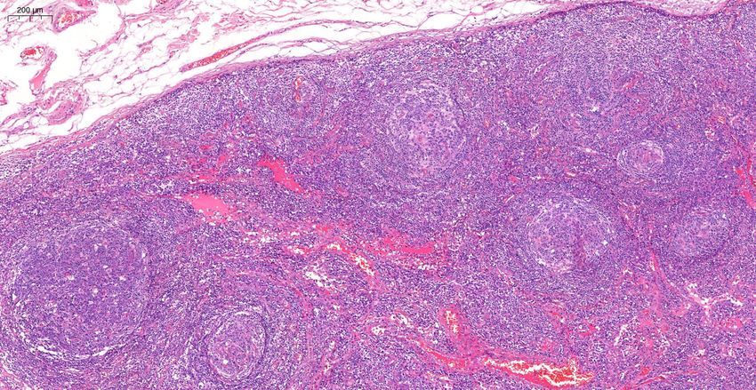
follicle structures, a concentric arrangement in the mantle PEL presents as serous effusion in body cavities (peritoneal,
zone areas and vascular structure penetrating into the pleural and pericardial) or solid tumour without effusion
germinal center were noteworthy (Figure 5). Occasionally, (“solid” PEL) and occurs in immunodeficient patients with
Figure 1: Microscopic examination of lymph node, lymphoid Figure 2: Hyalinized germinal center including plasmablast-like
follicle structures of varying shapes and sizes (H&E stain, x4). cells (H&E stain, x20).
Figure 3: HHV-8 positive cells in the germinal center and mantle Figure 4: EBV positive cells in the germinal center and mantle
zone (HHV-8 Immunohistochemistry, x5). zone by in situ hybridization (CISH EBER, x5).
2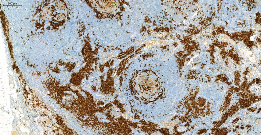
BÜLBÜL G et al: HHV8 & EBV Positive Lymphoproliferative Disease                                      Turkish Journal of Pathology

Figure 5: Concentric arrangement in the mantle zone areas and     Figure 6: CD38 positive plasma cells forming large aggregates in
vascular structure penetrating into the germinal center (H&E      interfollicular areas (CD38 Immunohistochemistry, x5).
stain, x10).

HIV infection. Infected patients have systemic symptoms           Although MCD and GLPD are two distinct diseases,
and prognosis is poor (5). That patients have HHV-8 and           similar/overlapping histopathological features can be seen
EBV positive immunoblasts with plasmacytoid cytoplasm             in these two entities (Table II).
and pleomorphic nuclei. PEL differs from GLPD in the
                                                                  As MCD progresses with systemic involvement, the
absence of cytoplasmic immunoglobulin expression (6).
                                                                  multiple lymph node involvement and systemic symptoms
Castleman disease (CD) describes 4 diseases: unicentric           in our patient primarily directed us to a diagnosis of
CD, HHV-8 associated MCD, POEMS associated MCD,                   MCD. Although GLPD usually presents as localized and
idiopathic -who are negative for HHV-8 and HIV-MCD                sometimes multifocal lymphadenopathy (12), a few cases
(7,8). MCD is characterized by enlarged lymph nodes in            with symptoms such as mild splenomegaly and systemic
multiple regions and spleen involvement. It is a systemic         symptoms have been reported (10,11).
disease and involves hepatomegaly, splenomegaly,
                                                                  Since GLPD is mostly seen in HIV-negative
constitutional symptoms and cytopenias (9). HHV-8
                                                                  immunocompetent patients, we may consider the HIV
associated MCD occurs in most commonly HIV positive
                                                                  negativity in favor of GLPD in our patient. However
patients but HIV negative patients have also been reported
(10). Histopathology is prominent, includes hyperplastic/         there is also a 58-year-old HIV-positive patient who was
atrophic germinal centers and hypervascularization;               diagnosed with GLPD in the literature (11). In addition,
plasmablasts generally located in mantle zones (11,12).           an HIV-negative HHV-8 positive subgroup of MCD,
                                                                  which occurs mostly in immunosuppressive patients, has
GLPD is a rare HHV-8 associated lymphoproliferative               also been identified (10). Therefore, the HIV status of the
disorder, first described in 3 cases in 2002 by Du et al. and     patient is not a reliable criterion in distinguishing these two
followed by 15 more case reports (12) (Table I). It presents      diseases.
as localized lymphadenopathy and on histopathological
examination it is characterized by an infiltration of germinal     Some features described in microscopic findings (plasma-
centers by plasmablastic cells, which are coinfected by           blast-like cells, atrophic germinal centers, decreased lym-
HHV-8 and EBV. Migration of neoplastic B-lymphocytes              phoid cells, hyalinization etc.) overlap with both entities
into germinal centers may be the origin of plasmablasts           but the presence of a concentric arrangement in the mantle
in GLPD. The presence of the atypical plasma cells in the         zone strengthens the diagnosis of MCD.
mantle zone and interfollicular area supports this theory.        Another important point according to all published GLPD
In addition to plasmablastic cells, residual follicle centers     cases in the literature is that HHV-8 and EBV co-infection
can be seen. There are sometimes atrophic follicles similar       is one of the most significant criteria that differentiates
to MCD. GLPD responds well to chemotherapy and                    GLPD from MCD (11-16). However, in an article published
radiotherapy.                                                     by Nobel et al. in 2019, EBV positivity was detected in two
In keeping with these features, the possibilities of “HHV-        of two HHV-8 positive MCD patients included in the
8 Positive Multicentric Castleman Disease” and “HHV-8             study (17). This newly defined condition, the presence of
Positive Germinotropic Lymphoproliferative Disorder”              EBV positivity in MCD, will cause serious difficulties in
were considered in the differential diagnosis of our case.        distinguishing these two diseases, as in our case (18-19).

                                                                                                                                3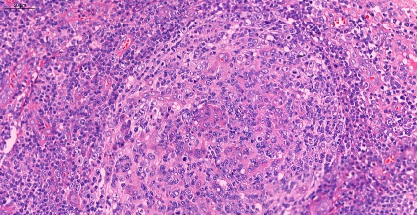
Turkish Journal of Pathology                                                    BÜLBÜL G et al: HHV8 & EBV Positive Lymphoproliferative Disease

 Table I: Clinicopathological features of patients diagnosed with germinotropic lymphoproliferative disorder
             Age/                   Clinical            HIV  Ig heavy/light                             Treatment and Prognosis
             Sex                  Presentation              chain expression
 Case 1     41y/M      Axillary and cervical lymph node  -       Lambda                                           CHOP
 (12)                       enlargement for 6 years            cIgM, cIgD                                 Complete remission
 Case 2     61y/M     Submandibular and inguinal lymph   -       Lambda                                 Excision and radiotherapy
 (12)                    node enlargement for 4 years              cIgA                                   Complete remission
                            Slightly enlarged spleen
 Case 3      63y/F                 Paresthesia           NI       Kappa                                               NI
 (12)                           Left leg swelling
                      Paraaortic lymph node enlargement
 Case 4     60y/M              Localized cervical        -        Kappa                                         Excision
 (12)                          lymphadenopathy                    cIgM                                   No evidence of relaps
 Case 5     65y/M         Right cervical lymph node      -        Kappa                               Without therapy, alive 7 years
 (16)                             enlargement                     cIgM
 Case 6     75y/M               Mass in the neck         -        Kappa                                         CHOP
 (20)                  Cervical lymph node enlargement                                                    19 months disease free
                           Cystic lymph node in left
                              submandibular area
 Case 7      49y/F     Right jugulo-cervical nodal mass  -       Lambda                                Excision and radiotherapy
 (15)                                                                                                     Complete remission
 Case 8      84y/F        Multifocal lymphadenopathy               -            None                             CHOP
 (11)                                                                                                     Complete remission
 Case 9     58y/M         Localized right axillary mass            +            None                            Resection
 (11)                              for 10 years                                                    One year later developed DLBCL,
                              Mild splenomegaly                                                    died due to his disease subsequent
 Case 10     72y/F      Palpable left cervical lymph node          -           Lambda                       Without therapy
 (21)                                                                                                    No evidence of relaps
 Case 11     63y/F      Autoimmune hemolytic anemia                -           Lambda                       Without therapy
 (1)                        Prominent mesenteric                                                    8 months later HHV8 + EBV +
                              lymphadenopathy                                                                  lymphoma
 Case 12 53y/M            Swelling of cervical nodes               -               μ                                  NI
 (22)
 Case 13 86y/M                  Localized cervical                 -            Kappa                       Without therapy
 (14)                           lymphadenopathy                                                           No evidence of relapse
 Case 14 52y/M               Inguinal lymph node                   -            None                             CHOP
 (14)                       enlargement for 3 years
 Case 15 47y/M           Generalized lymphadenopathy               +            None                                CHOP
 (14)                            B Symptoms
 Case 16 27y/M           Generalized lymphadenopathy               +            Kappa                             Rituximab
 (14)                            B Symptoms
 Case 17 30y/M           Generalized lymphadenopathy               +            Kappa                          R-DA-EPOCH
 (14)                            B Symptoms
 Case 18 42y/M           Generalized lymphadenopathy               +           Lambda                          R-DA-EPOCH
 (14)                            B Symptoms
NI: No information, DLBCL: Diffuse Large B Cell Lymphoma, CHOP: Rituximab, cyclophosphamide, doxorubicin, vincristine, prednisone,
EPOCH: Etoposide, prednisone, vincristine, cyclophosphamide, doxorubicin, R-DA-EPOCH: Rituximab, vincristine, adriamycin, cyclophosphamide,
methylprednisolone.
4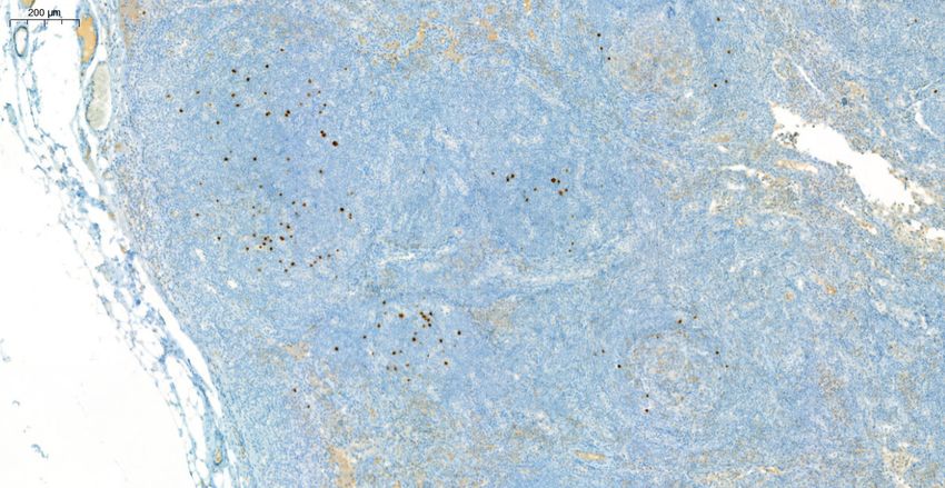
BÜLBÜL G et al: HHV8 & EBV Positive Lymphoproliferative Disease                                          Turkish Journal of Pathology

 Table II: Comparison of the clinical and pathological features of HHV-8 positive MCD and GLPD
                            HHV-8 Positive MCD                                HHV-8 Positive GLPD
 Clinical                   Mostly in HIV positive immunodeficient            Predominantly in HIV negative
 Presentation               patients                                          immunocompetent patients
                            Generalized lymphadenopathy,                      Often localized lymphadenopathy
                            splenomegaly, constitutional symptoms             Sometimes multifocal lymph node involvement
                                                                              and rarely systemic symptoms
 Prognosis                  Poor prognosis                                    Usually favorable response to chemotherapy and
                                                                              radiotherapy
 Microscopic                Abnormal follicle structures                      Residual follicle centers can be seen
 Findings                   Plasmablasts generally located in the mantle      Plasmablasts partially/completely invade germinal
                            zone but they may intrude into germinal           centers
                            centers
                                                                              Sometimes atrophic follicles similar to MCD
                            Atrophic or hyperplastic germinal centers
                            Prominent vascular proliferation
                            Concentric onion skin-like layering
                            Plasma cell hyperplasia in interfollicular area
 EBER                       Positive/Negative                                 Always positive
 HIV                        Usually positive, rarely negative                 Predominantly negative, rarely positive
 Cytoplasmic Ig             Elevated, only IgM                                Elevated, any heavy chain
 Heavy Chain
 Ig Light Chain             Monotypic lambda +                                Monotypic kappa or lambda +
 Clonality (Ig gene         Polyclonal                                        Polyclonal/Oligoclonal
 rearrangements)
 Mutated Ig Genes           Absent                                            Present
 Cell of Origin             A naive B cell                                    A germinal center B cell

Due to the reasons described above and the morphologically                                   REFERENCES
similar features, it is very difficult to distinguish between the    1. Courville EL, Sohani AR, Hasserjian RP, Zukerberg LR, Harris
two entities only by histopathological examination. At this             NL, Ferry JA. Diverse clinicopathologic features in human
point, the importance of clinicopathological correlation                herpesvirus 8-associated lymphomas lead to diagnostic problems.
becomes more evident, especially in the determination of                Am J Clin Pathol. 2014;142:816-29.
the treatment protocol applied to the patient. The physical          2. Guerrero C, Jain T, Kelemen K. HHV-8-associated
examination and laboratory findings should also be                      lymphoproliferative disorders and pathogenesis in an HIV-
                                                                        positive patient. Case Rep Hematol. 2019;2019:4536157.
evaluated in detail and carefully.
                                                                     3. Chen CH, Liu HC, Hung TT, Liu TP. Possible roles of Epstein-
                  CONFLICT of INTEREST                                  Barr virus in Castleman disease. J Cardiothorac Surg. 2009;4:31.

The authors declare no conflict of interest.                         4. Taris M, de Mascarel A, Riols M, Delwail V, Milpied N,
                                                                        Dubus P, Parrens M. KHSV/EBV associated germinotropic
            AUTHORSHIP CONTRIBUTIONS                                    lymphoproliferative disorder: A rare entity, case report and
                                                                        review of the literature. Ann Pathol. 2014;34:373-7.
Concept: GB, GG, Design: GB, GG, Data collection or
                                                                     5. Nador RG, Cesarman E, Chadburn A, Dawson DB, Ansari MQ,
processing: GB, GG, MAÖ, SÖ, Analysis or Interpretation:                Said J, Knowles DM. Primary effusion lymphoma: A distinct
GB, GG, MAÖ, SÖ, Literature search: GB, GG, Writing:                    clinicopathologic entity associated with the Kaposi’s sarcoma-
GB, GG, Approval: SÖ.                                                   associated herpes virus. Blood. 1996;88:645-56.

                                                                                                                                      5
Turkish Journal of Pathology BÜLBÜL G et al: HHV8 & EBV Positive Lymphoproliferative Disease
6. Dupin N, Diss TL, Kellam P, Tulliez Micheline, Du MQ, Sicard 16. Antonio D, Amedeo B, Maria A, Angel PM, Oscar N. KSHV-
D, Weiss RA, Isaacson PG, Boshoff C. HHV-8 is associated with and EBV-associated germinotropic lymphoproliferative
a plasmablastic variant of Castleman disease that is linked to disorder: A rare lymphoproliferative disease of HIV patient
HHV-8-positive plasmablastic lymphoma. Blood. 2000;95:1406- with plasmablastic morphology, indolent course and favourable
12. response to therapy. Leuk Lymphoma. 2007;48:1444-7.
7. van Rhee F, Greenway A, Stone K. Treatment of Idiopathic 17. Nabel CS, Sameroff S, Shilling D, Alapat D, Ruth JR, Kawano
Castleman Disease. Hematol Oncol Clin North Am. 2018;32:89- M, Sato Y, Stone K, Spetalen S, Valdivieso F, Feldman MD,
106. Chadburn A, Fosså A, van Rhee F, Lipkin WI, Fajgenbaum
8. Dispenzieri A, Fajgenbaum DC. Overview of castleman disease. DC. Virome capture sequencing does not identify active viral
Blood. 2020;135:1353-64. infection in unicentric and idiopathic multicentric Castleman
disease. PLoS ONE. 2018;14:1-13.
9. Waterston A, Bower M. Fifty years of multicentric Castleman’s
disease. Acta Oncol. 2004;43:698-704. 18. Zanelli M, Zizzo M, Bisagni A, Froio E, Marco LD, Valli R,
Filosa A, Luminari S, Martino G, Massaro F, Fratoni S, Ascani
10. Wang H, Pittaluga S, Jaffe ES. Multicentric Castleman disease:
S. Germinotropic lymphoproliferative disorder: A systematic
Where are we now? Semin Diagn Pathol. 2016;33:294-306.
review. Ann Hematol. 2020;99:2243-53.
11. Bhavsar T, Lee JC, Perner Y, Raffeld M, Xi L, Pittaluga S, Jaffe ES.
19. Nakaya Y, Ishii N, Kasamatsu Y, Shimizu K, Tatsumi N,
KSHV- and EBV-associated germinotropic lymphoproliferative
Tsutsumi M, Yoshida M, Yoshimura T, Hayashi Y, Nakao T,
disorder: New findings and review of the literature. Am J Surg
Inoue T, Yamane T. Human herpesvirus 8-positive multicentric
Pathol. 2018;41:795-800.
Castleman disease with germinotropic plasmablastic aggregates:
12. Du MQ, Diss TC, Liu H, Ye H, Hamoudi RA, Cabeçadas J, Dong Overlapping spectrum of human herpesvirus 8-associated
HY, Harris NL, Chan JKC, Rees JW, Dogan A, Isaacson PG. lymphoproliferative disorder. Pathol Int. 2020;70:574-80.
KSHV- and EBV-associated germinotropic lymphoproliferative
20. D’Antonio A, Addesso M, Memoli D, Liguori P, Cuomo R,
disorder. Blood. 2002;100:3415-8.
Boscaino A, Nappi O. Lymph node-based disease and HHV-8/
13. Swerdlow SH, Campo E, Harris N, Jaffe ES, Pileri SA, Stein H, KSHV infection in HIV seronegative patients: Report of three
Thiele J. World Health Organization Histological Classification new cases of a heterogeneous group of diseases. Int J Hematol.
of Tumors of Haematopoietic and Lymphoid Tissues. 4th ed. 2011;93:795-801.
IARC Press; 2017.
21. Zanelli M, Fraternali Orcioni G, Zizzo M, Marco LD, Martino
14. Gonzalez-Farre B, Martinez D, Lopez-Guerra M, Xipell M, G, Cerrone G, Cabras AD, Ascani S. HHV-8- and EBV-positive
Monclus E, Rovira J, Garcia F, Lopez-Guillermo A, Colomo L, germinotropic lymphoproliferative disorder. Ann Hematol.
Campo E, Martinez A. HHV8-related lymphoid proliferations: A 2019;98:2439-41.
broad spectrum of lesions from reactive lymphoid hyperplasia to
22. Papoudou-Bai A, Hatzimichael E, Kyriazopoulou L, Briasoulis E,
overt lymphoma. Mod Pathol. 2017;30:745-60.
Kanavaros P. Rare variants in the spectrum of human herpesvirus
15. Oh J, Yoon H, Shin DK, Jang MJ, Kim G, Chong SY, Oh D. A case 8/Epstein-Barr virus-copositive lymphoproliferations. Hum
of successful management of HHV-8 +, EBV + germinotropic Pathol. 2015;46:1566-71.
lymphoproliferative disorder (GLD). Int J Hematol. 2012;95:107-
11.
6You can also read