Technical Aspects And Early Results of Uniportal Video-Assisted Thoracoscopic Complex Segmentectomy: A 30 Case-Series Study - Research Square
←
→
Page content transcription
If your browser does not render page correctly, please read the page content below
Technical Aspects And Early Results of Uniportal Video-Assisted Thoracoscopic Complex Segmentectomy: A 30 Case-Series Study Xianglong Kong Harbin Medical University Third Hospital: Harbin Medical University Cancer Hospital https://orcid.org/0000-0002-5688-2888 Jun LU Harbin Medical University Third Hospital: Harbin Medical University Cancer Hospital Peng-Ju Li Harbin Medical University Third Hospital: Harbin Medical University Cancer Hospital Bo-Xiong Ni Harbin Medical University Third Hospital: Harbin Medical University Cancer Hospital Kai-Bin Zhu Harbin Medical University Third Hospital: Harbin Medical University Cancer Hospital Hai Xu ( xuhai18245159059@163.com ) Harbin Medical University Cancer Hospital https://orcid.org/0000-0002-2914-3270 Shi-Dong Xu Harbin Medical University Third Hospital: Harbin Medical University Cancer Hospital Research article Keywords: Uniportal video-assisted thoracic surgery, complex segmentectomy, subsegmentectomy, three dimensional computed tomography Posted Date: September 17th, 2021 DOI: https://doi.org/10.21203/rs.3.rs-793031/v1 License: This work is licensed under a Creative Commons Attribution 4.0 International License. Read Full License
Technical aspects and early results of uniportal video-assisted thoracoscopic
complex segmentectomy: a 30 case-series study
Xiang-Long Kong, Jun LU, Peng-Ju Li, Bo-Xiong Ni, Kai-Bin Zhu, Hai Xu & Shi-Dong Xu

Department of Thoracic Surgery, Harbin Medical University Cancer Hospital, Harbin, China

Abstract
Background. With the advantages of better cosmetic incision and faster recovery, uniportal video-assisted
thoracoscopic surgery (UP-VATS) has developed rapidly worldwide in recent decades and indications for
UP-VATS were further expanded as those for conventional VATS. Complex segmentectomy that makes
several, or intricate intersegmental planes, with more a complex procedure, continues to be a difficulty in
minimally invasive techniques. However, there are few reports as yet on UP-VATS complex
segmentectomy. In this report, we describe the perioperative clinical data and operative techniques and
present our early results of UP-VATS complex segmentectomy in our hospital.
Methods. The records of a total of 30 patients who underwent UP-VATS complex segmentectomy by a
single surgeon between January 2021 and June 2021 were retrospectively reviewed. We defined cases as
complex segmentectomy if they required resection of segment 9, 10, combined segmentectomy,
segmentectomy+subsegmentectomy, subsegmentectomy, or combined subsegmentectomy.
Results. The mean age was 52.8±9.9 years old; mean nodule size was 0.84±0.36 cm; the mean margin
width was 2.307±0.309 cm; median operative time was 229.0±58.06 minutes; mean operative hemorrhage
was 56.60±17.95 mL; 5.58±1.74 lymph nodes dissected had not metastasized; mean duration of
postoperative chest tube drainage was 4.7±1.4 days; and mean postoperative hospital stay was 6.5±3.0
days. Although 1 patient experienced a prolonged air leak, the other 29 recovered uneventfully. Another
patient failed to reach the 2cm safe margins who received completion lobectomy later.
Conclusions. UP-VATS complex segmentectomy is a safe and effective procedure in the treatment of lung
cancers, sparing more pulmonary parenchyma and ensuring safe margins, the disadvantage being lengthy
operative times during early acquisition of skills.
Keywords: Uniportal video-assisted thoracic surgery; complex segmentectomy; subsegmentectomy; three
 dimensional computed tomography.

Correspondence
Hai Xu, Department of Thoracic Surgery, Harbin Medical University Cancer Hospital, No. 150, Hapin Road,
 Harbin 150081, China.
Tel: +86 182 4515 9059 Email: xuhai18245159059@163.com
Introduction
For benign lesion and ground glass opacity (GGO) -dominant early stage lung cancer, the current trend is
precise resection with sufficient surgical margin and more preservation of pulmonary function[1][2][3][4].
Anatomic segmentectomy in treating early stage non-small cell lung cancer (NSCLC) is chosen more
frequently because of the same oncological outcomes as lobectomy and a sufficient surgical margin[5][6].
    A study based on high-resolution computed tomography reported that more than 33% of c-T1aN0M0
pulmonary nodules involved multiple segments[7]. To ensure an adequate surgical margin, complex
segmentectomy has been applied in certain clinical circumstances. Complex segmentectomy creates
several, or intricate intersegmental planes, with more a complex procedure[7][8][4][9]. Currently, few
articles have addressed complex segmentectomy, a procedure that may be particularly challenged in the
setting of UP-VATS, as the dexterity and fineness of instrument operation under UP-VATS are
limited[10][11][12].
    Our main goal is to evaluate the technical feasibility of UP-VATS complex segmentectomy and to
analyze the early clinical results of using this procedure.

Materials and methods

Patient Population

The Ethics Committee of Harbin Medical University Cancer Hospital approved this study because of the
retrospective design. From January 2021 and June 2021, 30 UP-VATS complex segmentectomies were
performed for benign lesion and ground glass opacity (GGO) -dominant early stage lung cancer by a team
at the Thoracic Surgery Department of Harbin Medical University Cancer Hospital.
    Complex segmentectomy was defined as segmental resection of the segment 9, 10, combined
segmentectomy, segmentectomy+subsegmentectomy, subsegmentectomy, or combined
subsegmentectomy.
    All 30 patients fulfilled the followed criteria: (1) The maximum diameter of the pulmonary nodule
detected on CT reconstruction lung window imaging was required to meet ≤ 2 cm with a ≥ 50% ground
glass opacity appearance on CT. (2) The lesion is far away from the visceral pleura, so wedge resection is
difficult to ensure the safe margin. (3) Intraoperative frozen negative lymph nodes were found in 10, 11,
12 and 13, and the estimated margin width was ≥2cm. (4) Compromise surgery or benign tumors may be
involved.
    Clinical data of patients undergoing UP-VATS complex segmentectomy were retrospectively reviewed
on age, sex, resected region, operative time, operative blood loss, final pathologic diagnosis, duration of
chest tube placement, hospitalization, intraoperative and postoperative complications. Each patient had
undergone enhanced thin-section (1-mm) thoracic CT. The reconstruction software “Mimics” was used to
reconstruct images of the pulmonary bronchi and blood vessels.

Operative Techniques

Complex segmentectomies were planned and performed under the guidance of 3D navigation. Tumor
locations, segmental structures and all bronchi, arteries, and veins involved were confirmed. To achieve an
adequate surgical margin, we designed the complex segmentectomy based on a nodule centered surgical
planning with parenchymal resection margins ≥2 cm and subsegment as a surgical unit (Fig 1). An incision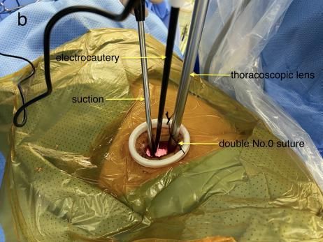
about 3.0 cm in length was made at the 5th intercostal space between the anterior-axillary line and
posterior-axillary line and protected with a wound retractor (Fig 2a,b). The specific location of the incision
depends on the anatomic location of the hilar of the target segment. It is best to strive for a 45 degree
angle between the operation angle of the surgical instrument and the hilar of the segment.
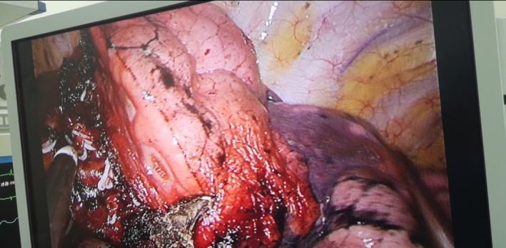
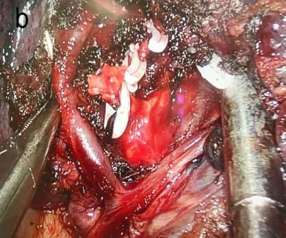
A electric hook with two curvature and ultrasonic scalpel were used for bronchovascular dissection
and No. 10, 11, 12 and 13 lymph nodes sampling (Fig 3a,b). The target bronchus was transected using a
stapler or Hem-o-lok (Fig 4). The target vessels can be stapled, ligated, or clipped according to the
specific conditions. A modified “inflation-deflation” technique was used to identify the intersegmental
border thereafter (Fig 5), enabling dissection of intersegmental planes using electrocautery, ultrasonic
scalpel, or staple along the intersegmental veins (Fig 6). Air leak test was done, and fibrin glue was applied
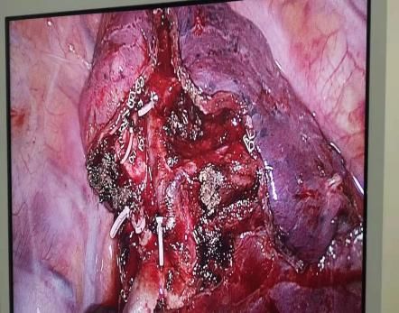
to reduce postoperative air leak. The margin width (from the resection margin to the nodule detected in the
removed specimen) was measured by the surgeon intraoperatively (Fig 7).
Figure 1 Complex segmentectomy based on a nodule centered surgical planning with parenchymal resection
margins ≥2 cm and subsegment as a surgical unit with the guidance of three dimensional (3D) navigation.
Figure (a) Body surface location of surgical incision. (b) Assistant stands on the opposite side of the
operator and holds the thoracoscopic lens, which is limited by the double No.0 suture.
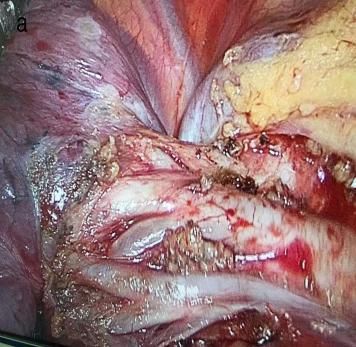
Figure 3 (a) Arteriovenous dissection and lymph nodes sampling of right upper lobe. (b) Arterial dissection
and lymph nodes sampling of right lower lobe. (c) Bronchovascular dissection and lymph nodes sampling
of left upper lobe.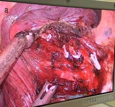
Figure 4 The target bronchus was transected using a stapler or Hem-o-lok.

Figure 5 A modified “inflation-deflation” technique was used to identify the intersegmental border.

Figure 6 Combine application of energy devices and staplers to the management of intersegmental plane.

Figure 7 The safe margin width ≥2 cm.
Results

The clinical characteristics of the 30 patients (9 men and 21 women, mean age 52.8±9.9, range: 32-71
years), the details of the nodule locations and surgical procedures in this study are presented in Table 1.
The mean nodule size was 0.84±0.36 cm. The lesions were located in the right upper lobe (13, 43.3%), the
left upper lobe (11, 36.6%), and the lower lobe (6, 20.0%).
    Evaluation of intraoperative and postoperative factors were shown in Table 2. No patients required
conversion to thoracotomy. Median operative time was 175 minutes (range, 75 to 294 minutes); mean
operative hemorrhage was 56.60±17.95 mL; 5.58±1.74 lymph nodes dissected had not metastasized;
mean duration of postoperative chest tube drainage was 4.7±1.4 days; and mean postoperative hospital
stay was 6.5±3.0 days. Aside from one instance of prolonged air leak ( >7 days ), all patients recovered
uneventfully. Another patient failed to reach the 2cm safe margins who received completion lobectomy
later. No deaths occurred. The tumor pathological diagnoses included: invasive adenocarcinoma (10 cases),
minimally invasive adenocarcinoma (9 cases), adenocarcinoma in situ (6 cases), atypical adenomatous
hyperplasia (3 cases), and benign (2 cases).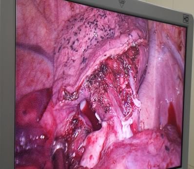
Table 1 Clinical characteristics of the patients, nodule locations and surgical procedures

Factor                                   Complex Segmentectomy

Age

      Mean (range)                       52.8±9.9 (32-71 years)
Gender

      Male                               9

      Female                             21

Mean nodule size (cm)                                0.84±0.36
Nodule location

RUL                                       13
       1     2
      S +S a                                 3
       2     3
      S +S a                                 1
       3     1
      S +S b                                 3
       1     2            3
      S +S +S a                              1
       1
      Sb                                         1
       3
      Sb                                         1
       2          3
      S b+S a                                2
       1     2            3
      S +S +S ai+bi                              1

LUL                                          11
       1+2
      S                                          1
       1+2
      S a+b                                  2
       1+2                    3
      S a+b+S c                              2
       1+2
      S b                                        1
       1+2            3
      S c+S a+b                              1
       1+2            3
      S c+S a                                    1
       3     1+2
      S +S a                                     2
       3          4
      S a+S a                                    1

RLL                                           2
       6          8               9
      S b+S a+S a                            1
       10
      S                                      1

LLL                                          4
       6     8
      S +S a                                     1
       8          9
      S a+S a                                    1
       7     8
      S +S                                       1
       9     10
      S +S                                       1

RUL, right upper lobe; LUL, left upper lobe; RLL, right lower lobe; LLL, left lower lobe.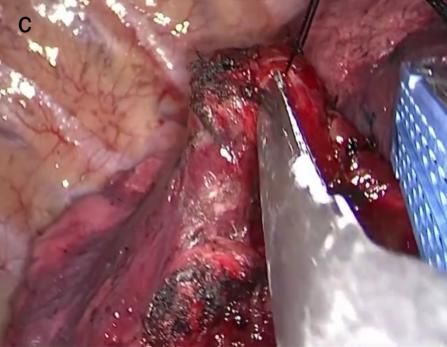
Table 2 Evaluation of intraoperative and postoperative factors

Factor                                 Complex Segmentectomy

Mean margin width (cm)                      2.307±0.309
Average operative duration (minutes)       229.0±58.06
Operative hemorrhage (mL)                   56.60±17.95
Number of lymph nodes dissected                       5.58±1.74
Duration of postoperative chest tub drainage (days)   4.7±1.4
Postoperative hospital stay (days)                    6.5±3.0
Pathological diagnoses

    Benign                                 2

    AAH                                    3

    AIS                                     6

    MIA                                    9

    IAC                                    10

AAH, atypical adenomatous hyperplasia; AIS, adenocarcinoma in situ; IAC, invasive adenocarcinoma; MIA, minimally

invasive adenocarcinoma.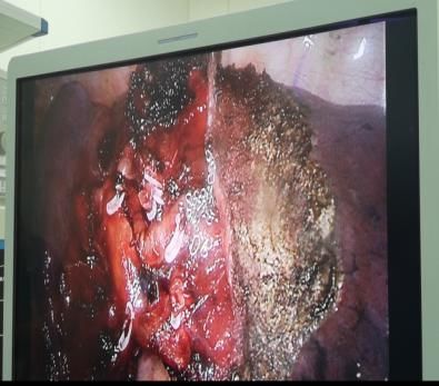
Discussion

Segmentectomy could be subdivided into simple and complex segmentectomy according to condition of
the intersegmental boundaries[8]. The ability to obtain a ≥2 cm safe surgical margin is the key factor that
makes complex segmentectomy superior to simple segmentectomy[7][13][11]. With the use of
reconstruction software “Mimics”, we designed the complex segmentectomy based on a nodule centered
surgical planning with parenchymal resection margins ≥2 cm and subsegment as a surgical unit.
    In our study, the margin width (2.307±0.309 cm) met the surgical requirement, except one patient who
received completion lobectomy later. The advantage of complex segmentectomy is achieving a sufficient
margin for the great majority of nodules.
    It is generally considered that it can be more difficult to divide complex segments when instruments
are operated in a single direction or are limited by angle changes in UP-VATS[14][15][11]. However, with
accumulated experience and familiarity with anatomy and operation techniques, the UP-VATS complex
segmentectomy could be performed safely[15][16][17]. The following tips of technical aspects can help if
we have some difficulties during the UP-VATS complex segmentectomy.
1. Further dissect the segmental hilar structures
    It is important to dissect the surrounding lung parenchyma and lymph nodes away from the root of
target segmental bronchus and vessels as completely as possible so that the bronchovascular bundle can
be further identified in targeted segments.
2. Apply rotatable stapler and adjust the angle by pulling the lung
    It is better to staple target segmental blood vessels and bronchi with rotatable stapler and adjust the
angle by pulling the lung to compensate the limitation in access angle for stapler. However, when
segmental hilar structures are right below the field of vision, stapler is difficult to be applied. In this case,
the better choice is ligation and clipping directly.
3. Combine application of energy devices and staplers to the management of intersegmental plane
    It is better to directly use electrocautery or ultrasonic scalpel to separate the inflation-deflation
interface along the intersegmental veins up to the outer one third of the pulmonary parenchyma. Once the
distal stumps of transected bronchus and vessels are dragged distantly from the hilum, the residual
intersubsegmental parenchyma is tailored by staplers easily. This method can keep the residual lung
extending and reduce air leakage.
    In our study, 1 patient experienced a prolonged air leak and stayed in hospital for 20 days after
operation. Air leakage was one of the major complications after segmentectomy[18]. The main causes of
air leakage are: ① injury of visceral pleura when dissecting segmental vessels, bronchus and lymph
nodes; ② injury of pulmonary parenchyma when energy devices are used to the management of
intersegmental plane; ③ air leakage at nail holes of stapler in patients with emphysema; ④ Injury of
bronchus. In order to reduce the occurrence of postoperative air leakage, it is necessary to separate the
inflation-deflation interface along the intersegmental veins when separating the intersegmental plane. If
bronchial injury is found, it must be sutured and repaired.
    Preoperative 3D lung reconstruction can clarify the anatomical structures, pre-judge the variations
and plan the surgical approach. Realtime 3D navigation can clearly identify the targeted bronchi and
vessels and localize the nodules, which improves the accuracy of the surgical technique[19][20].
    The study has several limitations. First, this was a single institute, retrospective review. Second, the
sample size was small, so statistical analysis was not robust. Finally, definition of complex segmentectomy
continues to be controversial. With the improvement of surgical experience and techniques, it is easy to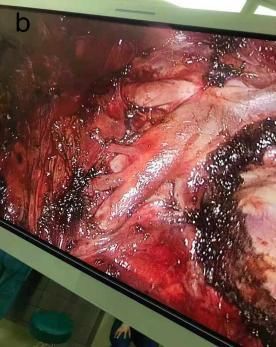
resect the lung segments with their own anatomical structure except the segment 9, 10. So we propose our
own definition of complex segmentectomy, which makes several intersegmental planes or involves multiple
target bronchi, arteries and veins. Our sample was limited and did not fully reflect the complexity of
complex segmentectomy. More various resection methods for complex segmentectomy should be
assessed further in future studies.

Conclusion

In conclusion, our limited experience indicates that UP-VATS complex segmentectomy is a safe and
effective procedure in experts’ hands, sparing more pulmonary parenchyma, ensuring safe margins and
providing the benefit of minimal invasiveness.

Abbreviations

VATS: Video-Assisted Thoracoscopic Surgery; UP-VATS: uniportal video-assisted thoracoscopic surgery;
GGO: ground glass opacity; CT: Computer Tomography; 3D: three dimensional; RUL: right upper lobe; LUL:
left upper lobe; RLL: right lower lobe; LLL: left lower lobe; AAH: atypical adenomatous hyperplasia; AIS:
adenocarcinoma in situ; MIA: Minimal Invasive Adenocarcinoma; IAC: Invasive Adenocarcinoma.

Acknowledgements

None.

Authors’ contributions

Hai Xu contributions to the conception and design of the work. Xianglong Kong contributions to the
acquisition, analysis, interpretation of data and the drafting and revision of the work. Jun Lu contributions
to the interpretation of data. The other authors read and approved the final manuscript.

Funding

This work was funded by the Wu Jieping Foundation (320675018551).

Availability of data and materials

The data and materials during the current study are available from the corresponding author on reasonable
request.

Ethics approval and consent to participate

The Ethics Committee of Harbin Medical University Cancer Hospital approved this study because of the
retrospective design. All patients obtained written consent on surgical technology and data use agreement
before surgery.
Consent for publication

Consent form from Harbin Medical University Cancer Hospital was obtained from patients included in the
research.

Competing interests

No authors report any conflict of interest.

References

1. Yamashita, S., et al., Thoracoscopic segmentectomy for T1 classification of
non-small cell lung cancer: a single center experience. Eur J Cardiothorac Surg, 2012.
42(1): p. 83-8.

2. Tsutani, Y., et al., Appropriate sublobar resection choice for ground glass
opacity-dominant clinical stage IA lung adenocarcinoma: wedge resection or
segmentectomy. Chest, 2014. 145(1): p. 66-71.

3. Fiorelli, A., et al., Sublobar resection versus lobectomy for stage I non-small cell lung
cancer: an appropriate choice in elderly patients? Surg Today, 2016. 46(12): p.
1370-1382.

4. Olland, A. and P.E. Falcoz, Complex segmentectomy in the treatment of stage IA
non-small-cell lung cancer. Eur J Cardiothorac Surg, 2020. 57(1): p. 122-123.

5. Yoshimoto, K., et al., Combined subsegmentectomy: postoperative pulmonary
function compared to multiple segmental resection. J Cardiothorac Surg, 2011. 6: p. 17.

6. Altorki, N.K., et al., Anatomical Segmentectomy and Wedge Resections Are
Associated with Comparable Outcomes for Patients with Small cT1N0 Non-Small Cell
Lung Cancer. J Thorac Oncol, 2016. 11(11): p. 1984-1992.

7. Wu, W.B., et al., Three-dimensional computed tomography bronchography and
angiography in the preoperative evaluation of thoracoscopic segmentectomy and
subsegmentectomy. J Thorac Dis, 2016. 8(Suppl 9): p. S710-S715.

8. Handa, Y., et al., Surgical Outcomes of Complex Versus Simple Segmentectomy for
Stage I Non-Small Cell Lung Cancer. Ann Thorac Surg, 2019. 107(4): p. 1032-1039.

9. Handa, Y., et al., Oncologic Outcomes of Complex Segmentectomy: A Multicenter
Propensity Score-Matched Analysis. Ann Thorac Surg, 2021. 111(3): p. 1044-1051.
10. Zhang, G., et al., Uniportal video-assisted thoracoscopic S(8) segmentectomy and
S(1a) subsegmentectomy for synchronous multiple primary lung cancers. J Thorac Dis,
2018. 10(7): p. 4475-4480.

11. Chen, Y.Y., et al., Uniportal versus Multiportal Thoracoscopic Complex
Segmentectomy: Propensity Matching Analysis. Ann Thorac Cardiovasc Surg, 2020.

12. Okamoto, K. and J. Hanaoka, Surgical outcome of combined subsegmentectomy in
the right upper lobe for GGO -dominant early stage lung cancer: Analysis of 7 cases.
Respir Med Case Rep, 2019. 26: p. 123-125.

13. Abdelsattar, Z.M. and S.H. Blackmon, Using novel technology to augment complex
video-assisted thoracoscopic single basilar segmentectomy. J Thorac Dis, 2018.
10(Suppl 10): p. S1168-S1178.

14. Yang, W., et al., Comparison of the perioperative efficacy between single-port and
two-port video-assisted thoracoscopic surgery anatomical lung resection for non-small
cell lung cancer: a systematic review and meta-analysis. J Thorac Dis, 2019. 11(7): p.
2763-2773.

15. Chang, C.C., et al., Single-port video-assisted thoracoscopic surgery
subsegmentectomy: The learning curve and initial outcome. Asian J Surg, 2020. 43(5): p.
625-632.

16. Wang, Y., Z. Wang, and F. Yao, The safety and feasibility of three-dimension
single-port video-assisted thoracoscopic surgery for the treatment of early-stage lung
cancer. J Thorac Dis, 2020. 12(12): p. 7257-7265.

17. Qu, J.C., K.M. Soultanis, and L. Jiang, Surgical techniques and outcome analysis of
uniportal video-assisted thoracic surgery complex sleeve lung resection: a 20
case-series study. J Thorac Dis, 2021. 13(4): p. 2255-2263.

18. Wang, J., et al., Technique for tailoring complex           demarcation   in   lung
segmentectomy. Thorac Cancer, 2018. 9(11): p. 1562-1564.

19. Wu, W.B., et al., Thoracoscopic Pulmonary Sub-Subsegmentectomy Based on
Three-Dimensional Images. Ann Thorac Surg, 2016. 102(5): p. e389-e391.

20. Wu, W.B., et al., Three-dimensional navigation-guided thoracoscopic combined
subsegmentectomy for intersegmental pulmonary nodules. Thorac Cancer, 2019. 10(1):
p. 41-46.
You can also read