Biology of Bone Repair - Giselle M. Hernandez, MD
←
→
Page content transcription
If your browser does not render page correctly, please read the page content below
Biology of Bone Repair
                                                Giselle M. Hernandez, MD
                           Original Author: Timothy McHenry, MD March 2004
                            Revision: J. Scott Broderick, MD November 2005
                                      Editors: Henry A. Boateng MD
                                            Regis Renard M.D.

*Xrays are from personal patients unless otherwise indicated                 Core Curriculum V5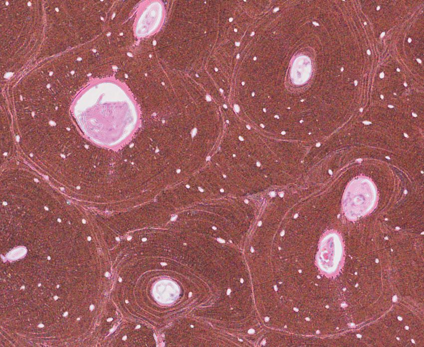
Objectives

• Bone Composition
• Bone Types
• Bone Healing
• Stages of Fracture Healing
• Factors that affect Bone Healing

                                     Core Curriculum V5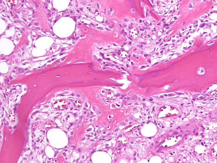
Bone Composition
• Cells
   • Osteocytes
   • Osteoblasts
   • Osteoclasts
• Extracellular Matrix
   • Organic Portion (35%)
       • Collagen Type 1 90%
       • Osteocalcin, Osteonectin
       • Proteoglycans, glycosaminoglycans
   • Inorganic Portion (65%)
       • Calcium Hydroxyapatite
       • Calcium Phosphate                   Normal Cortex.
                                             Courtesy of Andrew Rosenberg, MD

                                                                                Core Curriculum V5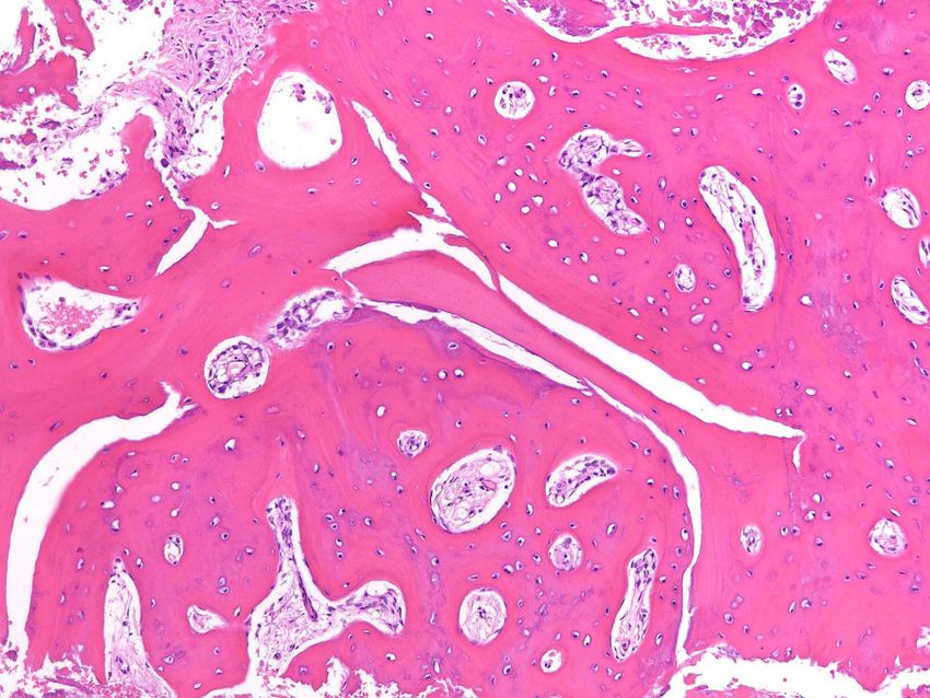
Osteocytes
• About 90% of cells in the mature skeleton
• Osteocytes live in lacunae
• Previous osteoblasts that get surrounded by the
  new formed matrix
• Control extracellular calcium and phosphorus
  concentration
• Stimulated by calcitonin
• Inhibited by PTH                                  Electron microscope picture of osteocyte.
                                                    Courtesy of Andrew Rosenberg, MD

                                                                               Core Curriculum V5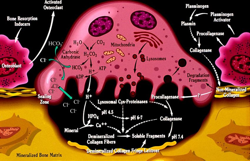
OsteoBlasts
• Derives from undifferentiated Mesenchymal Stem Cells
   • RunX2 directs mesenchymal cells to osteoblast lineage
• Line the surface of bone and produce osteoid
• Functions:
   • Form Bone
   • Regulate osteoclastic activity                          Courtesy of Andrew Rosenberg, MD

• Osteoblasts produce Type 1 collagen, RANKL, and Osteoprotegrin
• Osteoblasts are activated by intermittent PTH levels
• Inhibited by tumor necrosis Factor (TNF-α)

                                                                        Core Curriculum V5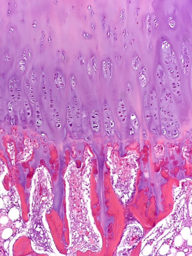
Osteoclasts
• Derived from Hematopoietic stem cell (monocyte precursor cells)
• Multinucleated cells
• Function to resorb bone and release calcium
• Parathyroid Hormone stimulates receptors on osteoblasts that activate
  osteoclasts

                                                Courtesy of Andrew Rosenberg, MD

                                                                             Core Curriculum V5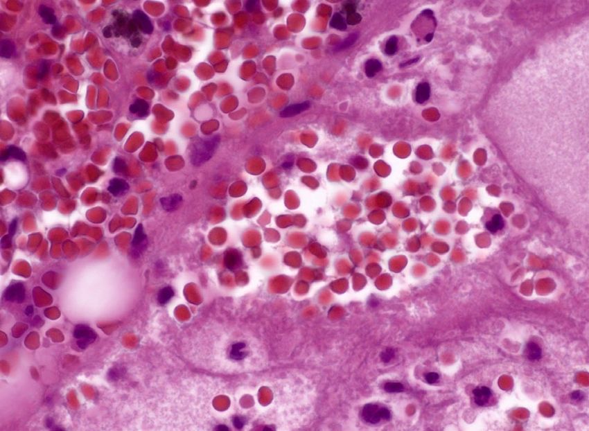
Osteoclasts
• Found in bone resorption craters called
  Howship Lacunae
    • Uses ruffled borders which increases
      surface area
    • Produces hydrogen ions through
      carbonic anhydrase
    • The lower pH increases the solubility
      of hydroxyapatite crystals                Above- Osteoclast in Howship
                                                Lacuna (blue arrow)
                                                Left- Electron Microscope of
                                                the same.
                                                Courtesy of Andrew Rosenberg, MD

                                                          Core Curriculum V5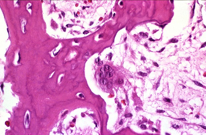
Osteocyte Network
• Osteocyte lacunae are connected by canaliculi
• Osteocytes are interconnected by long cell processes that project
  through the canaliculi
• Preosteoblasts also have connections via canaliculi with the
  osteocytes
• Network facilitates response of bone to mechanical and chemical
  factors
                                      Osteocyte

                                    Courtesy of Andrew Rosenberg, MD
                                                                       Core Curriculum V5
Osteon
                                                                     • Basic unit of bone
                                                                     • Consists of
                                                                         • Lamella- extracellular matrix
                                                                           made up of collagen fibers.
                                                                           Parallel to each other
                                                                         • Osteocytes in their lacunae
                                                                         • Vessels in the center in the
                                                                           Haversian Canal

Image from Rockwood and Green’s Fractures in Adults. Fig 1-12

                                                                                               Core Curriculum V5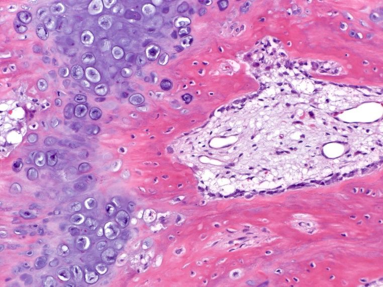
Extracellular Matrix
• Organic Components
  • Collagen- mostly Type 1 Collagen which provides tensile strength
  • Proteoglycans
  • Matrix proteins
     • Osteocalcin-most abundant noncollagenous protein
     • Growth Factors
     • Cytokines
• Inorganic Components
  • Calcium hydroxyapatite
  • Calcium Phosphate

                                                                       Core Curriculum V5
Blood Supply
• About 5-10% of a person’s cardiac output gets sent to
the skeletal system
• Long Bones Receive blood from three sources
• Nutrient artery system
• Metaphyseal-epiphyseal system
• Periosteal system
• Blood flow is one the most important factors in bone Acute Fracture Callus with Red Blood
healing along with stability Cell and Neutrophil infiltration.
• During fracture healing blood flow peaks at two weeks Courtesy of Andrew Rosenberg, MD
Core Curriculum V5Nutrient Artery
• Artery enters the nutrient
foramen in the diaphysis
• Branches into ascending and
descending arteries through
medullary canal
• This extends to the endosteum
and supplies about 2/3 of the
bone
Nutrient artery entering cortex
(Long arrow artery, short arrow cortex)
Courtesy of Andrew Rosenberg, MD Core Curriculum V5Metaphyseal Vessels
• Arise from the periarticular vessels (ex. Geniculate arteries)
• Penetrate the metaphyseal region and anastomose with the
medullary blood supply
Core Curriculum V5Periosteal Vessels
• Capillaries that supply the outer portion of the bone
• Arise from the periosteum which surrounds the cortex
• Supplies outer 1/3 of bone
• Can supply greater amount if endosteal supply is damaged.
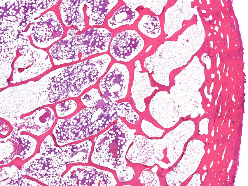
Core Curriculum V5Types of bone
• Lamellar
• Collagen fibers are arranged in parallel layers
• Normal adult bone
• Cortical
• Cancellous
• Woven Cancellous Bone Cortical Bone
• Collagen fibers are oriented randomly Courtesy of Andrew Rosenberg, MD
• Seen in remodeling bone or ligament/tendon insertion
• Pathological conditions
Core Curriculum V5Lamellar Bone
• Stress Oriented formation – highly organized
• Consists of Osteons and Interstitial lamellae (fibrils between osteons)
• Osteons communicate through Volkmann’s canals
• Cortical Bone
Above- Lamellar bone with
• Constitutes 80 % of bone osteocytes.
• Slow turnover rate
Left-Osteons
• Cancellous Bone
• Spongy or Trabecular bone Courtesy of Andrew Rosenberg, MD
• Higher turnover rate
• Less dense than cortical bone
Core Curriculum V5Woven Bone
• Immature or Pathologic Bone
• Random orientation of collagen
• Has more osteocytes
• Not stress oriented
• Weaker
Fracture with Reactive Woven Bone
Courtesy of Andrew Rosenberg, MD
Core Curriculum V5Mechanism of Bone Formation
• Bone Remodeling
• Wolff’s Law
• Bone will adapt according to the stress or load it endures
• Longitudinal Load will increase density of bone
• Compressive forces inhibit growth
• Tensile forces stimulates growth
• Types of Bone Formation
• Appositional
• Intramembranous (Periosteal) Bone Formation
• Endochondral Bone Formation
Core Curriculum V5Appositional Ossification
• Increase in diameter of bone by osteon formation on existing bone
• Osteoblasts align on existing bone surface and lay down new bone
• Periosteal bone increases in width
• Bone formation phase of bone remodeling
• Seen as bone grows in diameter and strength secondary to stress
• Remodeling due to forces on the bone
Core Curriculum V5Intramembranous Bone Formation
• Mostly seen in flat bones like cranium and clavicle
• Osteoblasts differentiate directly from preosteoblasts
and lay down osteoid
• There is no cartilage precursor
• Direct bone healing
Courtesy of Andrew Rosenberg, MD
Core Curriculum V5Endochondral Bone Formation
• Seen in embryonic bone formation, growth plates, and fracture callus
• Cartilaginous matrix is laid down osteoprogenitor cells come to the area
through vascular system- Osteoclasts resorb the cartilage Osteoblasts make
bone
• The Chondrocytes hypertrophy, degenerate and calcify
• Vascular Invasion of the cartilage occurs followed by ossification
• Cartilage is not converted to bone
• Bone Grows in Length
• Indirect bone healing
Core Curriculum V5Endochondral Bone Formation
Resting Zone
Proliferative Zone
Hypertrophic Zone
Calcification Zone
Fig 4-1. Ossification of the cartilage scaffold
in endochondral ossification. Ossification Zone
Image from Rockwood and Green’s Fracture’s In Adults.
Normal Growth plate.
Courtesy of Andrew Rosenberg, MD
Core Curriculum V5Stages of Fracture Healing
• Inflammatory Phase
• Repair
• Early Callus Phase
• Mature Callus Phase
• Remodeling Phase
Core Curriculum V5Inflammatory Phase
• Begins as soon as fracture occurs when a hematoma forms
• It lasts about 3-4 days
• Proinflammatory markers are released into the area
• IL-1, IL-6, TNF alpha
• This attracts cells like fibroblasts, mesenchymal cells and
osteoprogenitor cells
Fracture with hematoma.
Courtesy of Andrew Rosenberg, MD
Core Curriculum V5Inflammatory Phase
Image from Rockwood and Green’s Fractures in Adults. Fig 4-2
Core Curriculum V5Early Callus Phase
• Starts a few days after fracture
and lasts weeks
• Vascularization into the area
takes place
• Mesenchymal Cells in the area
differentiate into Chondrocytes
• Cartilage Callus is formed and
provides initial mechanical
stability
Image from Rockwood and Green’s Fractures in Adults. Fig 4-3
Core Curriculum V5Mature Callus Phase
• Cartilaginous Matrix is
mineralized
• Cartilage is degraded
• Bone is laid done as woven
bone through endochondral
ossification
• Fracture is considered
healed in this stage
Image from Rockwood and Green’s Fractures in Adults. Fig 4-4.
Core Curriculum V5Remodeling Phase
• Happens several months after fracture
• Woven Bone becomes Lamellar bone
• Previous shape of bone begins to be formed through Wolff’s Law
• This phase can continue for a year or more
• Fracture healing is complete when marrow space is reconstituted
Core Curriculum V5Cutting Cones
• Primary method of bone remodeling
• Osteoclasts are in the front of the cone
and remove the disorganized woven
bone
• Osteoblasts trail behind to lay down
new bone
• Blood vessel is in the center of the core
Image from Rockwood and Green’s Fractures in Adults Fig 4-6.
Core Curriculum V5Rockwood & Green’s Fractures in Adults Core Curriculum V5
Clinical Fracture Healing

• Direct (Primary) Bone Healing
   •   Cutting Cones
   •   Absolute Stability
   •   Rigid Fixation
   •   No callus formation
• Indirect (Secondary) Bone Healing
   •   Endochondral Ossification
                                      A.                                   B.
   •   Relative Stability                  A. Patient treated with fracture brace using secondary bone healing
   •   Comminution                         B. Patient with Compression plating and primary bone healing.

   •   Callus Formation

                                                                                   Core Curriculum V5
Direct (Primary) Bone Healing
• There is no motion at the fracture site
• Cutting cone crosses the fracture site
• Contact healing- there is direct contact between the
two fracture ends which allows for healing to start
with lamellar bone formation
• Gap Healing- if < 200-500 microns woven bone that
is formed can be remodeled into lamellar bone
• Examples: Compression Plating, lag screws and
neutralization plate
Core Curriculum V5Indirect (Secondary) Bone Healing
• Some motion at the fracture site
• Relative Stability
• Endochondral Ossification
• Large fracture gaps
• Comminution
• Example: Intramedullary nail,
Casting/bracing, Bridge plating
Right femoral shaft fracture treated with IMN.
Post OP 1 month, 8 months, 12 months.
Core Curriculum V5Strain
• Strain= change in fracture gap length/ length of fracture gap
• Strain < 2% promotes primary bone healing
• Strain 2-10% promotes secondary bone healing
• Multifragmentary fractures share strain
• Fracture creates mechanical instability and decreased
oxygenation. To promote healing the instability needs to be
decreased.
Core Curriculum V5Vascularity and Strain
• Vascularity helps create the scaffold for bone formation
• Strain and Vascularity have the most influence in type of bone healing
• Pericytes are stem cells that differentiate into osteoblasts or chondroblasts.
They come from the vasculature of the periosteum and endosteum.
• Pericytes become osteoblasts in low strain and high oxygen environment
and become chondrocytes in moderate strain and moderate vascularity
• When strain is reduced at the fracture site by stabilization of soft callus
formation, then endothelial cells migrate there in response to VEGF
• VEGF is released by chondrocytes and osteoblasts
Core Curriculum V5Direct (Primary) Bone Healing
• Bone healing with compression
• Bone formation with no cartilage cells.
• Osteoblasts and Osteoclasts working to
create new bone
Lamellar Bone formation in fracture site.
Courtesy of Andrew Rosenberg, MD
Core Curriculum V5Indirect (Secondary) Bone Healing
A. B.
C.
A. Fracture with Callus
B. High power view of fracture
C. Endochondral Ossification
Courtesy of Andrew Rosenberg, MD
Core Curriculum V5Factors affecting Healing
Biological Mechanical
• Comorbidities • Soft Tissue Attachments
• Nutritional Status • Stability
• Cigarette Smoking • High vs low energy mechanism
• Hormones • Extent of bone loss
• Growth Factors
• NSAIDs
Core Curriculum V5Biological Factors:
Comorbidities/Behavioral
• Comorbidities
• Diabetes- associated with collagen defects
• Vascular Disease- decreased blood flow to fracture site
• Nutritional Status
• Poor protein intake/ Albumin and prealbumin
• Vit D deficiency
• Cigarette Smoking
• Inhibits osteoclasts
• Causes Vasoconstriction decreasing blood flow to fracture site
Core Curriculum V5Biological Factors:
Hormones
• Growth Hormone: Increases gut absorption of calcium, Increases callus volume
• Calcitonin: Secreted from parafollicular cells in thyroid, Inhibits osteoclasts,
decreases serum calcium levels
• PTH: Chief cells of parathyroid gland, stimulates osteoclasts
• Corticosteroids: Decrease gut absorption of calcium, Inhibits collagen synthesis
and osteoblast effectiveness
Core Curriculum V5Biological Factors:
Growth Factors
• Bone Morphogentic Protiens (BMP): Stimulates bone formation by increasing
differentiation of mesenchymal cells into osteoblasts.
• Transforming growth factor Beta (TGF-β): Stimulates mesenchymal cells to
produce type II collagen and proteoglycans, stimulate osteoblasts to make
collagen
• Insulin like Growth Factor 2 (IGF-2): Stimulates collagen I formation, cartilage
matrix synthesis and bone formation
• Platelet-derived growth factor (PDGF): Attract inflammatory cells to fracture
sites
Core Curriculum V5Bone Morphogenetic Proteins
• Osteoinductive proteins initially isolated from demineralized bone matrix
• Noncollagenous glycoproteins that are part of the TGF-β family
• Induce Cell differentiation
• BMP-3 (osteogenin) is an extremely potent inducer of mesenchymal tissue
differentiation into bone
• Promote Endochondral ossification
• BMP-2 is FDA approved for open tibia fractures
• BMP 7 is FDA approved only for recalcitrant nonunion of long bones
• Regulate extracellular matrix production
Core Curriculum V5Insulin Growth Factors
• Two Types: IGF -1 and IGF II
• Synthesized by multiple tissues
• IGF-1 production in the liver is stimulated by Growth Hormone
• Stimulates bone collagen and matrix synthesis
• Stimulates replication of osteoblasts
• Inhibits bone collagen degradation
Core Curriculum V5Transforming Growth Factors
• Super-Family of growth factors (-34 factors)
• Acts on serine/threonine kinase cell wall receptors
• Promotes proliferation and differentiation of mesenchymal
precursors for osteoblasts, osteoclasts and chondrocytes
• Stimulates both endochondral and intramembranous bone
formation
• Induces synthesis of cartilage-specific proteoglycans and type II collagen
• Stimulates collagen synthesis by osteoblasts
Core Curriculum V5Platelet-Derived Growth Factors
• Large polypeptide that has two chains of amino acids
• Stimulates bone cell growth
• Mitogen for cells of mesenchymal origin
• Increases Type 1 Collagen synthesis by increasing the number of
osteoblasts
• PDGF-BB stimulates bone resorption by increasing the number of
osteoclasts
Core Curriculum V5Summary of Healing Molecules Table from Rockwood and Green’s Fractures in Adults Core Curriculum V5
Biological Factors:
 Non steroidal anti inflammatories (NSAIDs)
• NSAIDS work by binding to COX 1 or COX 1 and COX2 which
  decreases prostaglandin (PG) production. PGs assist in cell
  recruitment during fracture healing
• Both selective and non selective NSAIDs have been linked to
  decreased bone healing and nonunion formation
• Some studies suggest that COX 2 inhibitors do not effect healing as
  much
• Effects of NSAIDs on PG are reversible and levels return to normal at
  1-2 weeks when the drug is stopped.

                                                               Core Curriculum V5
Mechanical Factors
Soft tissue
• Periosteal Stripping
• Disruption of local blood supply
• Decrease ability of angiogenesis
• Decrease formation of soft callus or bone formation
• Interposition of fat or soft tissue in fracture site
• Increase fracture gap
• Inability to build upon a scaffold
Core Curriculum V5Mechanical Factors
Energy of injury
• High Energy
• GSW
• Crush Injury
• Motor Cycle or Motor Vehicle Accident
• More soft tissue injury and greater risk of nonunion
• Low Energy
• Fall from Standing Height
• Twisting Injury Mangled foot and open pilon from a
Motorcycle Crash
• Less soft tissue damage
Nondisplaced Lateral Tibial
Plateau Fracture Core Curriculum V5Mechanical Factors
Stability
• Absolute stability
• No movement between fracture fragments Stability Spectrum
• Anatomic Reduction of Fracture
• Intermembranous Ossification
• Relative Stability
• Controlled motion between fracture fragments
• Restoration of length, alignment, and rotation
• Endochondral Ossification
From left to right: Unstable, Casting, External fixation, Intramedullary
• Instability Nail, and Plate fixation
• Gross movement at the fracture site
• Cannot make callus or increase stability due to constant motion
• Leads to nonunion
Core Curriculum V5Failure of Stability
Instability Results in Nonunion Not Enough Stability Results in Hardware
failure and nonunion
Core Curriculum V5Absolute Stability
• Articular Fractures
• Pilon
• Tibial Plateau Injury Xray Open Pilon Fracture
and Post Op Xray about 12mo
• Distal Humerus
• Anatomic Reductions
• Fibular Fractures
• Humeral Shaft
• Radial and Ulnar Shafts
Injury Xray Both Bone Forearm fracture and Post Op Xray 6 mo
Core Curriculum V5Relative Stability
• High Comminution
Injury Xray and Post Op 12 months
• Long Bone Fractures
• Tibia Midshaft Fractures
• Femur Midshaft Fractures
• Metaphyseal fractures
• Distal Femur Fractures
• Proximal Femur fractures
Injury Xray and Post op 6 months
Core Curriculum V5Summary
• Two main types of bone cells are osteoblasts and osteoclasts
• Two main pathways of bone healing are intramembranous and
endochondral ossification
• There are many molecules that play a part and effect bone healing
• Stability of the fracture and blood flow to the region are the most
important factors in having a successfully healed fracture
Core Curriculum V5You can also read