Adversarial cycle-consistent synthesis of cerebral microbleeds for data augmentation
←
→
Page content transcription
If your browser does not render page correctly, please read the page content below
Adversarial cycle-consistent synthesis of cerebral
                                                     microbleeds for data augmentation

                                                         Khrystyna Faryna                                Kevin Koschmieder
                                                   Diagnostic Image Analysis Group                  Diagnostic Image Analysis Group
                                                  Radboud UMC, Nijmegen, 6525 GA                   Radboud UMC, 6525 GA Nijmegen
arXiv:2101.06468v1 [eess.IV] 16 Jan 2021

                                                 khrystyna.faryna@radboudumc.nl                  kevin.koschmieder@radboudumc.nl

                                                        Marcella M. Paul                             Thomas van den Heuvel
                                                  Diagnostic Image Analysis Group                   Dept. Of Medical Imaging
                                                 Radboud UMC, Nijmegen, 6525 GA                 Radboud UMC, Nijmegen, 6525 GA
                                                 marcella.paul@radboudumc.nl                  thomas.vandenheuvel@radboudumc.nl

                                                     Anke van der Eerden                             Rashindra Manniesing
                                                    Department of Radiology                      Diagnostic Image Analysis Group
                                                Erasmus MC, Rotterdam, 3015 CN                 Radboud UMC, Nijmegen, 6525 GA
                                                a.vandereerden@erasmusmc.nl                 rashindra.manniesing@radboudumc.nl

                                                                                 Bram van Ginneken
                                                                            Diagnostic Image Analysis Group
                                                                           Radboud UMC, Nijmegen, 6525 GA
                                                                          bram.vanginneken@radboudumc.nl

                                                                                        Abstract
                                                    We propose a novel framework for controllable pathological image synthesis for
                                                    data augmentation. Inspired by CycleGAN, we perform cycle-consistent image-
                                                    to-image translation between two domains: healthy and pathological. Guided
                                                    by a semantic mask, an adversarially trained generator synthesizes pathology on
                                                    a healthy image in the specified location. We demonstrate our approach on an
                                                    institutional dataset of cerebral microbleeds in traumatic brain injury patients.
                                                    We utilize synthetic images generated with our method for data augmentation in
                                                    cerebral microbleeds detection. Enriching the training dataset with synthetic images
                                                    exhibits the potential to increase detection performance for cerebral microbleeds in
                                                    traumatic brain injury patients.

                                           1   Introduction
                                           Clinical outcome for patients with traumatic brain injury (TBI) is associated with cerebral microbleeds
                                           (CMBs) [1]. CMBs are the result of leakages of small blood vessels, where hemosiderin deposits lead
                                           to focal dephasing of the MRI signal [2]. On MR-Susceptibility Weighted Imaging (SWI), CMBs
                                           appear as “spherical hypointense lesions”[3], with a diameter of less than ten millimeters. In TBI
                                           cases, CMBs can also have an elongated shape. Detection of CMBs, particularly their number and
                                           location, can provide useful information for the clinical prognosis of patients with TBI. However,
                                           due to their small size and visual similarity to blood vessels in a 2D view, manual annotation can
                                           be prohibitively time-consuming (≥ 1 h/scan) for clinical diagnosis of patients with moderate to
                                           severe TBI [4]. While a number of methods for automatic detection of CMBs have been proposed

                                           34th Conference on Neural Information Processing Systems (NeurIPS 2020), Vancouver, Canada.
[5, 6], a scarce availability of training samples with corresponding voxelwise annotations hampers
the detection performance of these algorithms. In this study, we aim to generate synthetic TBI images
(and the corresponding pathology mask) to augment the training dataset for a CMB detection system.
Synthesizing high-resolution images from random noise requires a vast number of training samples,
thus, in this work we focus on synthesizing pathology on healthy images. We propose a data
augmentation framework for synthesis of 3D medical images through pathology factorization and
adversarial cycle-consistent learning. Our approach is an extension on CycleGAN [7] by introducing
pathology masks as additional input and “abnormality mask” [8] loss, which encourages preservation
of identity in regions outside of the pathology mask. We perform pathological-to-healthy and healthy-
to-pathological image synthesis, factorizing pathology into a binary mask, to address the one-to-many
problem which is axiomatic in the healthy-to-pathological synthesis task. We additionally modify the
identity preservation pathway of the CycleGAN encouraging the lesions to be synthesized (in-painted)
exclusively in the area specified by the annotation provided. While the majority of methods in the
literature focus on synthesis of unlabeled data [9], patch in-painting [10] or images belonging to
restricted classes, our method is capable of synthesizing lesions of a size and location specified by a
binary mask; it is not restricted to a particular imaging modality or abnormality [11], neither does it
require any additional surrounding tissue annotation [12].
2 Materials and methods
Data The dataset used in this study consisted of 67 SWI scans from patients with moderate to
severe TBI and 18 healthy subject SWI scans, collected at the Radboud University Medical Center,
Nijmegen, The Netherlands. Scans have been acquired with the following parameters: TR:27 ms,
TE:20 ms, voxel size: 0.98 × 0.98 × 1.00 mm3 . A subset of 10 patient scans has been manually
annotated by 6 medical observers, to be used as a comprehensive testing set. The remaining scans
have been annotated by a single trained expert, subsequently split into training (46 pathological and
14 healthy scans) and validation (11 pathological and 4 healthy). First, we clip image intensities
between 0 and 99.5 percentile and then rescale the range between 0 and 1. From each scan, we
extract patches of 160 × 146 × 32, capturing the whole axial plane of the brain, with a 50% overlap
in z direction.
Lesion synthesis framework The schematic of the proposed approach along with training losses
is shown in Fig.1. Inspired by CycleGAN, the configuration comprises of two cycles: healthy-to-
pathological-to-healthy (HPH) synthesis and pathological-to-healthy-to-pathological (PHP) synthesis.
Here, we denote a healthy sample xhi with an empty pathology mask yhi , where xhi belongs to the
healthy data distribution, xhi ∼ H. A pathological sample is denoted as xpi with the corresponding
pathology mask ypi , where xpi belongs to the pathological data distribution, xpi ∼ P. Our objective
is two-fold: given xhi and a semi-random pathology mask ypi , generate a synthetic image x̃pi such
that x̃pi ∼ P, and given xpi (and a suitable pathology mask ypi ), generate a synthetic image x̃hi such
that x̃hi ∼ H. The index i specifies a particular subject.
LCC1 LCC2
xh1 x~p1 LAM
x^ h1 ~
xp2 xh2 x^ p2
G HP G PH
G PH G HP
yp1 yp1
yp2 yp2
LID
1
LID2
xh1 x'h1
xp2 x'p2
LGAN1 DP
G PH LGAN2 DH
G HP
xp2 yh
xh1 yh
Figure 1: The schematic of our proposed method: HPH cycle (right), PHP cycle (left). For visualization
purposes, we use brain tumour samples from BraTS2018 [13] dataset for this figure because their larger size
compared to CMBs better displays the concept of semantic maps and their results.
In the HPH cycle, the healthy-to-pathological generator GHP receives as input the healthy image xh1
and a semi-random pathology mask yp1 . The objective of GHP is to synthesize a pseudo-pathological
2image x̃p1 , such that the pathology would be located in the areas provided by the mask yp1 , while
the rest of the image should remain unchanged. The goal of the discriminator DP is to distinguish
between real (xp2 ) and fake (x̃p1 ) pathological samples. The obtained pseudo-pathological image x̃p1
is then concatenated with its pathology mask yp1 and passed to the pathological-to-healthy generator
GP H . The task of GP H is to reconstruct the input image x̂p2 . Additionally, to encourage preservation
of identity and assure that GP H does not modify a healthy image the xh1 is concatenated with yh and
passed through GP H . The losses used in this cycle are: cycle-consistency (LCC ), identity (LID ), and
adversarial loss (LGAN ). The latter is the Wasserstein loss with gradient penalty[14]. The reverse
is happening in PHP cycle, with only one exception–the use of abnormality mask loss (LAM )[8] to
encourage preservation of brain structures outside of the pathology region.
The architectures of GP H , GHP , DP and DH are inspired by [15], however we expanded on the
method by switching to a 3D approach, exchanging all 2D layers with their 3D counterparts. The
model was trained using the Adam[16] optimizer with β1 =0.5, β2 =0.99, learning rate of 0.0001 and
a batch size of 4.
Lesion detection The detection model used in this study consists of two stages: candidate proposal
with Fast Radial Symmetry Transform [17] and a classification CNN. The experiment is composed
of three pipelines, the detection network is trained with only real, synthetic and combined real and
synthetic data. We perform additional experiment comparing performance using also classical data
augmentation (cDA), in particular flipping in the axial plane, shearing, scaling, rotation, and intensity
(scaling, shifting) augmentations.
3 Results and discussion
Examples of synthetic healthy and pathological images produced with the proposed method are shown
in Fig. 2. Detection performance of models in different settings was evaluated using a Free-response
Receiver Operating Characteristic (FROC) curve, bootstrapping was used to compute 95% confidence
intervals (CI). Results of the experiments with synthetic data augmentation are shown in Fig. 3 and
Table 1.
Real pathological
Real healthy
Pathology mask
Pathology mask
Synthetic pathological
Synthetic healthy
(a) (b)
Figure 2: Synthetic SWI images generated with the proposed method: (a)–generation of synthetic pathological
images, generation of synthetic healthy images. The red bounding boxes indicate the place where the lesion is
generated/in-painted.
3Table 1: Sensitivity of models
trained on different data distributions
compared at a fixed, clinically relevant
rate of 10 false positives (FPs) per TBI
patient.
Training data Sensitivity (%)
Real 77.8
Synthetic 71.8
Real+Synthetic 78.9
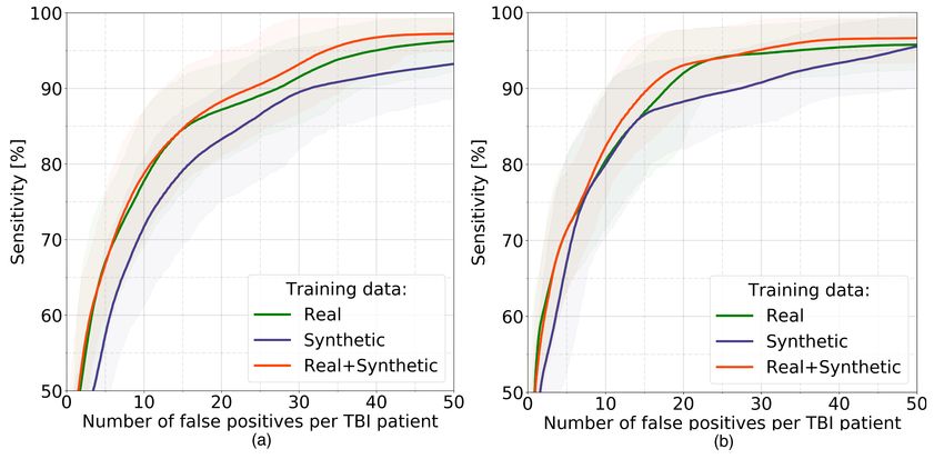
Real +cDA 80.5 Figure 3: FROC performance with 95% CI on an independent test
Synthetic +cDA 80.1 set of models trained with different (real, synthetic, real+synthetic)
Real+Synthetic +cDA 82.4
data: (a)–without cDA, (b)–with cDA.
The models trained on synthetic data are capable of producing meaningful predictions when tested on
real samples, demonstrating comparatively lower performance: 90% sensitivity is reached at 27.4
and 31.4 FPs/patient with and without cDA, respectively. The models trained with real data achieve a
sensitivity of 90% at 17.9 and 27.3 FPs/patient with and without cDA. Combining the real samples
with synthetic ones with 1:1 ratio, we observe a marginal yet consistent improvement in performance
with respect to the models trained on real data only: the models reach 90% sensitivity at 23.7 and
15.5 FPs/patient. We note here that intersections between CIs of the evaluated methods are relatively
high, nevertheless we envision the prospective value of using the trained system for generating CMB
lesions on external SWI data, potentially reducing the burden of additional manual annotation while
dealing with data coming from different institutions.
In conclusion, we proposed a method for synthesis of pathology on healthy medical images guided by
a pathology mask and demonstrated its use in data augmentation for CMB detection in TBI patients.
4 Broader impact
Detection of CMBs in TBI patients is a prime example of a medical application that is heavily afflicted
by data scarcity and variations between MR scanners. The proposed approach is aimed at reducing
the burden of manual annotation while shifting between various vendors and imaging protocols.
5 Acknowledgements
Khrystyna Faryna was supported by Erasmus Mundus Joint Master Degree in Medical Imaging and
Applications (MAIA) grant. We wish to thank Prof. Xavier Llado for his valuable feedback. We
would like to express our gratitude to T.M.J.C. Andriessen and P. Vos for collecting the data, and to
T.M.J.C. Andriessen, T. Vande Vyvere, L. van den Hauwe, B. Geurts and B.M. Goraj for their work
in annotating the Cerebral Microbleeds.
References
[1] David John Werring. Cerebral microbleeds: Pathophysiology to clinical practice. In Cambridge
University Press, Cambridge, UK, 2011.
[2] G. Roob, R. Schmidt, P. Kapeller, A. Lechner, H.-P. Hartung, and F. Fazekas. Mri evidence of
past cerebral microbleeds in a healthy elderly population. Neurology, 52(5):991–991, 1999.
[3] Steven M Greenberg, Meike W Vernooij, Charlotte Cordonnier, Anand Viswanathan, Rustam
Al-Shahi Salman, Steven Warach, Lenore J Launer, Mark A Van Buchem, and Monique MB
Breteler. Cerebral microbleeds: a guide to detection and interpretation. The Lancet Neurology,
8(2):165 – 174, 2009.
[4] T.L.A. van den Heuvel, A.W. van der Eerden, R. Manniesing, M. Ghafoorian, T. Tan, T.M.J.C.
Andriessen, T. vande Vyvere, L. van den Hauwe, B.M. ter Haar Romeny, B.M. Goraj, and
B. Platel. Automated detection of cerebral microbleeds in patients with traumatic brain injury.
NeuroImage: Clinical, 12:241–251, January 2016.
4[5] Saifeng Liu, David Utriainen, Chao Chai, Yongsheng Chen, Lin Wang, Sean K. Sethi, Shuang
Xia, and E. Mark Haacke. Cerebral microbleed detection using susceptibility weighted imaging
and deep learning. NeuroImage, 198:271 – 282, 2019.
[6] K. Standvoss, T. Crijns, L. Goerke, D. Janssen, S. Kern, T. V. Niedek, J. V. Vugt, N. Burgos,
E. Gerritse, J. Mol, D. V. Vooren, M. Ghafoorian, T. L. A. V. D. Heuvel, and R. Manniesing.
Cerebral microbleed detection in traumatic brain injury patients using 3d convolutional neural
networks. In Medical Imaging, 2018.
[7] J. Zhu, T. Park, P. Isola, and A. A. Efros. Unpaired image-to-image translation using cycle-
consistent adversarial networks. In 2017 IEEE International Conference on Computer Vision
(ICCV), pages 2242–2251, 2017.
[8] Liyan Sun, Jiexiang Wang, Yue Huang, Xinghao Ding, Hayit Greenspan, and John Paisley. An
adversarial learning approach to medical image synthesis for lesion detection. IEEE journal of
biomedical and health informatics, 24(8):2303—2314, August 2020.
[9] J. Wei, A. Suriawinata, L. Vaickus, Bing Ren, X. Liu, Jason Wei, and Saeed Hassanpour.
Generative image translation for data augmentation in colorectal histopathology images. In
ML4H@NeurIPS, 2019.
[10] Anant Gupta, Srivas Venkatesh, Sumit Chopra, and Christian Ledig. Generative image
translation for data augmentation of bone lesion pathology. In M. Jorge Cardoso, Aasa
Feragen, Ben Glocker, Ender Konukoglu, Ipek Oguz, Gozde Unal, and Tom Vercauteren,
editors, Proceedings of The 2nd International Conference on Medical Imaging with Deep
Learning, volume 102 of Proceedings of Machine Learning Research, pages 225–235, London,
United Kingdom, 08–10 Jul 2019. PMLR.
[11] M. Salem, S. Valverde, M. Cabezas, D. Pareto, A. Oliver, J. Salvi, À. Rovira, and X. Lladó.
Multiple sclerosis lesion synthesis in mri using an encoder-decoder u-net. IEEE Access,
7:25171–25184, 2019.
[12] Hoo-Chang Shin, Neil A. Tenenholtz, Jameson K. Rogers, Christopher G. Schwarz, Matthew L.
Senjem, Jeffrey L. Gunter, Katherine P. Andriole, and Mark Michalski. Medical image synthesis
for data augmentation and anonymization using generative adversarial networks. In Ali Gooya,
Orcun Goksel, Ipek Oguz, and Ninon Burgos, editors, Simulation and Synthesis in Medical
Imaging, pages 1–11, Cham, 2018. Springer International Publishing.
[13] B. H. Menze, A. Jakab, and S. Bauer et. al. The multimodal brain tumor image segmentation
benchmark (brats). IEEE Transactions on Medical Imaging, 34(10):1993–2024, 2015.
[14] Ishaan Gulrajani, Faruk Ahmed, Martin Arjovsky, Vincent Dumoulin, and Aaron Courville.
Improved training of wasserstein gans. In Proceedings of the 31st International Conference
on Neural Information Processing Systems, NIPS’17, page 5769–5779, Red Hook, NY, USA,
2017. Curran Associates Inc.
[15] Tian Xia, Agisilaos Chartsias, and Sotirios A. Tsaftaris. Pseudo-healthy synthesis with pathology
disentanglement and adversarial learning. Medical Image Analysis, 64:101719, 2020.
[16] Diederik P. Kingma and Jimmy Ba. Adam: A method for stochastic optimization. CoRR,
abs/1412.6980, 2015.
[17] Gareth Loy and Alexander Zelinsky. Fast radial symmetry for detecting points of interest. IEEE
Transactions on pattern analysis and machine intelligence, 25(8):959–973, 2003.
5You can also read