Minimally invasive treatment of acute Achilles tendon rupture with endoscopic flexor hallucis longus transfer
←
→
Page content transcription
If your browser does not render page correctly, please read the page content below
DOI: https://doi.org/10.30795/scijfootankle.2019.v13.933

ORIGINAL ARTICLE

Minimally invasive treatment of acute Achilles tendon
rupture with endoscopic flexor hallucis longus transfer
Tratamento minimamente invasivo de rupturas agudas do tendão calcâneo
com transferência endoscópica do flexor longo do hálux
Thiago Coelho Paim Lima1, Rodrigo Gonçalves Pagnano1, Gustavo Eiji Nodu Sato1, Carolina Lins1,
Mauro Cesar Mattos e Dinato1,2
1. Faculdade de Ciências Médicas, Universidade Estadual de Campinas, Campinas, SP, Brazil.
2. Instituto Vita, São Paulo, SP, Brazil.

ABSTRACT
Objective: To evaluate the clinical and functional outcomes of acute Achilles tendon rupture or rerupture repaired with minimally invasive
surgery and reinforcement with flexor hallucis longus transfer via posterior ankle arthroscopy in patients with poor compliance with follow-up.
Methods: A retrospective study was conducted that evaluated five patients with more than 24 months of postoperative follow-up using the
American Orthopaedic Foot and Ankle Society (AOFAS) scale, Victorian Institute of Sport Assessment-Achilles (VISA-A) scale, Achilles tendon total
rupture score (ATRS), and visual analog scale (VAS) for pain, as well as the range of motion and flexion strength.
Results: The mean scores on the VAS, AOFAS scale, and VISA-A scale and the ATRS were 0.6, 98, 98.2, and 100, respectively. The mean dorsiflexion
range of motion was 4.8° on the operated side and 7.6° on the contralateral side. The mean plantar flexion strength was 24.02 kgf on the operated
side and 24.64 kgf on the contralateral side. The flexion strength of the interphalangeal joint of the hallux was 13.94 kgf on the operated side and
17.6 kgf on the contralateral side. The patients had no functional complaints.
Conclusion: The proposed surgical treatment had good clinical and functional outcomes in the evaluated patients. The surgical technique
described may be a good alternative for treating patients with poor compliance diagnosed with acute tendon rupture or cases of rerupture.
Level of Evidence IV; Therapeutic Studies; Case Series.

Keywords: Rupture, Spontaneous; Achilles tendon; Arthroscopy.

RESUMO
Objetivo: Avaliar os resultados clínicos e funcionais de pacientes de perfil pouco colaborativo com ruptura aguda ou rerruptura do tendão
calcâneo tratados com reparo minimamente invasivo e reforço com transferência do tendão flexor longo do hálux por meio de artroscopia
posterior do tornozelo.
Métodos: Estudo retrospectivo em que foram avaliados cinco pacientes com mais de 24 meses de seguimento pós-operatório, por meio dos
questionários AOFAS, VISA-A, ATRS, escala visual analógica de dor, amplitude de movimento e força.
Resultados: Foram obtidas as médias de escala visual analógica de dor: 0,6; AOFAS: 98; VISA-A: 98,2 e ATRS: 100. A média de amplitude de
movimento de dorsiflexão no lado operado foi de 4,8º e no contralateral de 7,6º. A média da força de flexão plantar no lado operado foi de 24,02 kgf e
no contralateral foi de 24,64 kgf. A força de flexão da interfalangeana do hálux foi de 13,94 kgf no lado operado e 17,6 kgf no contralateral, porém
os pacientes não apresentaram queixas funcionais.

Work performed at the Hospital de Clínicas da Universidade Estadual de Campinas, SP, Brazil.
Correspondence: Mauro Cesar Mattos e Dinato. Rua Tessália Vieira de Camargo, 126 - Cidade Universitária Zeferino Vaz, Campinas, SP, Brazil
CEP: 13081-970. E-mail: mcmdinato@hotmail.com
Conflicts of interest: none. Source of funding: none.
Date received: March 11, 2019. Date accepted: May 02, 2019. Online: June 30, 2019

Copyright © 2019 SciJFootAnkle

                                                                                                             Sci J Foot Ankle. 2019;13(2):129-34   129
Lima et al. Minimally invasive treatment of acute Achilles tendon rupture with endoscopic flexor hallucis longus transfer
Conclusão: O método de tratamento cirúrgico proposto teve bons resultados clínicos e funcionais nos pacientes avaliados. A técnica cirúrgica
apresentada pode ser uma boa alternativa para tratamento de pacientes pouco colaborativos com diagnóstico de ruptura aguda do tendão
calcâneo ou casos de rerruptura.
Nível de Evidência IV; Estudos Terapêuticos; Série de Casos.
Descritores: Ruptura espontânea; Tendão do calcâneo; Artroscopia.
How to cite this article: Lima TCP, Pagnano RG, Sato GEN, Lins C, Dinato MCM. Minimally invasive treatment of acute Achilles tendon rupture with
endoscopic flexor hallucis longus transfer. Sci J Foot Ankle. 2019;13(2):129-34.
INTRODUCTION The inclusion criteria were age older than 18 years, non-
Acute rupture of the Achilles tendon causes significant compliant behavior at preoperative assessment, follow-up
functional loss. Treatment can be conservative or surgical, period of more than 24 months, and acute rupture or re-
with three possibilities for repair: open, minimally invasive, rupture of the Achilles tendon. The exclusion criteria were
or percutaneous(1). open injuries and loss to follow-up. The criteria used to
define noncompliant behavior were noncompliance with
Historically, open surgery is associated with severe
postoperative follow-up due to limited access to transpor-
complications, including deep infection and skin and ten-
tation or residing in an area with limited access to rehabi
don necrosis(1). Percutaneous and minimally invasive sur-
litation facilities.
geries were developed to decrease the frequency and se-
verity of these complications. The patients were evaluated using the visual analog
scale (VAS) for pain, American Orthopaedic Foot and Ankle
One of the criticisms of percutaneous surgery is the in-
Society (AOFAS) scale, Achilles tendon total rupture score
creased risk of rerupture because the suture is not visuali-
(ATRS), and Victorian Institute of Sport Assessment-Achilles
zed as in open surgery(2). Patients with poor compliance,
(VISA-A) scale, and function was assessed using the range of
those who are immunocompromised, smokers, and those
motion and plantar flexion strength. A goniometer was used
with difficulty in postoperative follow-up could benefit
to measure the range of motion, and a Lafayette® hand-held
from minimally invasive surgery. A strategy to increase the
dynamometer was used to measure flexion strength(4).
strength of repair in this type of surgery is reinforcement
with tendon transfer, such as with the flexor hallucis longus
Surgical technique
(FHL) or peroneus brevis tendon(3). The present study per-
formed a clinical and functional assessment of patients The patients were subjected to spinal anesthesia and
with acute Achilles tendon rupture treated with minimally sedation and placed in a prone position. A pneumatic tour-
invasive calcaneal tendon repair and reinforcement with niquet was placed on the thigh but was not initially infla-
FHL transfer via posterior ankle arthroscopy. ted. A 3-cm horizontal dissection was performed at the
site of the tendon rupture. Blunt dissection was performed
with the index finger, detaching the skin from the paraten-
METHODS don proximally and distally, without the opening the ten-
This study was approved by the Research Ethics Com- don (Figure 1).
mittee, with registration in the Brazil Platform (Plataforma After securing and holding the proximal stump with
Brazil) under CAAE No. 03875218.3.0000.5404. holding (Collin) forceps, two Vicryl® 2-0 sutures were pas-
This retrospective study evaluated five patients who sed percutaneously from lateral to medial, to avoid damage
underwent surgery between 2013 and 2014, were non- to the sural nerve, while ensuring that the needle passed
compliant with follow-up, suffered acute Achilles tendon through the two rings of the forceps and crossed the thick-
rupture or rerupture, and underwent minimally invasive ness of the Achilles tendon (Figure 2).
surgery for FHL transfer via posterior ankle arthroscopy. The forceps were inserted through the access route, ex-
The diagnosis was based on the medical history and pal- posing the opposite ends of each suture, one medial and
pation during a physical examination, and the Achilles one lateral to the Achilles tendon. At this time, the pneuma-
tendon rupture was between 2 and 8 cm from the inser- tic tourniquet was inflated, and posterior ankle arthroscopy
tion site. was performed as described by van Dijk(5). The posterola-
130 Sci J Foot Ankle. 2019;13(2):129-34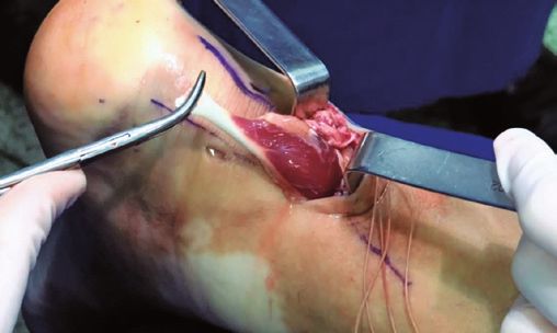
Lima et al. Minimally invasive treatment of acute Achilles tendon rupture with endoscopic flexor hallucis longus transfer

teral and posteromedial portals were constructed close to                       sutures were anchored to the tip of the FHL to allow pulling
the tendon. The FHL, which passes medially to the Stieda                        of the FHL with greater ease.
process of the talus, was located. The FHL retinaculum was                          The FHL was pulled through the posteromedial arthros-
opened, and the ankle and hallux were positioned in maxi-                       copic portal. A tunnel was made across the Achilles tendon
mal plantar flexion to perform tenotomy of the FHL using ar-                    from medial to lateral using Halsted mosquito forceps,
throscopic scissors as distally as possible to obtain a longer                  through which the graft was passed from medial to late-
graft (Figure 3). A small opening was made in the fascia at                     ral and exited through the posterolateral portal. Two addi-
the rupture site between the posterior superficial and deep                     tional Vicryl® 2-0 sutures were passed through the distal
compartments, and the muscle belly of the FHL was pulled,                       stump of the Achilles tendon in the same manner as pre-
exposing it through the access route (Figure 4). Vicryl® 2-0                    viously described for the proximal stump. The sutures were
                                                                                removed through the access route as described above.
                                                                                   With the ankle in plantar flexion, manual suturing was
                                                                                performed between the proximal ends of both stumps of
                                                                                the Achilles tendon through the rupture access route in the
                                                                                same manner used for the distal ends of each stump, com-
                                                                                pleting the percutaneous suture (Figure 5).
                                                                                   The FHL tendon was pulled by the transverse route
                                                                                and closed with Vicryl® 2-0 sutures between the stumps.

Figure 1. View of the access route.
Source: Author’s personal archive.

                                                                                Figure 4. Flexor hallucis longus tendon retrieved endoscopically.
                                                                                Source: Author’s personal archive.
Figure 2. Passage of the needle through the tendon and holding
forceps.
Source: Author’s personal archive.

                                                                                Figure 5. Tenorrhaphy of the proximal and distal stumps of the
Figure 3. Arthroscopy with posteromedial and posterolateral portals.            Achilles tendon.
Source: Author’s personal archive.                                              Source: Author’s personal archive.

                                                                                                                    Sci J Foot Ankle. 2019;13(2):129-34      131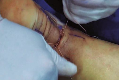
Lima et al. Minimally invasive treatment of acute Achilles tendon rupture with endoscopic flexor hallucis longus transfer
Patient 2
The patient was a 31-year-old man who was unemplo
yed, a smoker, and had no comorbidities or previous pain
in the Achilles tendon. The left Achilles tendon ruptured
on 09/23 2013 during a soccer game and was repaired 7
days after the trauma. Functional rehabilitation was per-
formed. The patient had no complications.
Patient 3
The patient was a 40-year-old man who was unemployed,
had no comorbidities and experienced a rupture of the right
Achilles tendon 4 months prior, which was treated con-
servatively. The tendon ruptured on 12/14/2013 during a
Figure 6. Reinforcement using the flexor hallucis longus tendon. fishing activity and was repaired 3 days after the trauma.
Source: Author’s personal archive. Functional rehabilitation was performed. The patient had
no complications. Eight months after the surgery, a partial
Double stitches were used through the two arthroscopic rupture of the contralateral Achilles tendon occurred, which
portals to reinforce the suture (Figure 6). The subcuta- was treated conservatively.
neous tissue was closed with clear Vicryl® 3.0 sutures, and
the skin was closed with simple nylon 4-0 stitches. Patient 4
During the postoperative period, the patients wore a The patient was a 36-year-old man who was an active
plaster cast in a mild equinus position for 2 weeks, without worker and had no comorbidities or previous pain in the
weight-bearing. An orthopedic boot with a heel was used Achilles tendon. The tendon ruptured on 01/25/2014 du-
from the third to the sixth week for maintenance of the ring a soccer game and was repaired 9 days after the trauma.
equinus and weight-bearing. Physical therapy rehabilita- Functional rehabilitation was performed. The patient had
tion began in the third week with passive exercises and no complications.
more intense exercises starting at the sixth week, when
the orthopedic boot was removed, and a silicone heel was Patient 5
used until four months after surgery.
The patient was a 72-year-old retired man who was a
former smoker (stopped smoking 20 years prior) and had
RESULTS no comorbidities or previous pain in the Achilles tendon.
The tendon ruptured on 09/16/2014 while pushing a car
Patient 1
and was repaired surgically 28 days after the trauma. Func-
The patient was a 51-year-old retired man who had no tional rehabilitation and physical therapy were not perfor-
comorbidities or previous pain in the right Achilles tendon. med. The patient presented paresthesia in the sural nerve
The patient presented a history of rupture of the contra- but without neuroma, pain, or hypersensitivity.
lateral Achilles tendon 12 years prior. He was underwent
The mean VAS, AOFAS scale, and VISA-A scale scores and
surgery at another hospital, with good progression and
the ATRS were 0.6, 98, 98.2, and 100, respectively (Table 1).
without complaints. The right Achilles tendon ruptured
The mean dorsiflexion range of motion was 4.8º on the
on 12/21/2013 during a soccer game and was repaired 20
days after the trauma. Functional rehabilitation was per-
formed, and a splint in the equinus position was used for 3 Table 1. Mean VAS, AOFAS, and VISA-A scores and the ATRS.
weeks; after the sutures were removed, physical therapy was Clinical evaluation method
started. An orthopedic boot was used for 1 week with ele- Patient VAS AOFAS VISA-A ATRS
vation of the calcaneus starting with partial weight bea 1 3 90 97 100
2 0 100 100 100
ring. Total weight bearing was started in the fourth week,
3 0 100 95 100
keeping the splint in equinus until 6 weeks postoperatively.
4 0 100 99 100
Then, the patient wore shoes with heels for another 3 months. 5 0 100 100 100
He had good progression throughout the postoperative pe- Mean 0,6 98 98,2 100
riod, with only mild pain during the first 3 months. Source: Prepared by the author based on the results of the research.
132 Sci J Foot Ankle. 2019;13(2):129-34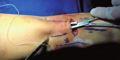
Lima et al. Minimally invasive treatment of acute Achilles tendon rupture with endoscopic flexor hallucis longus transfer
Table 2. Mean range of motion of the ankles (in degrees). The rates of severe complications are reduced using mi-
Range of motion nimally invasive surgeries, which result in rupture rates si-
Dorsiflexion Plantar flexion milar to those of open surgery and higher patient satisfac-
Patient
Operated Contralateral Operated Contralateral tion(10,11). Studies using specific instruments for minimally
1 8 4 40 34 invasive procedures reported a mean postoperative AOFAS
2 2 10 20 20 score of 93.00 to 96.81(12,13), VISA-A score of 92, rerupture
3 4 4 20 20 rate of 3.2% and 2%(12,13), and rate of sural nerve injury of 0
4 4 10 20 30 to 3.3%(12-14). A recent Brazilian study found no significant dif
5 6 10 30 30
ferences in isokinetic functional outcomes between open
Mean 4,8 7,6 26 26,8
and percutaneous surgery(15). In the present series, no cases
Source: Prepared by the author based on the results of the research.
of sural nerve injury or rerupture occurred, and the mean
AOFAS score, VISA-A score, and ATRS were 98, 98.2, and
100, respectively, indicating satisfactory clinical and func-
Table 3. Mean plantar flexion strength (in kgf ) of the ankle and
interphalangeal joint of the hallux. tional results, with minimal pain (VAS of 0.6).
Plantar flexion force FHL transfer has been shown to decrease the flexion
Ankle Interphalangeal joint strength of the interphalangeal joint; however, the func-
Patient
Operated Contralateral Operated Contralateral tion of this joint is not affected(16). The patients in our se-
1 30.2 27.1 18 19.9 ries had an absolute decrease in flexion of the interpha-
2 22.8 27.3 9.8 18.7 langeal joint of the hallux, but a statistical analysis was
3 24.9 23.8 13.5 19.3 not performed. The patients had no complaints related to
4 31 35.2 14.6 15.8 hallux or ankle mobility and no difficulty in performing
5 11.2 9.8 13.8 14.3 sports activities. Furthermore, the function of the repaired
Mean 24.02 24.64 13.94 17.6
tendon was not affected in any patient.
Source: Prepared by the author based on the results of the research.
The limitations of this study were its retrospective and
cross-sectional design, the heterogeneity of the evaluated
cases, and an inability to perform a statistical analysis be-
cause of the small sample size. With respect to the descrip-
operated side and 7.6º on the contralateral side (Table 2). tive analysis, patients 3 and 5 represented the main indi-
The mean plantar flexion strength of the ankle was 24.02 kgf cation of this technique. Patient 3 was a case of rerupture
on the operated side and 24.64 kgf on the contralateral with the need for tendon graft transfer, and the chosen
side. The mean flexion strength of the interphalangeal technique was minimally invasive surgery, which had an
joint of the hallux was 13.94 kgf on the operated side and excellent outcome (AOFAS score, 100; VISA-A score, 95;
17.6 kgf on the contralateral side (Table 3). The reduction ATRS, 100). Patient 5 was an elderly man with difficulty
of the flexion strength of the hallux on the operated side attending follow-up visits, a previous history of smoking,
did not cause any complaints or difficulty in performing and low socioeconomic status, and he did not undergo
physical activities. functional rehabilitation or physical therapy. Minimally
invasive surgery and strengthening with FHL transfer may
have been critical for achieving favorable results.
DISCUSSION
A prospective study with control conditions consisting
The repair of acute Achilles tendon injuries is contro- of conservative treatment and open surgery and inclu-
versial. The Clinical Practice Guideline of the American sion of only cases of rerupture or cases of only noncom-
Academy of Orthopedic Surgeons makes a minor re- pliant patients with comorbidities (diabetes and a history of
commendation for conservative or surgical treatment(6). smoking) may better demonstrate the results of the new
Although controversial, a meta-analysis demonstrated surgical technique described.
that the rerupture rates of surgical repair (2.7-3.6%) were
lower than those of conservative treatment (4.2-13.0%)(7,8).
However, suture infection and dehiscence occurred in CONCLUSION
2.4-4.7% of operated patients and increased to 10.4% in The described surgical technique may be effective for
the presence of risk factors, including diabetes, smoking, treating noncompliant patients with a diagnosis of acute
and the use of steroids(9). rupture or rerupture of the Achilles tendon.
Sci J Foot Ankle. 2019;13(2):129-34 133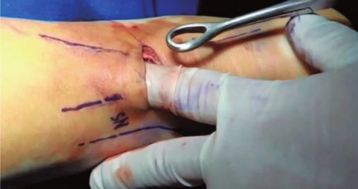
Lima et al. Minimally invasive treatment of acute Achilles tendon rupture with endoscopic flexor hallucis longus transfer

Authors’ contributions: Each author contributed individually and significantly to the development of this article: TCPL *(https://orcid.org/0000-0003-1238-
2475) analysis of patient records, clinical examination, formatting of the article; RGP *(https://orcid.org/0000-0002-6064-2027) conceived and planned the
activities that led to the study; participated in the review process and formatting of the article; GENS *(https://orcid.org/0000-0003-2717-3609) participated in
the review process and formatting of the article; CL *(https://orcid.org/0000-0002-7595-3303) data collection and participated in the review process; MCMD
*(https://orcid.org/0000-0001-6572-1771) conceived and planned the activities that led to the study, participated in the review process, formatting of the
article and approved the final version. *ORCID (Open Researcher and Contributor ID).

REFERENCES                                                                                     et al. Wound complications after open Achilles tendon repair: an
                                                                                               analysis of risk factors. Clin Orthop Relat Res. 2004;(427):63-66.
1.   Molloy A, Wood EV. Complications of the treatment of Achilles
     tendon ruptures. Foot Ankle Clin. 2009;14(4):745-59.                                10.   McMahon SE, Smith TO, Hing CB. A meta-analysis of randomised
                                                                                               controlled trials comparing conventional to minimally invasive
2.   Maes R, Copin G. Is percutaneous repair of the Achilles tendon a safe
                                                                                               approaches for repair of an Achilles tendon rupture. Foot Ankle Surg.
     technique? A study of 124 cases. Acta Orthop Belg. 2006;72(2):179-83.
                                                                                               2011;17(4):211-17.
3.   Lui TH. Minimally invasive flexor hallucis longus transfer in management            11.   Wu, Y, Mu, Y, Yin, L, Wang, Z, Liu, W, Wan, H. Complications in the
     of acute Achilles tendon rupture associated with tendinosis: a case
                                                                                               management of acute achilles tendon rupture: a systematic review
     report. Foot Ankle Spec. 2012;5(2):111-4.
                                                                                               and network meta-analysis of 2060 patients. Am J Sports Med. 2019
4.   Bohannon RW. Test-retest reliability of hand-held dynamometry                             Feb 19:363546518824601.
     during a single session of strength assessment. Physical Therapy. 1986;             12.   Assal M, Jung M, Stern R, Rippstein P, Delmi M, Hoffmeyer P. Limited
     66(2):206-9.                                                                              open repair of Achilles tendon ruptures: a technique with a new
5.   van Dijk CN, Scholten PE, Krips R. A 2-portal endoscopic approach for                     instrument and findings of a prospective multicenter study. J Bone
     diagnosis and treatment of posterior ankle pathology. Arthroscopy.                        Joint Surg Am. 2002;84(2):161-70.
     2000;16(8):871-6.                                                                   13.   Jung HG, Lee KB, Cho SG, Yoon TR. Outcome of Achilles tendon
6.   Chiodo CP, Glazebrook M, Bluman EM. American Academy of                                   ruptures treated by a limited open technique. Foot Ankle Int. 2008;
     Orthopaedic Surgeons clinical practice guideline on treatment of                          29(8):803-7.
     Achilles tendon rupture. J Bone Joint Surg Am. 2010;92(14):2466-68.                 14.   Keller A, Ortiz C, Wagner E, Wagner P, Mococain P. Mini-open
7.   Khan RJK, Fick D, Keogh A, Crawford J, Brammar T, Parker M. Treatment                     tenorrhaphy of acute Achilles tendon ruptures: medium-term
     of acute Achilles tendon ruptures: a meta-analysis of randomized,                         follow-up of 100 cases. Am J Sports Med. 2014;42(3):731-36.
     controlled trials. J Bone Joint Surg Am. 2005;87(10):2202-10.                       15.   Lazaroni PSO, Baumfeld TS, Magalhães JMB, Lopes FAS, Amaral GM,
8.   Wilkins R, Bisson LJ. Operative versus nonoperative management of                         Baumfeld DS. Isokinetic functional results of open and percutaneous
     acute Achilles tendon ruptures: a quantitative systematic review of                       Achilles tendon repair. Sci J Foot Ankle. 2018;12(1):55-60.
     randomized controlled trials. Am J Sports Med. 2012;40(9):2154-60.                  16.   Coull R, Flavin R, Stephens MM. Flexor hallucis longus tendon transfer:
9.   Bruggeman NB, Turner NS, Dahm DL, Voll AE, Hoskin TL, Jacofsky DJ,                        evaluation of postoperative morbity. Foot Ankle Int. 2003;24(12):931-34.

134      Sci J Foot Ankle. 2019;13(2):129-34
You can also read