Modified gastrostomy feeding tubes in patients with oesophageal cancer: our experience from Northern Tanzania
←
→
Page content transcription
If your browser does not render page correctly, please read the page content below
Journal of Surgical Case Reports, 2021;5, 1–5

                                                                             doi: 10.1093/jscr/rjab221
                                                                             Case Series

CASE SERIES

Modified gastrostomy feeding tubes in patients

                                                                                                                                                         Downloaded from https://academic.oup.com/jscr/article/2021/5/rjab221/6285833 by guest on 17 July 2021
with oesophageal cancer: our experience from
Northern Tanzania
Jay Lodhia1 ,2 ,*, Jamil Suleiman1 , Hillary Chipongo1 , Mathayo Shadrack1 ,
David Msuya1 ,2 and Kondo Chilonga1 ,2
1
Department of General Surgery, Kilimanjaro Christian Medical Centre, Moshi, Tanzania and 2 Faculty of
Medicine, Department of General Surgery, Kilimanjaro Christian Medical University College, Moshi, Tanzania
*Correspondence address. Department of General Surgery, Kilimanjaro Christian Medical Centre, PO Box 3010, Moshi, Tanzania.
Tel: +255754759345; E-mail: jaylodhia06@gmail.com

Abstract
Surgeons in resource-limited settings have adapted to overcome the challenges of the limitations of resources using different
available methods and inventions from the local environment. We report four cases of oesophageal cancer palliatively treated
with improvised gastrostomy feeding tubes by using 24Fr urinary catheters, to optimize their nutritional status to withstand
chemotherapy/radiotherapy. Two patients managed to begin chemo and radiotherapy, but only one out of the four survived.
The aim of this report is to appraise the methods used by surgeons to overcome the challenges they face in clinical practice.

BACKGROUND                                                                   CASE PRESENTATIONS
Surgical practice in sub-Saharan Africa is faced with many chal-             Case 1
lenges that pose a barrier to meet the desired goals of treatment            A 46-year-old female presented with progressive grade-six dys-
[1]. Surgical practice in Tanzania as a whole, where the majority            phagia, vomiting and significant unintentional weight loss. She
of the patients are from rural areas and have limited access to              was wasted and had a BP of 82/53 mmHg, pulse rate of 80 beats
proper health-care facilities and specialist care [2, 3]. A lot of           per minute, temperature of 36.4◦ C, saturating at 98% on room air.
the referral hospitals in resource poor countries lack required              Her full blood picture was normal with haemoglobin of 12.3 g/dl,
equipment due to various socio-economic factors needed to                    Creatinine 41 μmol/l, and urea was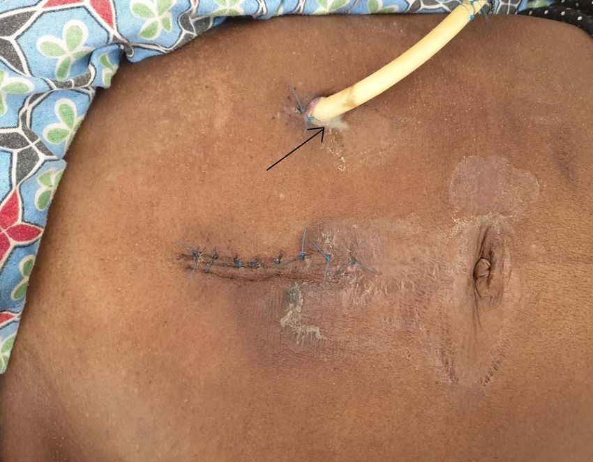
2 J. Lodhia et al.
Downloaded from https://academic.oup.com/jscr/article/2021/5/rjab221/6285833 by guest on 17 July 2021
Figure 1: OGD showing a fungating tumour at 20 cm from the upper incisors with
almost complete luminal obstruction.
Figure 3: Peristomal surgical site infection (arrow).
Figure 2: 24Fr catheter gastrostomy in Stamm technique.
obstruction (Fig. 1) and biopsies taken revealed oesophageal well
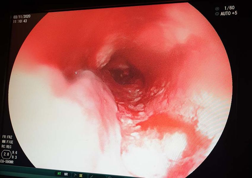
differentiated invasive squamous cell carcinoma. Figure 4: OGD showing a fungating mass partially obstructing esophageal lumen.
The patient was scheduled for GFT insertion, whereby a 24Fr
urinary catheter was inserted via the paramedian incision into
the stomach and secured using the Stamm technique (Fig. 2). Case 2
Intraoperatively the liver had no lesions. The patient was sent A 62-year-old male, a smoker and excessive alcohol consumer,
to the general ward and semi-solid feeds were initiated through non-hypertensive, presented with progressive grade-6 dysphagia
the gastrostomy feeding tube. The patient was discharged on that started gradually 7-month ago, associated with uninten-
the second day and was instructed to continue with semi-solid tional loss of weight. His biological brother was diagnosed
feeds, and wound dressing at a nearby health centre. The care- with oesophageal cancer 3-year back and had an improvised
taker was clearly instructed to spigot the catheter after use and GFT inserted but passed away shortly thereafter. On general
to flush the food contents with water after every meal to avoid examination he was weak, cachexic, severely pale, not jaundiced,
blockage. not cyanosed with bilateral pitting lower limb edema with
Two weeks after the GFT insertion, the patient presented vitals within normal range. Full blood picture initially showed
to the outpatient clinic with complaints of pus discharge per microcytic hypochromic anaemia of 6.4 g/dl, other parameters
GFT site. A diagnosis of peristomal surgical site infection was were essentially normal. Chest X-ray and abdominal ultrasound
made (Fig. 3), the wound was dressed thoroughly and the patient were essentially normal. Patient did an OGD which revealed
was sent home with oral antibiotics and had to continue with at 20 cm a fungating mass partially obstructing lumen (Fig. 4), a
dressing at a nearby health facility. biopsy was taken but the scope could not pass through. Histology
One-week later, the patient presented to the outpatient results for biopsy showed oesophageal invasive squamous cell
clinic with leakage per GFT site. The diameter of the stoma had carcinoma grade 2.
become wider and a diagnosis of peristomal leakage was made. Patient was scheduled for GFT insertion, blood transfusion
A purse-string suture was put under local anaesthesia using and hematemics were given and control haemoglobin obtained
Nylon 2–0. The patient was then discharged with analgesics and was 13.4 g/dl. Surgery was done and the findings were a
antibiotics. collapsed stomach and a non-nodulated liver, a 24Fr urinary
The patient was referred to the oncology centre for radiother- catheter was inserted, ballooned and secured using the Stamm
apy ∼4 months after GFT insertion, but she passed away 2 days technique (Fig. 5). Patient was transferred to the general ward
after the initiation of the first cycle. and was discharged on the second day post-surgery.
Modified gastrostomy feeding tubes in patients with oesophageal cancer 3
Downloaded from https://academic.oup.com/jscr/article/2021/5/rjab221/6285833 by guest on 17 July 2021
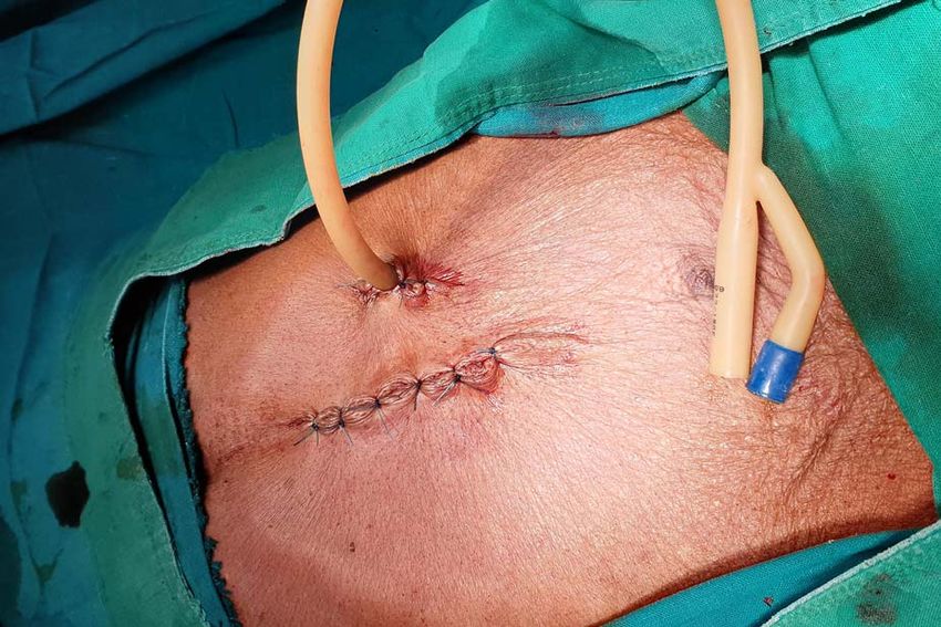
Figure 5: 24Fr catheter as gastrostomy tube.
Figure 6: Peristomal leakage (arrow).
The patient was reviewed at the outpatient clinic 2-week later
and was found to be emaciated, but vitally stable. The patient’s but no lower limb edema. His vitals were blood pressure of
relatives were counselled on the correct nutritional plan and had 139/71 mmHg, Temperature of 35.8◦ C, oxygen saturation of 95%
to be seen in the Oncology unit 3-week later but they were lost to on room air, respiratory rate of 18 cycles per minute and a pulse
follow up. A phone call was made to the relatives that reported rate of 73 beats per minute. He had a scaphoid abdomen with
that the patient had passed away 16 days after the final visit. tenderness on the right lumbar region. Fine crepitations were
heard with reduced air entry on the right side of his chest. Full
Case 3 blood picture was done which was normal with a haemoglobin
A 62-years-old male who presented with grade-5 dysphagia for of 11.3 g/dl, other investigations including renal function tests
9-month associated with significant unintentional weight loss. and liver enzymes were normal. Chest X-ray done showed signs
He had a positive history of chronic alcohol use and cigarette of metastasis (Fig. 8). CT-scan of the abdomen done at the refer-
smoking. No family history of oesophageal cancer. Had an OGD ring hospital with both oral and intravenous contrast reported
done 2-week prior to admission, which showed a mass obstruct- structures above the diaphragm showing dilated oesophagus
ing the LES and the scope could not pass through. A biopsy measuring 41 × 38 mm due to distal retro-cardiac concentric
was taken which revealed well differential adenocarcinoma. The oesophageal wall thickening amounting to 18 mm with luminal
lab work up done during admission showed microcytic anaemia narrowing with possibility oesophageal fistula tract. The gas-
of 8.4 g/dl and 400 ml of whole blood was transfused. Other tric cavity was normal without focal lesions. OGD revealed the
parameters including electrolytes were within normal ranges oesophageal mucosa was hyperemic with a blocking mass at
(Sodium-134.8 mmol/l and potassium-4.66 mmol/l). 29 cm from the upper incisors. The scope was able to pass beyond
He was planned for an improvised GFT insertion. Gastros- the lesion. The cardiac, fundus and body mucosa was atrophic.
tomy was done at the body of the stomach and a 24Fr Foley’s There were metaplastic changes at the antrum and pylorus. Mul-
urinary catheter was inserted and secured. Around 4 l of amber tiple biopsies were taken. Histology results revealed oesophageal
coloured ascitic fluid was drained. The liver was not nodulated well differentiated invasive squamous cell carcinoma.
and the mesenteric and paragastric lymph nodes were not palpa- The patient was scheduled for GFT insertion. Under aseptic
ble. The patient was then discharged after 24-h postoperatively technique, with the patient in supine position, 24 Fr urinary
with counselling of wound care and use of semi-solid foods only, catheter as an improvised GFT was inserted in Stamm manner,
through the tube. haemostasis achieved, abdomen closed in layers and dressed
The patient was re-admitted 12-day post discharge com- (Fig. 9). There was no ascites and mesenteric lymph nodes were
plaining of leak per GFT insertion site. There was significant not palpable. After the procedure the patient was discharged
peristomal leakage (Fig. 6). He was in a gasping state with cold home on the second day with analgesics and was booked for a
extremities. The blood pressure and saturation was unrecordable surgical outpatient clinic appointment after 2 weeks.
with a respiratory rate of 28 breaths per minute and a weak pulse The patient was then seen at the clinic after 2 weeks, she was
rate of 50 beats per minute. The chest had basal crepitations on clinically and vitally stable and the GFT was functioning well.
the right lung. Abdominal X-ray showed features of small bowel The wound had healed, the tube was patent and there was no
obstruction with pneumoperitoneum and right sided pleural leakage from the GFT site. Midline sutures were removed and
effusion was noted (Fig. 7). He was supported with high flow the patient was discharged.
oxygen and resuscitated with IV fluids but 3-h post admission The patient presented to the surgical OPD 3-month later with
the patient succumbed. complaints of GFT blockage at the tip. Under local anaesthesia,
the previous tube was removed and another 24 Fr catheter
was inserted in a sterile manner and secured and patency was
Case 4
tested. The patient was then discharged and care takers were
A 63-year-old male, 10-day post completion of anti-tuberculosis counselled on use of semi-solid food and to flush the tube with
regimen for pulmonary tuberculosis, known smoker and alco- water after every meal.
holic, presented with grade-5 dysphagia associated with food He was reviewed 3-month later, at the OPD, free from com-
regurgitation, cough and weight loss. On general examination, plaints and no leakage with a patent tube. He had already begun
he was ill looking, cachexic, dehydrated and pale, not jaundiced his first cycle of radiotherapy.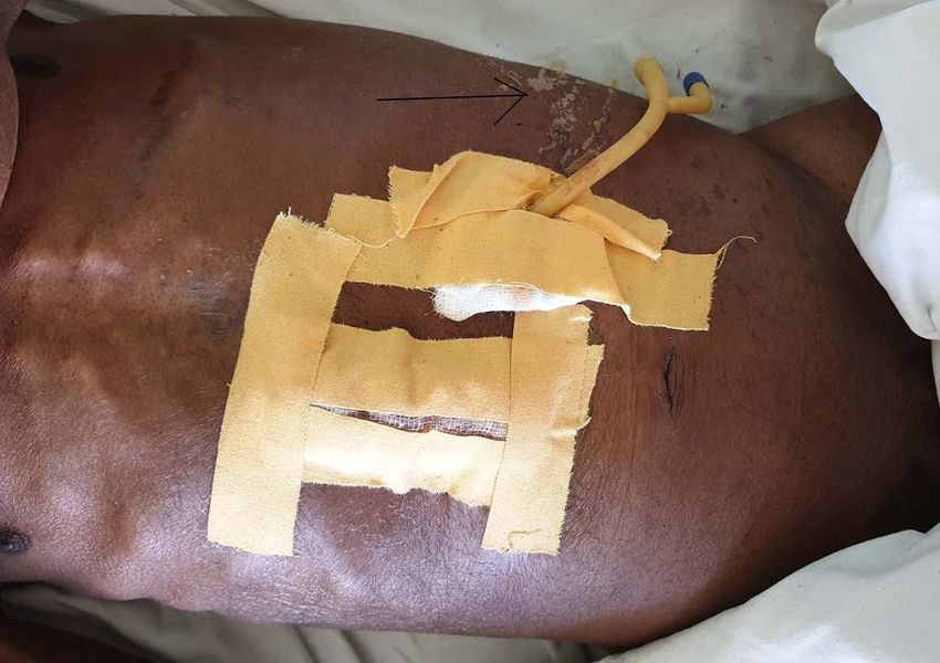
4      J. Lodhia et al.

                                                                                                                                                                          Downloaded from https://academic.oup.com/jscr/article/2021/5/rjab221/6285833 by guest on 17 July 2021
Figure 7: Supine (A) and erect (B) abdomen X-rays show dilated small bowel loops with multiple air fluid levels. Free air seen underneath the hemidiaphragm bilaterally
and outlining both sides of the small bowel walls (Rigler sign) suggestive of pneumoperitoneum.

                                                                                      Figure 9: 24Fr urinary catheter as an improvised gastrostomy feeding tube.

                                                                                      of tube insertion and use of laparoscopic techniques [4]. GFTs
                                                                                      have been used in supportive care, in patients with severe head
                                                                                      injury who are on ventilators, as well as in palliative care patients
                                                                                      with oesophageal cancer like in our cases [5].
                                                                                           The complications of using a urinary catheter as an impro-
                                                                                      vised GFT were peristomal surgical site infection, peristomal
Figure 8: Supine chest X-ray shows patchy infiltrates in the right mid and lower
                                                                                      leakage and malfunctioning tube due to blockage. Similar com-
zone with micronodular infiltrates. Micronodules seen in the left lower zones.
                                                                                      plications have been reported using percutaneous endoscopic
                                                                                      gastrostomy tubes, together with colonic perforation, tube dis-
                                                                                      lodgement, buried bumper syndrome and a non-healing stoma
DISCUSSION                                                                            [6].
Improvisation is vital to surgical practice in Tanzania and other                          After the improvised GFTs were inserted successfully, the
sub-Saharan countries due to the resource-limited medical facil-                      patients were referred back to the oncologists for palliative
ities hence have led to the optimization of the locally available                     care using chemotherapy/radiotherapy. Tube feeding enables
equipment. This has made the cost of surgery affordable along                         patients to have a high calorie intake to withstand the compli-
with satisfaction to the surgeon [1]. Many patients have to bear                      cations of chemotherapy/radiotherapy such as loss of fat mass
the costs of the hospital bills and a lot of the time consumables                     and skeletal muscle mass leading to overt malnutrition [7, 8].
too [1].                                                                                   Other types of improvisation reported are underwater seal
    Enteral feeding is a common method of nutritional support                         apparatus for thoracostomy tube whereby water bottles are
when oral intake is inadequate. There has been an increase in                         modified, similar to our cases, large foley catheters are used
the use of GFTs due to the introduction of percutaneous methods                       as gastrostomy tube, the use of urine bags in place of silo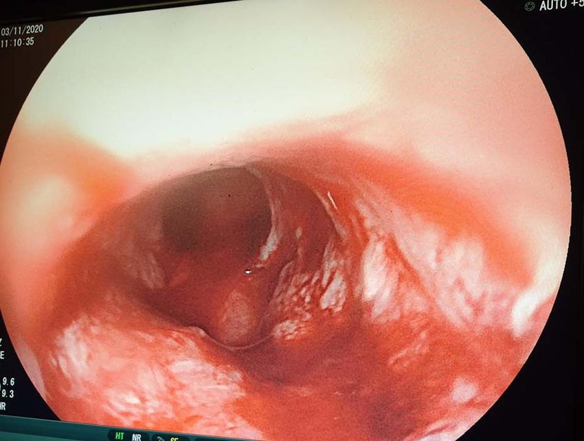
Modified gastrostomy feeding tubes in patients with oesophageal cancer         5

for omphalocele closure and usage of nasogastric tubes as                 infrastructure: a strategy towards achieving universal health
ventriculoperitoneal shunts to manage hydrocephalus [1].                  coverage in Tanzania. BMC Health Serv Res 2020;20:1–4.
                                                                     3.   Penoyar T, Cohen H, Kibatala P, Magoda A, Saguti G, Noel L,
                                                                          et al. Cherian M. Emergency and surgery services of primary hospi-
CONCLUSION                                                                tals in the United Republic of Tanzania, BMJ Open 2012;2:e000369.
The scarcity of surgical equipment in Tanzania has given room        4.   Carey TS, Hanson L, Garrett JM, Lewis C, Phifer N, Cox CE, et al.
for improvisation of such items using locally available materials,        Expectations and outcomes of gastric feeding tubes. Am J Med
hence come up with various techniques with local resources                2006;119:527-e11.
in overcoming the limitations imposed by scarcity of certain         5.   Chahal A, Malla S, Dash C, Gupta D, Gamanagatti S. Pull-
equipment/devices.                                                        type radiologically inserted gastrostomy: an improvised tech-
                                                                          nique using a frugal innovation. J Clin Intervent Radiol ISVIR.
                                                                          2019;3:007–11.
CONFLICT OF INTEREST STATEMENT                                       6.   Sealock R, Munot K. Common gastrostomy feeding tube
None declared.                                                            complications and troubleshooting. Clin Gastroenterol Hepatol

                                                                                                                                               Downloaded from https://academic.oup.com/jscr/article/2021/5/rjab221/6285833 by guest on 17 July 2021
                                                                          2018;16:1864–9.
                                                                     7.   Yip C, Goh V, Davies A, Gossage J, Mitchell-Hay R, Hynes O,
REFERENCES
                                                                          et al. Assessment of sarcopenia and changes in body com-
1. Adejumo AA, Adeosun OA, Omoregie PO, Alayande B.                       position after neoadjuvant chemotherapy and associations
   Improvisation of surgical equipment in the surgical ser-               with clinical outcomes in oesophageal cancer. Eur Radiol.
   vices of a developing country. Niger J Surg Res 2016;                  2014;24:998–1005.
   17:48.                                                            8.   Schizas D, Lidoriki I, Moris D, Liakakos T. Nutritional man-
2. Kapologwe NA, Meara JG, Kengia JT, Sonda Y, Gwajima D,                 agement of esophageal cancer patients. In: Jianyuan Chai, ed.
   Alidina S, et al. Development and upgrading of public pri-             Esophageal Abnormalities. IntechOpen, 2017. doi: 10.5772/inte-
   mary healthcare facilities with essential surgical services            chopen.69607.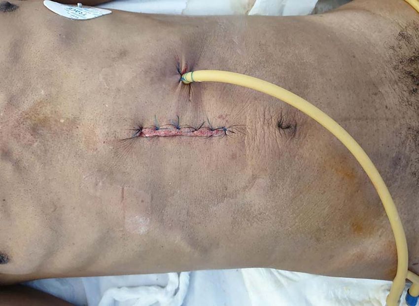
You can also read