CENTRAL ASIAN JOURNAL OF MEDICAL AND NATURAL SCIENCES - Central Asian ...
←
→
Page content transcription
If your browser does not render page correctly, please read the page content below
CENTRAL ASIAN JOURNAL OF MEDICAL AND NATURAL
                      SCIENCES

                  Volume: 02 Issue: 01 | Jan-Feb 2021 ISSN: 2660-4159
                               www. cajmns.centralasianstudies.org/index.php

                                             Effects of cellular cord blood
                                       on skin pathology in laboratory animals

                                                 ABSTRACT: In work studied character of the influence
                                   1
Khusanov Erkin Uktamovich                   of the cryopreserved nucleated cells of cord blood on the skin
Korzhavov Sherali Oblakulovich2             morphology was investigated on the different stages of skin
Kurbanova Latofat Murodullaevna3            cultivation in vitro and character of the influence stem cells
Lapasova Zebiniso Hidirovna4                of cord blood on morphofunctinal condition of a derma in
Kurbanova Gulbakhor Aslamovna5              conditions of an experimental hypothyroidism in vivo. The
                                            application of cord blood stem cells is perspectives as
    EMAIL : sherali.korjavov@gmail.com      therapeutic treatment of derma. The skin, as the largest organ
Received 26th December 2020,
                                            in the human and animal body, is not only a "battle arena"
Accepted 17th January 2021,                 against various microorganisms and harmful influences, but
Online 06th February2021                    also a mirror reflecting the general health of the body. The
                                            endocrine system plays a leading role in regulating the
1
 Associate Professor of the Department of   functioning of the skin, ensuring the metabolism in this organ,
Human Anatomy and OSTA                      its repair and restoration of lost elements, the functioning of
2,3
    assistants of the Department of Human   the glands and hair growth
Anatomy and OSTA
4,5
                                                 KEYWORDS: cord blood, skin, rats, epidermis,
    assistants of the Department of
                                            morphology, and hypothyroidism.
Pathological Physiology
Samarkand State Medical Institute,
Republic of Uzbekistan

       INTRODUCTION

       The skin, as the largest organ in the human and animal body, is not only a "battle arena" against
various microorganisms and harmful influences, but also a mirror reflecting the general health of the
body. The endocrine system plays a leading role in regulating the functioning of the skin, ensuring the
metabolism in this organ, its repair and restoration of lost elements, the functioning of the glands and
hair growth. Thyroid hormones have the most important effect on the functioning of the skin [2, 3].
Thyroid hormones play a major role in metabolism and are essential for normal skin growth and
development. The main mechanism of action of thyroid hormones is the stimulation of protein
synthesis in the cytoplasm of cells and an increase in the level of oxygen consumption by tissues.
Disturbances in the structure of the dermis that develop in chronic hypothyroidism are manifested by

    116         Published by “ CENTRAL ASIAN STUDIES" http://www.centralasianstudies.org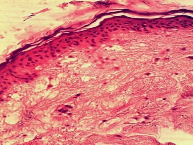
CAJMNS                           Volume: 02 Issue: 01 | Jan-Feb 2021

changes in the hairline, functional changes in the sweat and sebaceous glands, dry skin, increased
desquamation of the epidermis, violations of the basic abilities of the skin in the implementation of
physiological, immune and biochemical functions [3].
        It is known that biologically active preparations, such as the placenta, its extracts, and cell
suspensions, are increasingly used to stimulate the processes of repairing damaged or regenerating
aging skin that has lost its turgor and elasticity [7-10]. However, the mechanism of action of these
drugs is not well understood. Umbilical cord blood (CK) cells were originally used to treat diseases of
the blood system, but recently, due to the discovery of pluripotent stem cells (SC) and mesenchymal
stem cells in cord blood, cord blood is considered as a potential source for cell therapy in a wide range
of diseases [ 9]. In this regard, theoretical prerequisites arise for the use of CK preparations in the
processes of rat skin regeneration both in the in vitro system and in dermatopathology caused by
experimental hypothyroidism (EHT).
        The aim of the work was to study the effect of cryopreserved nuclear cells of human
umbilical cord blood on the morphology of rat skin in vitro and in vivo.
        Material and research methods. In vitro experiments were carried out on rat skin
samples. Skin fragments 0.3 to 0.3 cm in size were placed on solid agar, then covered with standard
culture growth medium in a volume of 0.5 ml. When setting up the experiment, the material was
divided into the following groups: 1st group - intact skin; 2nd - (control) - skin samples placed on the
surface of agar with culture medium; 3rd - (experiment) - skin samples placed on the surface of agar
with culture medium, to which 10% of the cord blood preparation was added. The umbilical cord
blood preparation "Stemcord" was a cryopreserved suspension of stem cells (SC) at a concentration of
(1-3) 105 in 1 ml of CC plasma, rich in biologically active substances, growth factors, hormones,
cytokines, and microelements [1]. Studies of nucleated cells (CD45 +) of umbilical cord blood,
including hematopoietic cells (CD34 +), were carried out before and after cryopreservation by flow
cytometry according to the international ISHAGE protocol [1]. Skin cultivation was performed in vitro
in a thermostat at a temperature of 37 C and a pH of 7.2. Skin fragments were examined on the 5th,
15th and 25th days of cultivation. The material for the study in in vivo experiments was 4-month-old
female outbred white rats weighing 110-120 g. Work with animals was carried out in compliance with
the provisions of the European Convention for the Protection of Vertebrate Animals and national
legislation on humane treatment of animals. Subtotal thyroidectomy (100% removal of the thyroid
gland) was performed according to the method [5]. The experiment involved animals that underwent
thyroidectomy and subsequent administration of the CA preparation into the tail vein [8]. All
experiments were carried out within the first 40 days from the moment of thyroidectomy, taking into
account the dynamics of the restoration of thyroid hormones in the blood of experimental animals [3,8]
        The animals were composed of the following experimental groups: group 1-EGT -
thyroidectomized animals;
        group 2-EGt - thyroidectomized animals, which were injected with the KK preparation.
        Each group consisted of 10 animals. Intact animals served as control. For the preparation of
histological preparations, sections of the skin excised to the full thickness from the back region and
cultured skin fragments were fixed in 10% formalin, washed with running water, dehydrated in
alcohols of increasing concentration, clarified in xylene, and embedded in paraffin-celloidin.

   117       Published by “ CENTRAL ASIAN STUDIES" http://www.centralasianstudies.org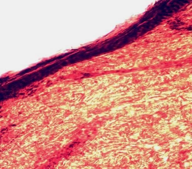
CAJMNS                           Volume: 02 Issue: 01 | Jan-Feb 2021

Microtome sections 5-7 microns thick obtained from paraffin-celloidin blocks were stained with
hematoxylin and eosin to obtain general histological preparations, and also with picrofuchsin
according to the Van Gieson method for studying connective tissue [6]. Differentiation of preparations
was carried out under a light microscope "Biolam" at a magnification of × 400. Statistical processing
of the data obtained was carried out by the Student-Fisher method [4].
        Research results and their discussion. In the first series of experiments in in vitro
culture on histological preparations, the structure of intact skin corresponded to the norm and was
represented by well-differentiated layers - the epidermis and dermis. After 5 days of cultivation of skin
fragments of the control group on agar growth medium, a decrease in the thickness of the epidermis
was observed due to a decrease in the number of cells in its layers (keratinization and cell migration).
Smoothness of the dermoepidermal border was noted, epidermal outgrowths expanded. The
proliferation of fibroblasts was observed in the papillary layer of the dermis, their number increased in
comparison with intact skin. Young collagen fibers appeared in the reticular layer of the dermis, which
were intensely stained, and the number of cells in the hair follicles and glandular structures increased
(Fig. 1, a).In skin fragments grown on agar nutrient medium to which the Stemcord preparation (group
3) was added, on the 5th day of cultivation, the structure of the epidermis was comparable to the intact
one. The basal layer contained one row of cells. The dermoepidermal border was generally well
contoured, but in some places there was a detachment of the epidermis from the dermis in the area of
the basement membrane. Collagen fibers formed a dense network, the number of fibroblasts increased
in comparison with groups 1 and 2 (Fig. 1, b).

       А                                                                B
           Figure: 1. Skin in vitro culture on day 5. a - on agar nutrient medium; b - on agar nutrient
                     medium with the addition of the drug "Stemcord". Coloring G.
                                               E. Uv. × 400.

   118        Published by “ CENTRAL ASIAN STUDIES" http://www.centralasianstudies.org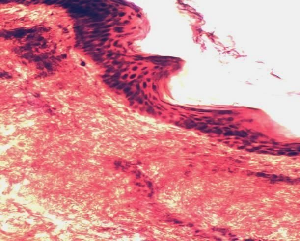
CAJMNS Volume: 02 Issue: 01 | Jan-Feb 2021
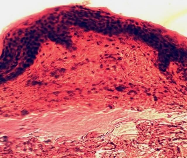
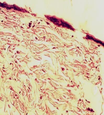
А B
Figure: 2. Skin in vitro culture on day 15. a - on agar nutrient medium; b - on agar nutrient
medium with the addition of the "Stemcord" preparation. Uv. × 400.
On the 15th day of cultivation of skin fragments without the preparation, the thickness of the
epidermis decreased in comparison with intact skin. The dermoepidermal border was not clearly
outlined. The structure of the connective tissue fibers of the dermis corresponded to intact skin. The
number of fibroblasts increased. In the hair follicles and sebaceous glands, the proliferation of
epithelial cells was observed, the size of which was increased (Fig. 2, a). In the presence of the
Stemcord preparation, on the 15th day of skin cultivation, the thickness of the epidermis increased as
compared to group 2, but the differentiation of its cell layers was difficult, and cells in a state of
mitosis were found in the basal layer. The cells of the basal layer of the epidermis had a pronounced
basophilia (hyperchromic nuclei, occupying almost the entire cell, the cytoplasm is acidophilic). The
dermoepidermal border was contoured. In the dermis, directly in the papillary layer, the number of
fibroblasts increased compared to their content in the skin of groups 1 and 2. The connective tissue
was represented by densely packed bundles of collagen fibers, fibroblasts are hyperchromic (Fig. 2, b).
А B
Figure: 3. Skin in vitro culture on day 25. a - on agar nutrient medium; b - on agar nutrient
medium with the addition of the "Stemcord" preparation. Uv. × 400.
119 Published by “ CENTRAL ASIAN STUDIES" http://www.centralasianstudies.org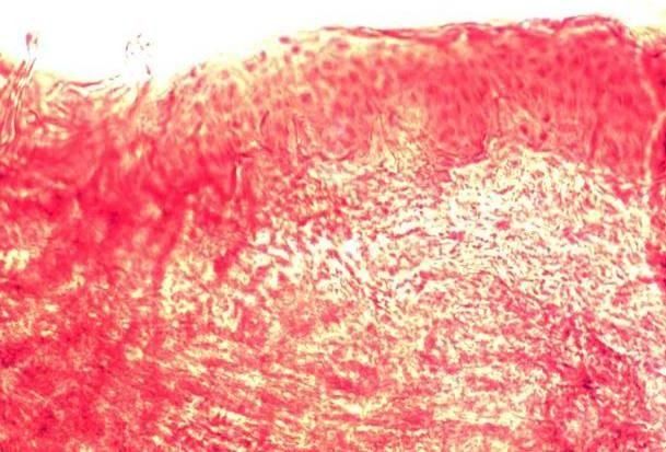
CAJMNS                           Volume: 02 Issue: 01 | Jan-Feb 2021

        А                                            B                            C

       Figure: 4. Skin of rats of experimental groups: a - intact animals; b - animals with simulated
    hypothyroidism after 1 month; c - thyroidectomized animals, which were injected with the KK
                               preparation. Coloring G.-E. Uv. × 400.

        On the 25th day of cultivation on agar medium without the drug, foci of micronecrosis
appeared in the skin. In the epidermis, the intercellular spaces increased, the layers of cells were
poorly differentiated. The nuclei of epithelial cells were pyknotic, and the cytoplasm was vacuolated.
The boundary between the epidermis and the dermis was blurred, and foci of cellular detritus were
observed. In the dermis, connective tissue disorganized in the form of homogenization and lumpy
disintegration of collagen and elastin fibers. The number of fibroblasts decreased. Edema and
destruction of glandular structures and hair follicles were observed (Fig. 3, a). During cultivation of
skin fragments in a medium with the addition of Stemcord on the 25th day, necrotic processes in both
the epidermis and the dermis were not observed. The thickness of the epidermis remained at the level
of 15 days, and the basal layer remained active, an increase in the number of merging horny scales was
noted.
        The border between the epidermis and the dermis was clearly defined. The dermis retained a
high cellularity, although the number of fibroblasts decreased in comparison with 15 days of
cultivation. In the reticular layer, collagen and elastin fibers were clearly contoured, which form the
reticular structure of the connective tissue; however, their density decreased compared to 15 days of
cultivation, and in some areas of the skin fragments the dense structure of the connective tissue
remained. Derivatives of the skin - hair follicles and sebaceous glands - retained their structure and
were well contoured. Separation of the epidermis and micro-foci of cell detritus were observed in
some areas of skin fragments (Fig. 3, b). The results obtained indicate that the Stemcord KK
preparation stimulates the proliferative activity of dermis and epidermal cells in vitro.
        In vivo experiments during histological examination of the skin of thyroidectomized animals
(group 1-EGt) 1 month after hypothyroidism modeling, it was found that the epidermis is very thinned,
flattened, and its normal folding is absent. The layers of the epidermis do not differentiate; some cells
have pyknotic nuclei. In the dermis proper, the papillary and reticular layers are also not differentiated.
The connective tissue part of the dermis is a loosened, fragmented and contoured bundle of collagen
and elastic fibers, among which there is a reduced, compared to the norm, the number of fibroblastic
cells with dense nuclei. Derivatives of the skin - sebaceous glands and hair follicles - are rare, they are
reduced in size, the nuclei of their cells are pyknotic (Fig.4b).

   120        Published by “ CENTRAL ASIAN STUDIES" http://www.centralasianstudies.org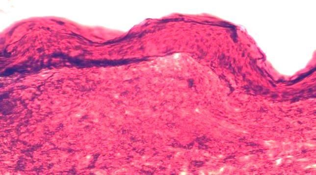
CAJMNS                           Volume: 02 Issue: 01 | Jan-Feb 2021

         The skin of thyroidectomized animals, which were injected with the KK preparation (group 2-
EGt) 1 month after the operation, was excised from the animals under ether anesthesia after 7 days.
Histological examination revealed a newly formed epidermis, which was thickened compared to the
norm. It showed folding and differentiation of cells with the formation of characteristic layers. The
cells of the fibroblastic row, the number of which was increased in comparison with the norm, in most
cases were located parallel to the newly formed bundles of collagen fibers that form the dermis; no
cracks or gaps between them were found (Fig. 4c). In the dermis itself, especially in its deep parts,
growing hair follicles were found. At the border of the dermis and subcutaneous tissue, the growth of
small blood vessels and capillaries was observed, as well as fibroblastic cellular elements, probably
derived from pericytes (perivascular cells of mesenchymal origin, which serve as the main precursors
of fibroblasts).
         The results of a histological study in an in vivo experiment show that the nature of the recovery
processes in the skin of animals with experimental hypothyroidism upon administration of the KK
preparation has a pronounced tendency to complete regeneration of the lost morphological and
functional properties of the skin. In our opinion, the main mechanism providing regenerative processes
in the skin, both in vitro and in vivo, is the introduction of biogenic stimulants present in the plasma of
CK, which are involved in the normalization of metabolism in the body, and a certain amount of stem
cells, including mesenchymal ones, cells that stimulate the growth of capillaries and fibroblasts.
         Conclusion. The KK "Stemcord" preparation, added to the agar culture medium, stimulates
the proliferative activity of the cells of the dermis and epidermis in vitro. In an in vivo experiment, the
nature of the recovery processes in the skin of animals with experimental hypothyroidism when
administered with the Stemcord CC preparation has a pronounced tendency to the complete
regeneration of the morphofunctional properties lost by the skin. The use of cord blood preparations
both in the cultivation of skin fragments in vitro and under conditions of experimental hypothyroidism
can be considered as a promising factor influencing the course and consequences of endocrine
disorders of the whole organism and skin in particular.

       LITERATURE:

1. Бабийчук Л. А. Новые перспективы в криоконсервировании ядросодержащих клеток
   пуповинной крови / Л. А. Бабийчук, О. В. Кудокоцева, В. В. Рязанцев // Гематологія і
   переливання крові. – 2008. № 34. – С. 17-21.
2. Калюжная Л., Дзюбак В. Старение кожи: патогенетические и лечебные аспекты / Л.
   Калюжная, В. Дзюбак // Укр. мед. часопис. – 2002. – № 2 (28). – С. 68-72.
3. Кандрор В. Физиологические эффекты тиреоидных гормонов и механизм их действия //
   Руководство по клинической эндокринологии: статья / В. Кандрор. - Питер, 1996. - С. 120-
   124.
4. Лакин Г. Ф. Биометрия / Лакин Г. Ф.– М. : Высш. школа, 1990. – 352 с.
5. Легач Е. И. Ретроградный способ тиреоидэктомии крыс как адекватная модель гипотиреоза
   / Е. И. Легач // Трансплантологія. - 2005. – Т. 8, № 2. - С. 92-94.

   121        Published by “ CENTRAL ASIAN STUDIES" http://www.centralasianstudies.org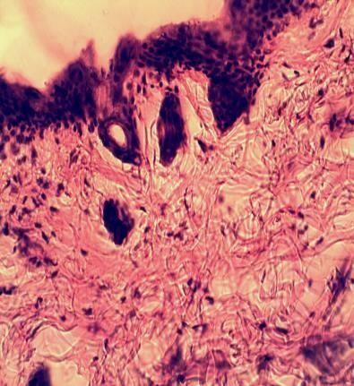
CAJMNS                        Volume: 02 Issue: 01 | Jan-Feb 2021

6. Меркулов Г. А. Курс патологогистологической техники / Меркулов Г. А. - Ленинград, 1961. –
   340 с.
7. Озерская О. Экспериментальные подходы к обоснованию применения клеточных
   композиций на основе фибробластов в дерматокосметологии/ О. Озерская, В. Щеголев //
   Клеточная трансплантология и тканевая инженерия. – 2008. - Т. 3, № 2. - С. 66-67.
8. Применение препаратов пуповинной крови и общей экстремальной аэрокриотерапии для
   восстановления нарушений дермы, вызванных хроническим гипотиреозом / В. Ю.
   Пурышева, И. И. Ломакин, О. В. Кудокоцева [и др.] // Трансплантологія. - 2008. - Т. 10, №
   1. - С. 100-103.
9. Стволовые клетки. Биология и потенциальное клиническое использование / Н. Я. Спивак, Г.
   Т. Сухих, В. В. Малайцев [и др.] // Трансплантологія. – 2005. – Т.8, №3. – С. 6-14.
10. Scott D. W. Miller and Kirk‘s small animal dermatology / Scott D. W., Miller W. H., Griffin C.
   E. - 5th ed. - Philadelphia etc.: W/B/Saunders Company, 1995. - 1213 p.
11. Uktamovich, Husanov Erkin, and Korjavov Sherali Oblakulovich. "Пилородуоденал соҳа
    махаллий бошкарув нерв аппаратининг микроанатомик асослари." //Journal of Biomedicine
    and Practice 6.5 (2020).
12. Хусанов Е.Ю., Коржавов Ш.О., Ортикбаева Н.Т. Морфологическая картина дегрануляции
    апудоцитов гастродуоденальной зоны при экспериментальном голоде // Международная
    научно-практическая конференция Мировая наука. - РОСТ, 2017. - Т. 5. - № 5. - С. 59-61.
13. Хусанов Е.У., Исмоилов О.И., Коржавов Ш.О. Влияние препаратов пуповинной крови на
    морфологию кожи. // Международное научное обозрение проблем естествознания и
    медицины, Бостон, США. 4-5 ноября 2019 г. П-383-394

  122       Published by “ CENTRAL ASIAN STUDIES" http://www.centralasianstudies.org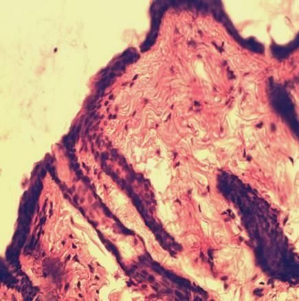
You can also read