Nevus Spitz - Everlasting Diagnostic Difficulties - The Review
←
→
Page content transcription
If your browser does not render page correctly, please read the page content below
Coll. Antropol. 32 (2008) Suppl. 2: 171–176
                                                                                                                                Review

Nevus Spitz – Everlasting Diagnostic Difficulties –
The Review
Mirna [itum1, @eljana Bolan~a1, Marija Buljan1, Davor Tomas2 and Marijana Ivan~i}3
1   University Department of Dermatovenerology, University Hospital »Sestre milosrdnice«, Zagreb, Croatia
2   University Department of Pathology, University Hospital »Sestre milosrdnice«, Zagreb, Croatia
3   Medical Center Zagreb, Zagreb, Croatia

ABSTRACT

    In 1910, Darier and Civatte described in details an unusual melanocytic tumor characterized by rapid growth on the
nose of a young child. They could not state whether the tumor was benign or malignant. In 1947, Sophie Spitz described
the same lesion as juvenile melanoma in which prognosis was frequently excellent. Later, the study was revised and it
was concluded that juvenile melanoma was a benign tumor and can affect adults. Although, the prognosis was mostly
excellent, Spitz reported in one of 13 cases fatal metastases from nevus Spitz. In 1999, Barnhill et al described one fatal
case of the patient for whom it was thought to have typical Spitz nevus. Nowadays, there is still a lack of consensus about
histopathology and also a terminology of the tumors that are neither typical nevus Spitz, neither malignant melanoma.
All histopathological, clinical and ancillary criteria must be weighed in the final interpretation of epitheloid/spindle cell
lesion. At the present, the final diagnosis remains pathohistological, with important emphasis given to clinical impres-
sion. Persistently changing lesion indicates malignancy potential of the lesion. Barnhill recommends that all Spitz tu-
mors are completely excised. Atypical tumors should be excised with wider margins up to 1 cm. Patient should be care-
fully monitored by regular examinations for recurrence and metastasis.

     Key words: nevus Spitz, atypical nevus Spitz, Spitzoid melanoma, pathohistological analysis, surgical excision

Introduction
   In 1910, Darier and Civatte1 described in details an            pediatricians recommended 1–2 mm margin of normal
unusual melanocytic tumor characterized by rapid growth            appearing skin around the nevus. They were also less
on the nose of a young child. They could not state                 likely to monitor patients whose Spitz nevi were com-
whether the tumor was benign or malignant. In 1947,                pletely excised. 74% of respondents believed that nevus
Sophie Spitz2 described the same lesion as juvenile mela-          Spitz was entirely benign lesions, 4% believed that it was
noma in which prognosis was frequently excellent. Later,           precursor of malignant melanoma, and 22% of the inter-
the study was revised and it was concluded that juvenile           viewed physicians were not sure.
melanoma was a benign tumor and can affect adults3. Al-
though, the prognosis was mostly excellent, Spitz re-
ported in one of 13 cases fatal metastases from nevus              Typical Nevus Spitz
Spitz. In 1999, Barnhill et al.4 described one fatal case of
the patient for whom it was thought to have typical Spitz              The prevalence of nevus Spitz (NS) is unknown in
nevus. Nowadays, there is still a lack of consensus about          general population, but they appear less often than ac-
histopathology and also a terminology of the tumors that           quired and congenital melanocytic nevi6. The incidence
are neither typical nevus Spitz, neither malignant mela-           of NS in general population is about less than 0.2% and
noma. The treatment of nevus Spitz is clear, but the sur-          in children about 1%. There is no sex prevalence and the
gical margins of the tumor remain unclear. Gelbard5 in-            majority of nevi occur in children (about 60% by the age
vestigated how American dermatologist and pediatrician             of 30). The majority of NS occur on the head and neck
manage nevus Spitz. The majority of dermatologist and              (37%), followed by 28% on lower extremities, 19% on the

Received for publication July 30, 2008

                                                                                                                                 171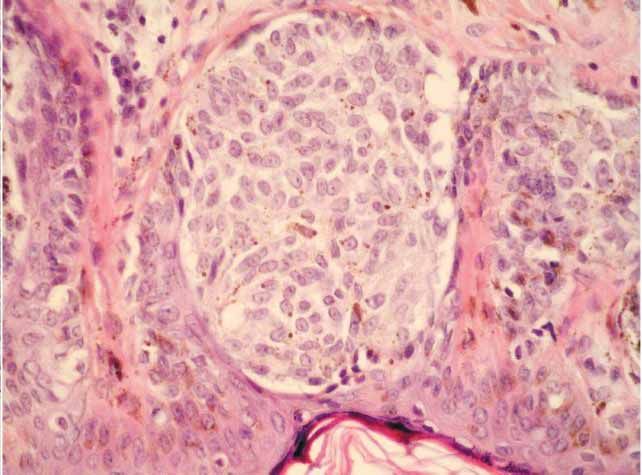
M. [itum et al.: Nevus Spitz, Coll. Antropol. 32 (2008) Suppl. 2: 171–176
upper extremities and 6% on the trunk. NS is usually sol-
itary papule, but could also be multiple, widespread,
grouped, agminated or eruptive and usually less than 1
cm in diameter. The color is usually pink to red but could
range from flesh colored to tan, dark brown and black de-
pending on amount of melanin present and the vascula-
rity of the lesion7.
There are four clinical types of NS:
1. the light colored soft form which is pink to light
tan, smooth, and flatters with dermatoscopy,
2. the light colored hard form, which appears like a
dermatofibroma or keloid and may have halo and
teleangiectases,
3. the dark form, which is variably pigmented and
smooth,
4. the multiple form, which includes the grouped and Fig. 1. b) Histological picture of typical nevus Spitz (100x HE).
widespread, disseminated, eruptive lesions8.
Clinical differential diagnosis includes acquired me- diagnostic confusion, nevus Spitz when typical can be
lanocytic nevus, dysplastic melanocytic nevus, congenital differentiated from malignant melanoma10. Spitz nevi
melanocytic nevus, blue nevus, pyogenic granuloma, der- are melanocytic nevi that are junctional (66%), com-
matofibroma, hemangioma, angiofibroma, scar, keloid, pound (11%) and dermal (18%). Melanocytic elements
fibroma, xanthogranuloma, verruca vulgaris, molluscum are usually arranged in well-circumscribed nests in epi-
contagiosum, epidermal nevus, histiocitoma, xanthoma, dermis and dermis. The epidermis is usually hyperpla-
anthropod bite reaction, lichen planus, lupus vulgaris, stic, with elongated and bulbous pegs and knobs extend-
granuloma faciale, eosinophilic granuloma, pseudolym- ing into the dermis (Figure 1b). Spindle cells preominate
phoma, seborrheic keratosis, chondrodermatitis, nodular in 45–54% of Spitz nevi, epitheloid cells in 21% and rela-
helicis, actinic keratosis, pale cell achantoma, granular tively equal combination of cell types in 24–34%. The pig-
cell tumor, leiomyoma, glomus tumor, squamous cell car- mented spindle cell nevus described by Reed et al.11 is
cinoma, basal cell carcinoma, Kaposi sarcoma and angio- usually sharply demarcated, uniformly darkly pigmented
sarcoma7. papule or plaque consisting of compact and aggregated
The typical appearance under the dermatoscope is spindle-shaped, pigment-producing melanocytes, distin-
pigmented spindle nevus with a striking starburst pat- guished from melanoma by its uniform nuclei, uniform
tern due to components that extend radially towards the cellular detail, and distinctive pattern of growth. Unlike
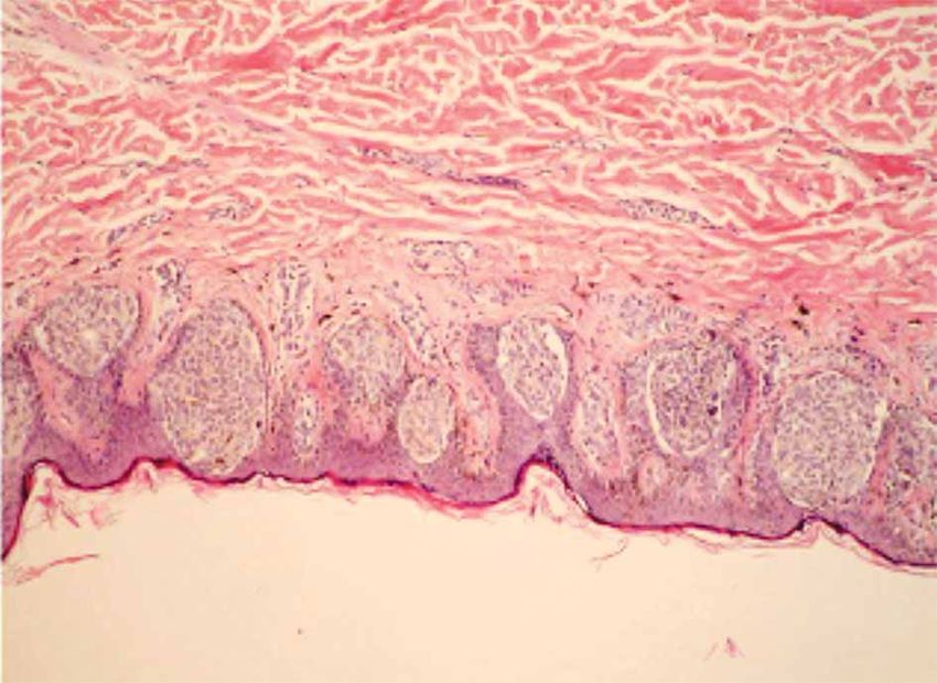
corona, leaving targetoid pattern (Figure 1a). The bran- ordinary nevi and melanomas, melanocytic cells in Spitz
ched streaks reach the surrounding skin only occasion- nevus are large, often twice the size of epidermal basal
ally, usually dissipating before or in the corona and being keratinocytes (Figure 1c). Mitoses, usually few in num-
replaced by pigmented globules and dots9. ber are nested in half the cases. The melanocytic cells in
Although the bizarre histopathological features and NS show progressive maturation with increasing depth,
frequent occurrence of dermal inflammation may cause becoming smaller and more similar to ordinary melano-
Fig. 1. a) Nevus Spitz under dermatoscope. Fig. 1. c) Histological picture of typical nevus Spitz (400x HE).
172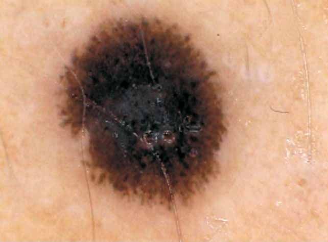
M. [itum et al.: Nevus Spitz, Coll. Antropol. 32 (2008) Suppl. 2: 171–176

cytes. Coalescent oesinophilic globules (Kamino bodies),                  Other helpful diagnostic features:
PAS positive have been reported in 60% of Spitz nevi.                     1. Mitotic rate
M. [itum et al.: Nevus Spitz, Coll. Antropol. 32 (2008) Suppl. 2: 171–176

individuals and were located on the head and neck, and              abundant opaque or glass cytoplasm18,19. The importance
extremities. Six patients had positive regional lymph               of differentiation between NS and melanoma is empha-
node metastases with involvement of the sinuses and pa-             sized in the literature. Recent data suggest that Spitz
renchyma by tumor identical to the primary cutaneous                nevi differ from melanomas in their immunohistochemi-
lesion. The older the patients, especially beyond 20–30             cal pattern of expression of cell cycle and apoptosis regu-
years, the likelihood of malignancy are greater. The loca-          lators (bax, Ki-67, Rb, p-16, cyclin A, cyclin B1, p-27,
tion of atypical tumors on sites less commonly involved             p-53) and more closely resemble common benign nevi20,21.
by Spitz tumor, such as the back, is also another factor            There are many authors who tried to find clear patho-
suggesting careful follow up of the patient14. However,             histological hallmarks of NS and Spitzoid melanoma.
there are some authors15 who disagree about the terms               Weedon D and Little JH22 find these features important;
»atypical« nevus Spitz »malignant« nevus Spitz, nevus               presence of some nevus cells maturity at the base, an ab-
Spitz and metastasizing nevus Spitz. They found out                 sence of atypical mitoses, no significant upward epider-
that an overwhelming majority of neoplasm that claimed              mal spread and the nuclear chromatin pattern. Crotty23
to be »atypical«, »metastasing« and »malignant« Spitz               reviewed several studies and concluded that histological
nevi were in fact melanomas. Complementary to these                 features that support diagnosis of malignant melanoma
facts are the results of study of 12 patients with »atypi-          rather than atypical nevi are deep and marginal mitoses,
cal« nevus Spitz who underwent sentinel node biopsy.                atypical mitoses, asymmetry, pleomorphisam and promi-
Nodal micrometastases were found in 33.3% of patients16.            nent epidermal involvement. The same author compared
    Histopatological criteria for atypical Spitz tumors from        clinical and histopathological features of 13 malignant
Barnhill at al4:                                                    melanomas in children under the age of 13 with 15 NS24.
                                                                    Histological features favoring malignancy were mitoses
   1. Diameter in mm (³10 mm considered abnormal),
                                                                    within 0.25 mm of the dermal margin of the melanoma, a
   2. Depth in mm (subcutaneous fat considered abnor-               dermal mitotic rate exceeding 2/mm2, ulceration, sur-
      mal),                                                         face, exudates, large pigmented granules and clear-cell
   3. Ulceration,                                                   differentiation. The median thickness of malignant mel-
   4. Poor circumscription,                                         anomas was 1.3 mm, but in 4 children, who died with
   5. Pagetoid melanocytes over a larger front,                     melanoma, median thickness was 2.9 mm. Absence of
                                                                    mitoses, predominance of spindle cells and diffuse matu-
   6. Prominent confluence of melanocytes,
                                                                    ration favored NS. The median thickness of the NS was
   7. Asymmetry,                                                    0.7 mm. The most frequent clinical features found in the
   8. Few or no dull pink (Kamino bodies),                          malignant melanoma were bleeding, ulceration, itching
   9. High cellular densits,                                        and black or variegated color. Kapur et al.25 compared ex-
   10.Lack of zoonation and maturation.                             pression of Ki-67, p21 and fatty acids synthesis by immu-
                                                                    nohistochemistry in 10 atypical NS, 28 typical NS, 19
   Proliferation criteria from Barnhill at al4:                     compound melanocytic nevi and 18 malignant melano-
                                                                    mas. There was a progressive increase in fatty acid syn-
   1. Significant mitotic rate ³2–6/mm2,
                                                                    thesis cytoplasmic expression with statistically signifi-
   2. Deep/marginal mitoses                                         cant differences observed between Ns and atypical NS
   3. Proliferation index-Ki-67 expression between 2–10%;           and between atypical NS and malignant melanoma Ki-67
      ³10%.                                                         nuclear staining was lower in both typical and atypical
   Cytological criteria from Barnhill at al4:                       forms of Spitz lesions than in malignant melanoma. The
   1.   Granular vs. ground glass cytoplasm,                        degree of p21 nuclear expression in atypical NS was not
                                                                    significantly different than in NS, but was significantly
   2.   High nuclear to cytoplasmic ratios,                         greater than expression in conventional nevi and ap-
   3.   Loss or delicate or dispersed chromatin patterns,           proached significance after multiple comparisons correc-
   4.   Thickening of nuclear membranes,                            tions for malignant melanoma. Thus, a high level of p21
   5.   Hyperchromatism,                                            expression makes a tumor more likely to be a typical or
                                                                    atypical NS than a malignant melanoma, especially when
   6.   Large nucleoli.
                                                                    coupled with a low Ki-67 index and weak expression of
                                                                    fatty acid synthase. These immunohistochemical obser-
Spitzoid Melanoma                                                   vations support the concept that atypical NS are distinct
                                                                    lesions of borderline biologic behavior residing between
    Spitzoid melanoma term should be used for mela-                 NS and malignant melanoma. The study also compared a
noma with morphological resemblance to nevus Spitz17.               large array of histological features of 16 cases of typical
The resemblance includes features: dome-shaped, pla-                NS in children with 12 typical NS in adults. The adult le-
que-like, wedge-shaped morphology, little or no asymme-             sions were significantly more likely to be intradermal
try, epidermal hyperplasia, clefting about intraepidermal           and to display dermal fibroplasia, but were histologically
nests of melanocytes, presence of dull Kamino bodies,               similar to their pediatric counterparts in all other re-
some evidence of zonation or maturation and population              spects. Vollmer et al.26 in his study used previously pub-
of enlarged epitheloid or/and spindelled melanocytes with           lished data, exponential and g probability density func-

174
M. [itum et al.: Nevus Spitz, Coll. Antropol. 32 (2008) Suppl. 2: 171–176
tions to model statistical distributions of proliferation TABLE 2
RISK FOR METASTASIS FOR SPITZ TUMORS
index (PI), respectively, in NS and melanomas and Bayes
rule to estimate the predictive probability that a lesion is
Parameter Score
a NS, given an observed PI. Results indicate that PIs
more than 10% favor a melanoma diagnosis and PIs less Age
than 2%, NS. PI values between 2% and 10% yield vari- 0–10 0
ous predictive values for NS, depending on the a priori 11–17 1
probability that the lesion is a NS. Diameter /mm/
0–10 0
>10 1
Conclusion Involvement of subcutaneous fat
All histopathological, clinical and ancillary criteria absent 0
must be weighed in the final interpretation of epithe- present 2
loid/spindle cell lesion. At the present, the final diagnosis Ulceration
remains pathohistological with important emphasis gi- absent 0
ven to clinical impression. Persistently changing lesion present 2
indicates malignancy potential of the lesion. Barnhill Mitotic activity /mm2/
recommends that all Spitz tumors are completely ex-
0–5 0
cised. Atypical tumors should be excised with wider mar-
6–8 2
gins up to 1 cm. Patient should be carefully monitored by
>19 5
regular examinations for recurrence and metastasis. The
approach should be individual with efforts to avoid over
treatment or suboptimal treatment. The need of proper
patient counseling cannot be overemphasized, especially 2. Application of all histopathological, clinical and ot-
considering the psychological aspect of coping with ma- her attributes for assessing abnormalities present,
lignant skin tumors27. 3. Seek consultation,
Protocol for Spitz tumor from Barnhill et al.4: 4. Placement into risk category28 (Table 2),
1. Examination of the entire lesion, 5. Management of the patient.
REFERENCES
1. DARIER J, CIVATTE, A Bull Soc Franc Derma Syph, 21(1910) 61. RMAN AB, Am J Dermatopathol, 26 (2004) 310. — 16. URSO C, BORGO-
— 2. SPITZ S, Am J Pathol, 24 (1947) 591. — 3. ALLEN AC, SPITZ S, GNONI L, SAIEVA C, FERRARA G, TINACCI G, BEGLIOMINI B,
Cancer, 13 (1953) 612. — 4. BARNHILL RL, ARGENVI ZB, FROM l, REALI UM, Hum Pathol, 37(2006) 816. — 17. WALSH N, CROTTY K,
GLASS LF, MAIZE JC, MIHM MC, RABKIN MS, RONAN SG, WHITE PALMER A, MCCARTHY S, Hum Pathol, 22 (2000) 489. — 18. MOOI
WL, PIEPKON M, Hum Pathol, 30 (1999) 513. — 5. GELBARD SN, WJ, Adv Anat Pathol, 9 (2002) 209. — 19. DAHLSTROM JE, SCOLYER
TRIPP JM, MARGHOOB AA, KOENIG KL, KIM JY, BART RS, J Am RA, THOMPSON JF, JAIN S, Pathology, 36 (2004) 452. — 20. STEFA-
Acad Dermatol, 47 (2002) 224. — 6. SULIT DJ, GUARDIANO RA, KRI- NAKI C, STEFANAKI K, ANTONIOU C, ARGYRAKOS T, PATERELI A,
VDA S, Cutis, 79 (2007) 141. — 7. BRAUN FALCO O, PLEWIG G, WOLF STRATIGOS A, KATSAMBAS A, J Am Acad Dermatol, 56 (2007) 815. —
HH, BURGDORF WHC, Dermatology (Springer Verlag, Berlin Heidel- 21. BATINAC T, HADZISEJDI} I, BRUMINI G, RUZI} A, VOJNIKOVI}
berg New York, 2000). — 8. MORGAN CJ, NYAK N, COOPER A, PEES B, B, ZAMOLO G, Coll Antropol, 31 (2007) 17. — 22. WEEDON D, LITTLE
FRIEDMANN PS, Clin Exp Dermatol, 31 (2006) 368. — 9. PERIS K, FE- JH, Cancer, 40 (1977) 217. — 23. CROTTY KA, Australas J Dermatol, 38
RRARI A, ARGENZIANO G, SOYER HP, CHIMENTI S, Clin Dermatol, (1997) 49. — 24. CROTTY KA, MCCARTHY SW, PALMER AA, NG AB,
20 (2002) 259. — 10. MOOI WJ, Curr Top Pathol, 94 (2001) 65. — 11. THOMPSON JF, GIANOUTSOS MP, SHAW HM, World J Surg, 16 (1992)
REED RJ, ICHINOSE H, CLARK W, Semin Oncol, 2 (1975) 119. — 12. 179. — 25. KAPUR P, SELIM MA, ROY LC, YEGAPPAN M, WEINBERG
DA FORNO PD, FLETCHER A, PRINGLR JH, SALDANHA GS, Br J AG, HOANG MP, Mod Pathol, 18 (2005) 197. — 26. VOLLMER RT, Am J
Dermatol, 158 (2008) 4. — 13. SMITH KJ, BARRETT TL, SKELTON HG, Clin Pathol, 122 (2004) 499. — 27. VURNEK M, BULJAN M, SITUM M,
LUPTON GP, GRAHAM JH, Am J Surg Pathol, 13 (1989) 931. — 14. Coll Antropol, 31 (2007) 53. — 28. SPATZ A, CALONJE E, HANDFIELD
HELM TN, HELM KE, Cutis, 3 (2007) 183. — 15. MONES JM, ACKE- JONES S, BARNHILL RL, Arch Dermatol, 135 (1999) 282.
@. Bolan~a
University Department of Dermatovenerology University Hospital »Sestre milosrdnice«, Vinogradska cesta 29,
10000 Zagreb, Croatia
e-mail: zeljana.bolanca@gmail.com
175M. [itum et al.: Nevus Spitz, Coll. Antropol. 32 (2008) Suppl. 2: 171–176
NEVUS SPITZ – PATOHISTOLO[KA DVOJBA
SA@ETAK
1910. godine Darier i Civatte su detaljno opisali brzorastu}u, melanocitnu promjenu na vrhu nosa djeteta. Nisu sa
sigurno{}u mogli re}i jeli promjena benigne ili maligne naravi. 1947. godine Sophie Spitz je opisala identi~nu leziju kao
juvenilini melanoma sa relativno odli~nom prognozom. Kasnije je napravljena revizija studije te je zaklju~eno da se radi
o juvelnilnom melanomu koji je benigan, a mo`e se pojaviti ~ak i u odrasloj dobi. Iako je prognoza odli~na, Spitz je
opisala jedan slu~aj sa fatalnim metastazama. Barnhill i sur. su tako|er opisali smrtni ishod bolesnika za kojeg se
vjerovalo da ima Nevus Spitz. I u dana{nje vrijeme, tako|er postoji nesuglasje oko histopatologije i terminologije pro-
mjena koje nisu ni atipi~ni nevus Spitz, ali ni melanom. Kod dono{enje kona~ne odluke, trebaju se uzeti u obzir sva
histolo{ka i klini~ka obilje`ja promjena. Promjene koje se neprestano mijenjaju upu}uju na maligni potencijal. Barnhill
i sur. predla`u da se svi tipi~ni Spitz nevusi trebaju operativno odstraniti u cijelosti. Za atipi~ne promjene predla`e
rubove do 1 cm. Sve bolesnike treba redovito kontrolirati radi lokalnog recidiva i/ili metastaza.
176You can also read