Case report METASTATIC CHOLANGIOCARCINOMA IN A BEARDED DRAGON (Pogona vitticeps) - Sciendo
←
→
Page content transcription
If your browser does not render page correctly, please read the page content below
Acta Veterinaria-Beograd 2020, 70 (2), 267-276
                                                                     UDK: 636.98.09:616-006-073
                                                                    DOI: 10.2478/acve-2020-0019
Case report
METASTATIC CHOLANGIOCARCINOMA IN
A BEARDED DRAGON (Pogona vitticeps)

KOMENDA Dominik1*, DOLENŠEK Tamara2, ŠVARA Tanja2,
KASTELIC Marjan3,4, PROKS Pavel1, NÝVLTOVÁ Ivana1, KVAPIL Pavel3

1
 Department of Diagnostic Imaging, Small Animal Clinic, Faculty of Veterinary Medicine, University
of Veterinary and Pharmaceuticals Sciences Brno, Brno, Czech Republic; 2Institute of Pathology, Wild
Animals, Fish and Bees, Veterinary Faculty, University of Ljubljana, Slovenia; 3Ljubljana Zoo, Ljubljana,
Slovenia; 4Veterinary clinic BUBA, d.o.o., Grosuplje, Slovenia

(Received 23 September 2019, Accepted 18 February 2020)

        A 6.5-year-old female bearded dragon (Pogona vitticeps) was presented with a swollen
        right pelvic limb. A tissue core biopsy from the swollen area was performed and a
        presumptive histopathological diagnosis of adenocarcinoma was made. This diagnosis
        was confirmed after limb amputation. Two months after amputation a sudden
        deterioration in the overall health of the patient occurred. Ultrasound examination of
        the coelomic cavity revealed hypoechoic lesions in the liver. The patient was euthanized
        and submitted for necropsy which revealed a severely enlarged liver with multiple
        coalescing yellowish nodules. Cholangiocarcinoma of the liver with metastases to the
        spleen, left mesovarium and right pelvic limb was diagnosed after histopathological
        examination.
        Key words: histopathology, necropsy, neoplasia, reptiles, X-ray, ultrasonography

                                         INTRODUCTION

Diagnoses of neoplastic diseases are increasing amongst reptiles due to their growing
popularity as pet animals [1].
Many different types of neoplasia have been described in bearded dragons (Pogona
vitticeps). Cases of gastric neuroendocrine carcinoma [2-6], intestinal leiomyosarcoma
[7], ovarian metastatic leiomyosarcoma [8], peripheral nerve sheath tumour
[9,10], oral fibrosarcoma [11], several periorbital tumours – adenocarcinoma [12],
cystadenoma [13], myxosarcoma [14] and squamous cell carcinoma [15] have been
published. Leukaemia has also been described in this species [16,17]. One bearded
dragon presented intrahepatic cholangiocarcinoma as well as in situ adenocarcinoma
and two adenomas of the gallbladder [18].

*Corresponding author: e-mail: komendad@vfu.cz

                                                                                                     267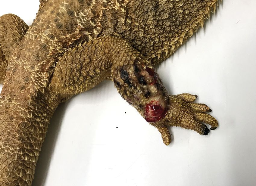
Acta Veterinaria-Beograd 2020, 70 (2), 267-276

Differential diagnoses of swelling in reptiles are bacterial granulomas, neoplastic
disorders, parasitic cysts, fungal granulomas, epidermal cysts, hematomas, aneurysms
and gout or pseudogout [19,20]. The finding of a mass on a reptile´s body does not
immediately mean that the lesion is neoplastic. The appropriate management of the
swelling without histological or cytological confirmation is not possible [21]. Therefore,
it is very useful to biopsy such lesions and perform further examinations.
The aim of this case report is to describe a metastatic cholangiocarcinoma of the liver
in a bearded dragon. During the review of available literature regarding neoplasia in
bearded dragons, the authors did not find any reported case of cholangiocarcinoma
with metastases to the spleen, mesovarium and pelvic limb.

                                  CASE PRESENTATION

A 6.5-year-old female bearded dragon (Pogona vitticeps), weighing 430 g, was presented
by zookeepers to the veterinary clinic in Ljubljana Zoo due to swelling of the right
pelvic limb. The bearded dragon was owned by the Zoo and was kept in a terrarium
with sandy substrate together with four other females. The food mainly consisted of
insects such as crickets, grasshoppers, flour worms and a minimal herbivorous diet,
mainly lettuce.
Clinical examination revealed a deteriorated nutritional status with a body condition
score of 2/5. The body orifices showed no discharge and the animal’s breathing was
calm. A marked swelling of the right shin with skin ulcerations was the main finding
(Fig. 1). The swelling was approximately 2.5 cm in diameter and the skin ulceration

Figure 1. The swollen pelvic limb with skin ulceration during the initial clinical presentation

268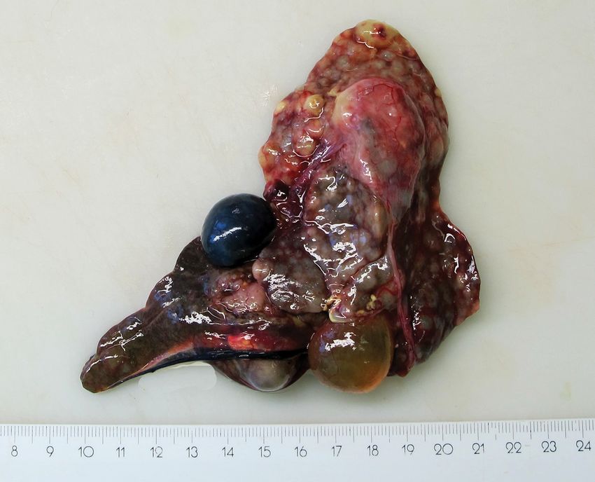
Komenda et al.: Metastatic cholangiocarcinoma in a bearded dragon (Pogona vitticeps)
measured about 4 mm in diameter. The skin and dermal derivatives on other parts of
the body were without abnormalities. The swollen area was stiff, slightly sensitive to
palpation and extended from the stifle to the tarsal joint. X-ray examination (HiRay
Plus X-ray Machine, Eickemeyer Veterinary Equipment Ltd., Sunbury-on-Thames,
UK) of the affected limb showed pronounced swelling of the soft tissues, whereas
no radiological abnormalities were detected on the bone structures. Due to the
assumption of an inflammatory process, anti-inflammatory and analgesic therapy with
meloxicam (0.5 mg/kg) (Meloxidyl 5 mg/ml, Ceva Santé Animale, Libourne, France)
(subcutaneously, q 24 h) and antibiotic therapy with marbofloxacin (10 mg/kg)
(Marbocyl 10%, Vetochinol, Cedex, France) (subcutaneously, q 48 h) was administered
empirically [22].
Slight improvement of the clinical status was noticed after 2 weeks of therapy. This
improvement was reflected in a reduction of the skin ulceration. Blood from the
coccygeal vein was collected for biochemical examination and the results were within
normal limits. Anti-inflammatory and antibiotic therapy was prolonged, and the
skin ulcerations were treated daily with antimycotic terbinafine (Lamisil 1% Cream,
Novartis s.r.o., Prague, Czech Republic). After an additional 2 weeks the clinical status
did not improve, therefore biopsy samples of the soft tissue from the affected area
were obtained for histopathological examination under local anaesthesia. Tramadol
(2 mg/kg) (Tramal 50 mg/ml, Stada Arzneimittel AG, Bad Vilbel, Germany) [22] and
meloxicam (0.5 mg/kg) were used as premedication and 4 mg/kg of lidocaine (Lidocain
2%, Egis Pharmaceuticals PLC, Budapest, Hungary) was administered into the muscles
proximal to the stifle. Two punch biopsies were obtained, fixed in 10% buffered
formalin and sent for histopathology. A presumptive diagnosis of adenocarcinoma
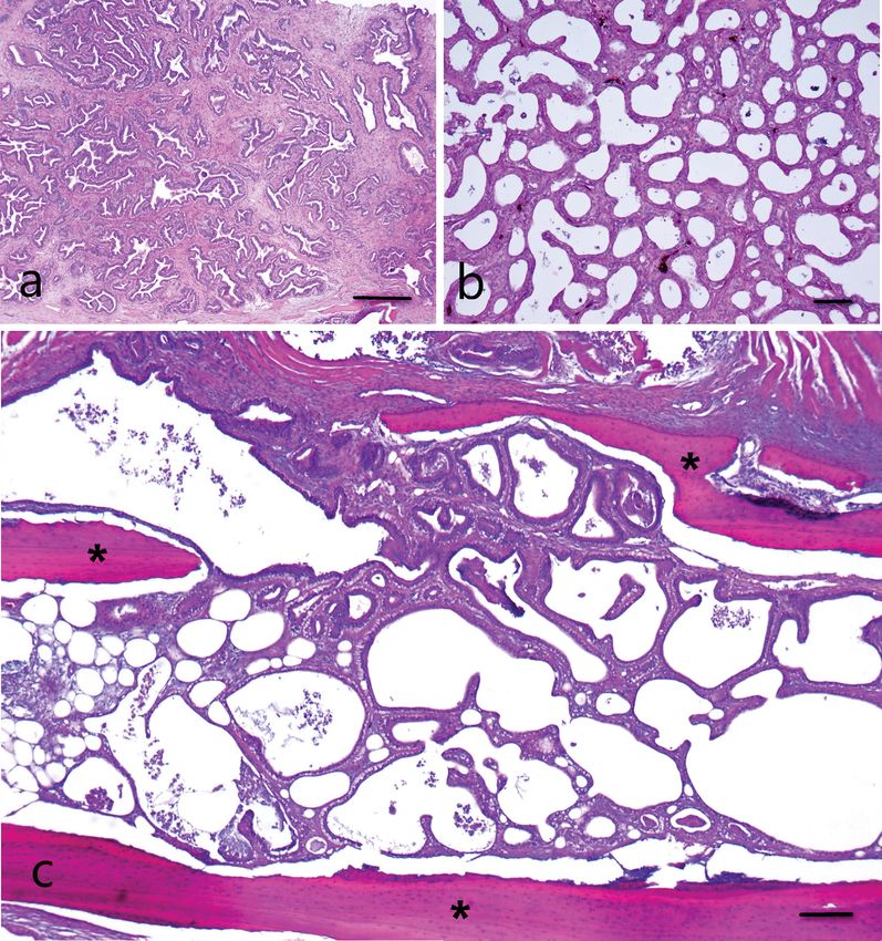
of unknown origin was the result of the histopathological examination (Fig. 2a). A
control X-ray after biopsy revealed a transverse single-stranded fracture of the fibula
(Fig. 3).
After receiving the histopathological diagnosis of adenocarcinoma, amputation of the
affected limb was performed and the limb was sent for histopathological examination.
The premedication chosen was the same as that administered when sampling for the
histopathological examination - tramadol (2 mg/kg) and meloxicam (0.5 mg/kg).
Induction of anaesthesia was performed by intravenous administration of propofol
(5 mg/kg) (Propofol 2% MCT/LCT Fresenius, Fresenius Kabi Deutschland GmbH,
Bad Homburg, Germany) into the ventral coccygeal vein. Subsequently, the patient
was intubated by means of an intravenous catheter (Vasofix Safety 16G, B. Braun
Melsungen, Melsungen, Germany) and the maintenance of anaesthesia was carried
out with a mixture of oxygen (2 L/min) and 3% of isoflurane (Aerrane, Baxter S.A.,
Lessines, Belgium). The limb was amputated at the stifle and the stump was stitched in
two layers with a horizontal mattress stitch (PDS 4-0; Johnson & Johnson Medical N.V.,
Belgium). A bolus of Ringer solution (20 ml/kg) (B. Braun Melsungen, Melsungen,
Germany) together with Duphalyte (20 ml/kg) (Pfizer Olot SLU, Vall de Bianya,
Spain) in a total volume of 10 ml was administered subcutaneously after the operation.
269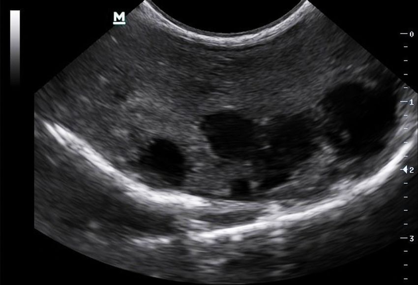
Acta Veterinaria-Beograd 2020, 70 (2), 267-276

Recovery from anaesthesia was without complications. Prokinetic medication with
metoclopramide (2 mg/kg) (Vomend 5mg/ml; Hyperdrug Pharmaceuticals Ltd,
Middleton-in-Teesdale, UK) was administered subcutaneously after the operation and
artificial feeding of the patient by oesophageal tube was conducted for 5 days (PD
Hill’s Urgent Care Canine/Feline a/d, Hill’s Pet Nutrition Inc., USA). Application of
meloxicam and marbofloxacin was prolonged in the above-mentioned doses for one
week after the operation.

Figure 2. Histopathological micrographs of liver cholangiocarcinoma and leg metastasis; a)
biopsy of the leg with variably sized tumorous tubular structures, predominantly composed
of a single layer of columnar cells that led to a presumptive diagnosis of adenocarcinoma of
unknown origin. H&E, Bar = 300 µm; b) liver cholangiocarcinoma with tumorous structures
similar to the structures previously found in the leg biopsy. H&E, Bar = 100 µm; c) metastasis
of cholangiocarcinoma with tumorous structures infiltratively growing into the leg bone
(asterisks). H&E, Bar = 200 µm.

270
Komenda et al.: Metastatic cholangiocarcinoma in a bearded dragon (Pogona vitticeps)

Histopathological examination of the amputated pelvic limb confirmed the initial
histopathological diagnosis of adenocarcinoma. The tumour infiltrated the dermis,
striated muscles and also bone tissue, where it extended into the medullary cavity (Fig.
2c).
Sudden deterioration of the overall health condition occurred two months after
amputation of the affected limb. The patient was apathetic and stopped eating.
Ultrasound examination of the coelomic cavity was performed by Mindray M7
(Mindray DS USA Inc., Mahwah, USA) with an 8 MHz microconvex transducer.
Significant enlargement of the liver with presence of multiple cystic anechoic structures
with clear distal signal amplification was observed (Fig. 4) and liver neoplasia was the
presumed diagnosis. Blood was obtained for haematological and control biochemical
examination. All biochemical and haematological parameters were within normal
range. Due to poor clinical status and the ultrasonographic finding, the animal was
euthanized. A combination of propofol (5 ml pro toto) and T61 (1.5 ml pro toto)
(Intervet International BV, Boxmeer, The Netherlands) was administered into the
ventral coccygeal vein. Successful euthanasia was verified by acoustic oscillometric
examination of the heart.

Figure 3. X-ray of right pelvic limb on unsedated animal. A single transverse fracture of the
right fibula and swollen soft tissues are evident.

                                                                                                   271
Acta Veterinaria-Beograd 2020, 70 (2), 267-276 Figure 4. Ultrasonographic examination of liver. Multiple anechoic cystic structures are visible. The rest of the liver parenchyma is slightly heteroechoic. Subsequently, the animal was referred for necropsy. A markedly enlarged liver with multiple coalescing yellowish nodules measuring 2 to 14 mm in diameter, which were multifocally cystic, was the major finding at necropsy. The affected liver measured 10 x 9 x 2 cm and occupied one third of the entire coelomic cavity. The gallbladder was significantly dilated and contained 5 ml of green, translucent bile (Fig. 5). There was 30 ml of slightly cloudy, reddish free fluid in the coelomic cavity. A single, slightly protruding, white-yellow nodule measuring 5 mm in diameter was found on the left part of the fat body and on the surface of the left lung lobe. The right ovary was not present in the coelomic cavity, whereas the left ovary contained several small follicles. All organs from the coelomic cavity and the brain were collected for histopathology. Most of the liver parenchyma was replaced by variably sized tumorous tubular structures, predominantly composed of a single layer of columnar cells, which expressed moderate anisocytosis, had a moderate amount of lightly basophilic cytoplasm and a round to oval, moderately anisokaryotic nucleus with one or two nucleoli, or the nucleolus was not visible (Fig. 2b). The mitotic index was 4 mitoses per 10 high power fields; rare mitoses were bizarre. Multifocally, there were groups of neoplastic cells in the spleen and fat tissue around the left ovary and the fallopian tube which were morphologically identical to the neoplastic cells in the liver. The macroscopically observed nodule on the left part of the fat body proved to be liponecrosis and the nodule on the left lung lobe was determined to be a chronic granuloma. Sewing thread with concurrent granulomatous inflammation was present in the fat under the left ovary. The kidneys showed moderate cholemic nephrosis. In other organs of the coelomic cavity and the brain no histopathological changes were found. 272
Komenda et al.: Metastatic cholangiocarcinoma in a bearded dragon (Pogona vitticeps)

Figure 5. Liver cholangiocarcinoma. Markedly enlarged liver with multiple coalescing yellowish
nodules and multifocal cystic changes.

                                        DISCUSSION

Many swellings in reptiles are caused by abscesses [23]. Because of this fact, antibiotic
and anti-inflammatory medication was the empirical first choice of therapy in this
case. Fungal infection is another frequent cause of limb swelling, including the fingers.
Surface ulceration in swelling caused by fungal infections is often observed [24,25].
Such ulceration was also observed in the described case, but there was no improvement
after daily antimycotic administration.
Due to a wide range of different diagnoses of swelling on reptiles´ bodies biopsy
is useful for externally palpable masses [21]. The best method for determining the
diagnosis of neoplastic disease is tissue biopsy [1,21]. Reavill (2004) suggests fine-
needle aspiration biopsy of the affected area [26]. We chose to perform punch biopsy
under local anaesthesia in our case so as to provide an adequate amount of tissue for
histopathological examination.
We encounter many types of neoplasia in reptiles which are already known in
mammals [26,27]. Cholangiocarcinoma of the liver was diagnosed after necropsy
in this case. Cholangiocarcinoma is a malignant neoplasia that originates from the
biliary epithelium. In domestic animals, such as dogs, cats, cattle, sheep and horses, it
is less frequent than hepatocellular tumours. In these animals cholangiocarcinomas are

                                                                                                   273
Acta Veterinaria-Beograd 2020, 70 (2), 267-276
known to metastasize to the drainage lymph nodes, lungs and also to the peritoneal
serosal surfaces [28].
Only swelling of soft tissues without detectable osteolysis of the bone structures was
detected on the primary X-ray. A fibula fracture, without osteolysis, was evident in the
affected area in the repeated X-ray examination after 4 weeks. It is possible that the
fracture was caused by frequent manipulation of the limb during treatment or during
biopsy (the control X-ray was performed after the punch biopsy).
We suppose that the swelling of the right pelvic limb was a result of metastasis of the
primary cholangiocarcinoma of the liver into the limb, because there is no primary
glandular tissue in this region from which the tumour could have originated. When the
overall clinical condition of the patient deteriorated and ultrasonographic examination
of the coelomic cavity revealed a severely enlarged liver, a primary tumour of the liver
was highly suspected. Diagnostic imaging methods were not performed initially since
the patient had no clinical symptoms except unilateral pelvic limb swelling. On the
basis of this experience, the authors recommend a thorough screening examination
of the animal’s body using imaging methods to determine whether it is a primary or
metastatic neoplastic process.
Acknowledgements
This paper was supported by Internal Creative Agency of University of Veterinary
and Pharmaceutical Sciences Brno (Project No. FVL/Crha/ITA2019).
Authors’ contributions
DK, MK and PK actively participated in clinical, diagnostic (X-ray, ultrasound, blood
tests) and surgical procedures of this clinical case. TD and TŠ did the necropsy of
the agama and histopathology of agama´s organs, DK, PP, TD, TŠ, MK, IN, PK
participated in writing this manuscript. All authors have approved the final version of
the manuscript.
Declaration of conflicting interests
The author(s) declared no potential conflicts of interest with respect to the research,
authorship, and/or publication of this article.
REFERENCES
1. Hernandez-Divers SM, Garner MM: Neoplasia of reptiles with an emphasis on lizards. Vet
Clin North Am Exot Anim Pract 2003, 6:251–273.
2. Ritter JM, Garner MM, Chilton JA, Jacobson ER, Kiupel M: Gastric neuroendocrine
carcinomas in bearded dragons (Pogona vitticeps). Vet Pathol 2009, 46:1109–1116.
274Komenda et al.: Metastatic cholangiocarcinoma in a bearded dragon (Pogona vitticeps)
3. Lyons JA, Newman SJ, Greenacre CB, Dunlap J: A gastric neuroendocrine carcinoma
expressing somatostatin in a bearded dragon (Pogona vitticeps). J Vet Diagn Invest 2010,
22:316–320.
4. Mooij TS, Martel A, Bosseles L, Chiers K, Pasmans F, Hellebuyck T: Atypische klinische
presentatie van een metastatisch gastrisch neuroendocrien carcinoom bij een baardagame
(Pogona vitticeps). Vlaams Diergen Tijds 2014, 83:293–298.
5. Anderson KB, Meinkoth J, Hallman M, Bailey K, Brandaos J: Cytological diagnosis of
gastric neuroendocrine carcinoma in a pet inland bearded dragon (Pogona vitticeps). J Exot
Pet Med 2019, 29:188–193.
6. Collins SN: Diagnostic challenge: Gastric neuroendocrine carcinoma in a bearded dragon
(Pogona vitticeps). J Exot Pet Med 2019, 30:7–11.
7. Łojszczyk-Szczepaniak A, Śmiech A, Chlebicka N, Szczepaniak K, Klimiuk P: First case
of intestinal leiomyosarcoma in a bearded dragon: Ultrasonographic findings. Med Weter
2016, 72:303–306.
8. Sonntag FD, Schroff C, Dietz J, Heckers KO: Metastatic leiomyosarcoma of the ovary in
an inland bearded dragon (Pogona vitticeps) – A case report. Prakt Tierarzt 2014, 95:518–523.
9. Mikaelian I, Levine BS, Smith SG, Harshbarger JC, Wong VJ: Malignant peripheral nerve
sheat tumor in a bearded dragon, Pogona vitticeps. J Herpetol Med Surg 2001, 11:9–12.
10. Lemberger KY, Manharth A, Pessier A: Multicentric benign peripheral nerve sheath tumors
in two related bearded dragons, Pogona vitticep. Vet Pathol 2005, 42:507–510.
11. Geczy C, Jakab C: Oral fibrosarcoma in a bearded dragon (Pogona vitticeps). Magy Allatorvosok
2014, 135:413–419.
12. Darrow BG, McLean NSJ, Russman SE, Schiller CA: Periorbital adenocarcinoma in a
bearded dragon (Pogona vitticeps). Vet Ophtalmol 2013, 16:177-182
13. Pryor SG, Cutler D, Yau W, Diehl KA: Adnexal cystadenoma in a bearded dragon (Pogona
vitticeps). J Exot Pet Med 2018, 27:85–89.
14. Gardhouse S, Eshar D, Lee-Chow B, Foster RA, Ingrao JC, Poirier VJ: Diagnosis and
treatment of a periocular myxosarcoma in a bearded dragon (Pogona vitticeps). Can Vet J
2014, 55:663–666.
15. Hannon DE, Garner MM, Reavill DR: Squamous cell carcinomas in inland bearded dragons
(Pogona vitticeps). J Herpetol Med Surg 2011, 21:101–106.
16. Tocidlowski ME, McNamara PL, Wojcieszyn JW: Myelogenous leukemia in a bearded
dragon (Acanthodraco vitticeps). J Zoo Wildl Med 2001, 32:90–96.
17. Jankowski G, Sirninger J, Borne J, Nevarez JG: Chemotherapeutic treatment for leukemia
in a bearded dragon (Pogona vitticeps). J Zoo Wildl Med 2011, 42:322–325.
18. Jakab C, Rusvai M, Szabó Z, Gálfi P, Marosán M, Kulka J, Gál J: Claudin-7-positive
synchronous spontaneous intrahepatic cholangiocarcinoma, adenocarcinoma and
adenomas of the gallbladder in a bearded dragon (Pogona vitticeps). Acta Vet Hung 2011,
59:99–112.
19. Jacobson ER: Reptile dermatology. In: Kirk’s Current Veterinary Therapy XI, Small Animal
Practice. Philadelphia. United States: WB Saunders; 1992, 1204-1210.
20. Schmidt V: Abscesses/Fibriscesses. In: Mader´s Reptile and Amphibian Medicine and Surgery. 3th
ed. St. Louise-Missouri, United States: Elsevier; 2019, 1288-1289.
21. Mayer J, Moore AS: Oncology. In: Mader´s Reptile and Amphibian Medicine and Surgery. 3th ed.
St. Louise-Missouri, United States: Elsevier; 2019, 827-834.
275Acta Veterinaria-Beograd 2020, 70 (2), 267-276
22. Gibbons PM, Klaphake E, Carpenter JW: Reptiles. In: Exotic Animal Formulary. 4th edn. St.
Louise-Missouri, United States: Elsevier; 2005, 83-182.
23. Barten S, Simpson S: Differential Diagnoses by Clinical Signs – Lizards. In: Mader´s Reptile
and Amphibian Medicine and Surgery. 3th ed. St. Louise-Missouri, United States: Elsevier; 2019,
1257-1265.
24. Johnson RSP, Sangster CR, L Sigler L, Hambleton S, Paré JA: Deep fungal dermatitis
caused by the Chrysosporium anamorph of Nannizziopsis vriesii in captive coastal bearded
dragons (Pogona barbata). Aust Vet J 2011, 89:515–519.
25. Schmidt V: Fungal infections in reptiles – an emerging problem. J Exot Pet Med 2015,
24:267–275.
26. Reavill DR: Neoplasia. In: BSAVA Manual of Reptiles, 2nd ed. Gloucester, United Kingdom:
British Small Animal Veterinary Association; 2004, 309-318.
27. Elkan E, Cooper JE: Tumours and pseudotumours in some reptiles. J Comp Pathol 1976,
86:337–348.
28. Head KW, Cullen JM, Dubielzig RR, Else RW, Misdorp W, Patnaik AK, Tateyama S, van
der Gaag I. Tumors of the alimentary system of domestic animals. Washington DC: AFIP, CL
Davis DVM Foundation and WHO Collaborating Center for Worldwide Reference on
Comparative Oncology; 2003.
METASTATSKI HOLANGIOKARCINOM KOD
BRADATE AGAME (Pogona vitticeps)
KOMENDA Dominik, DOLENŠEK Tamara, ŠVARA Tanja,
KASTELIC Marjan, PROKS Pavel, NÝVLTOVÁ Ivana, KVAPIL Pavel
Ženka bradate agame (Pogona vitticeps), starosti 6,5 godina, dovedena je sa otokom u
predelu desnog pelvisa. Izvedena je biopsija otečene regije i preliminarno postavljena
histopatološka dijagnoza adenokarcinoma. Dijagnoza je potvrđena nakon amputacije
ekstremiteta. Dva meseca nakon amputacije došlo je do naglog pogoršanja opšteg zdrav-
lja pacijenta. Ultrazvučni pregled celoma je pokazao hipoehogene lezije jetre. Pacijent
je eutanaziran, nakon čega je izvršena obdukcija tokom koje je ustanovljena značajno
uvećana jetra sa brojnim koalescirajućim žućkastim čvorićima. Histopatološkom pre-
tragom dijagnostikovan je holangiokarcinom jetre sa metastazama na slezini, levom
mezo-ovarijumu i desnom ekstremitetu.
276You can also read