Tinea Corporis Caused by Trichophyton Benhamiae Transmitted by a Dog: First Case Report in Portugal
←
→
Page content transcription
If your browser does not render page correctly, please read the page content below
CASOS CLÍNICOS

Tinea Corporis Caused by Trichophy-
ton Benhamiae Transmitted by a Dog:
First Case Report in Portugal
Tinea Corporis Causada por Trichophyton Benhamiae de
Transmissão Canina: Primeiro Relato em Portugal
Ermelindo Tavares1*, Raquel Sabino2, Helena Simões2, Cristina Veríssimo2,
Esperança Ussene3

                                                                                                              *Corresponding Author/Autor Correspondente:
                                                                                            Ermelindo Tavares [tavares.ermelindo@gmail.com]
                                                                 Estrada Carlos Lima Costa Nº2, Povos, 2600-009 Vila Franca de Xira, Portugal
                                                                                                         ORCID iD: 0000-0002-4590-3962

  ABSTRACT
  Dermatophytoses are superficial and contagious infections caused by dermatophyte fungi. They are the most fre-
  quent infectious dermatosis in clinical practice, and can affect the skin, hair and nails. Its correct diagnosis allows
  the understanding of clinical, ecological and epidemiological aspects associated with these microorganisms. Clinical
  presentation is variable and depends on the habitat (human, animal or soil origin), virulence of the fungus and on the
  host’s immune status.
  We report the clinical case of a 62-year-old man with tinea corporis, which stands out for its exuberant clinical pres-
  entation and by the isolated agent, Trichophyton benhamiae (formerly known as Arthroderma benhamiae), an etiologi-
  cal agent of dermatophytosis that has not been scientifically reported in Portugal yet.
  KEYWORDS: Adult; Animals, Domestic; Arthrodermataceae; Dermatomycoses; Dogs; Tinea

1. Department of Dermatology and Venereology, Vila Franca de Xira Hospital, Vila Franca de Xira, Portugal. 2. Department of Infectious Diseases, National Reference
Laboratory for Parasitic and Fungal Infections, National Institute of Health Dr. Ricardo Jorge, Lisbon, Portugal. 3. Department of Pathology, Vila Franca de Xira Hos-
pital, Vila Franca de Xira, Portugal.
Received/Recebido: 03/12/2020 - Accepted/Aceite: 07/01/2021 - Published online/Publicado online: 18/01/2021 - Published/Publicado: 31/03/2021
©
  Author(s) (or their employer(s)) and Gazeta Médica 2021. Re-use permitted under CC BY-NC. No commercial re-use. © Autor (es) (ou seu (s) empregador (es)) e Gazeta
Médica 2021. Reutilização permitida de acordo com CC BY-NC. Nenhuma reutilização comercial.
RESUMO
 As dermatofitoses ou tinhas (tinea do latim) são infeções superficiais e contagiosas causadas por fungos dermatófitos. São
 as dermatoses infeciosas mais frequentes na prática clínica, podendo afetar a pele, o cabelo e as unhas. O seu correto diag-
 nóstico permite compreender os aspetos clínicos, ecológicos e epidemiológicos relacionados com estes microrganismos.
 A apresentação clínica é variável e depende do habitat (origem humana, animal ou solo), da virulência do fungo e do estado
 imunológico do hospedeiro.
 Relata-se o caso clínico de um homem de 62 anos com tinea corporis, que se destaca pela sua apresentação clínica exuberan-
 te e pelo agente isolado, Trichophyton benhamiae (anteriomente designado Arthroderma benhamiae), um agente etiológico
 de dermatofitose ainda sem relato científico em Portugal.

 PALAVRAS-CHAVE: Adulto; Animais Domésticos; Arthrodermataceae; Cão; Dermatomicoses; Tinha

INTRODUCTION                                                      served in the dermatology consultation due to an ery-
                                                                  thematous and scaly dermatosis with three weeks of
Dermatophytes are keratinophilic filamentous fungi
                                                                  evolution. On March 20th, 2020 (day zero) he referred
that cause skin, nail and hair infections in animals and
                                                                  direct contact with a portuguese podengo dog (Canis
humans. Those infections are designated as dermato-
                                                                  lupus familiaris) that presented a wound on its left pos-
phytosis, ringworm (Latin - tinea) or even as epidermo-
                                                                  terior leg. On March 30th, 2020 (day 10 post-contact)
phytosis. In what concerns to taxonomy, dermatophytes
                                                                  he noticed the appearance of erythematous papules and
are now classified in seven genera: Trichophyton (T.), Ep-
                                                                  pustules on the upper and lower limbs that increased
idermophyton, Microsporum (M.), Nannizzia, Paraphyton,
                                                                  in diameter resulting in round and oval scaly cutaneous
Lophophyton and Arthroderma (A.). The genus Tricho-
                                                                  plaques with vesicles and pustules on the periphery and
phyton is the most frequently isolated in man. Accord-
                                                                  blisters that quickly ruptured releasing a purulent exu-
ing to their habitat, dermatophytes are classified into
                                                                  date. The lesions were pruritic, and the patient had no
anthropophilic fungi (infecting almost exclusively the
                                                                  complaints regarding other organs and systems. The
man, with mild or even absent inflammatory reaction),
                                                                  animal was not taken to the vet for clinical evaluation.
zoophilic (infecting animals and, accidentally, man,
                                                                  Its wound was washed at home with 0.4% sodium hy-
with moderate to severe inflammatory reaction) and
                                                                  pochlorite solution with complete resolution in 14 days.
geophilic (found predominantly in the soil, infecting hu-
                                                                  The patient denied contact with other pets.
mans and animals, with moderate to severe inflammato-
ry reaction).1,2                                                  By the day the patient was observed in our department
                                                                  (day 31 post-contact) the dermatological examination
A. benhamiae (basonym) is a zoophilic dermatophyte
                                                                  revealed, in the right leg, a dermatosis characterized
whose main reservoir is the guinea pig, in which it can
                                                                  by an erythematous, scaly plaque, with more than 10
cause hair and nail infections. The first case of dermato-
                                                                  cm diameter, with an annular border with vesicles and
phytosis in humans caused by this specie was reported
                                                                  pustules and slightly scaly center. More than ten round
in Japan in 2002. Since then, it has been isolated in skin,
                                                                  and oval plaques with 3 to 5 cm diameter with erythe-
hair and nail dermatophytosis in Northern Europe and
                                                                  matous and violet edges were also observed. The major-
the American continent. For many years, this fungus was
                                                                  ity of them are coalescent and cutaneous detachment
considered as a Trichophyton species and integrated in
                                                                  and blisters with purulent exudate content were also
the T. mentagrophytes complex.2-5 In the work published
                                                                  detected (Fig. 1). In the thighs and forearms, five erythe-
in 2017 by de Hoog et al,1 based on the sequencing of
                                                                  matous, round and oval plaques, with 1 to 2 cm diameter
the ribosomal DNA, this microorganism was renamed as
                                                                  were also present. No lesions were found in the scalp,
T. benhamiae.
                                                                  face and nails. The overall examination was normal. The
                                                                  hypothesis of diagnosis was tinea corporis.
CASE REPORT                                                       Incisional biopsy was performed in the right leg for his-
A 62-year-old man, Caucasian, carpenter, with obesi-              tological examination and direct immunofluorescence
ty and dyslipidemia treated with atorvastatin was ob-             (DIF). The first was compatible with tinea corporis, with
A A
B
B C
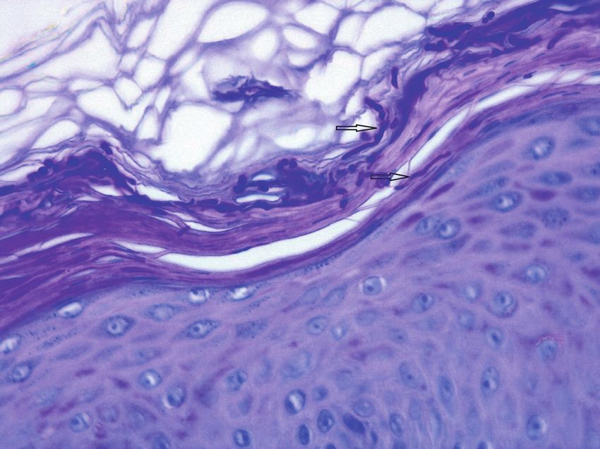
FIGURE 2. Tinea corporis: histopathological features. Acanthosis,
compact hyperkeratosis, orthokeratosis and neutrophils are ob-
served in the epidermis and perivascular lymphocytic inflamma-
tion is observed in the dermis (A) (hematoxylin and eosin staining,
10x). Hyphae (black arrows) are present in the stratum corneum
(B) (periodic acid-Schiff staining, 40x).
by MALDI-TOF MS (MAtrix-assisted laser desorption/
FIGURE 1. Tinea corporis: clinical features. Erythematous plaque
ionization time-of-flight mass spectrometry) with a
with desquamated center, annular and circular rim containing confidence level of 99.9%. Confirmation was done by
vesicles and pustules on the right leg (A, B). Blister with purulent molecular method. Total genomic DNA was extracted
exudate (B). Round and oval plate and skin detachment (A-C).
from purified colonies. The internal transcribed spacer
(ITS) region of ribosomal DNA (rDNA) was amplified
observation of acanthosis, compact hyperkeratosis, fo- using the primer set ITS1 and ITS4. Sequencing was
cal orthokeratosis, few neutrophils and hyphae (positive performed and nucleotide sequences were edited using
reaction with periodic acid-Schiff staining) in the stra- the program Chromas 2 and aligned using the program
tum corneum and a mild chronic superficial perivascular CLUSTAL X2. The obtained sequence was compared
lymphocytic inflammation with few eosinophils in the with sequences deposited in the NCBI and Westerdijk
dermis (Fig. 2). The DIF examination was negative. The Fungal Biodiversity Institute – KNAW databases, result-
bacteriological culture of the purulent exudate did not ing in identification of T. benhamiae (97% homology). The
reveal any pathogens. obtained sequence was deposited in GenBank with the
Skin scales were collected for mycological examination, accession number MT956947 and the isolated strain is
revealing spores in the direct examination with KOH referenced as Tb2020HVFX.
30%. Sample was inoculated onto Sabouraud dextrose Treatment was instituted using oral terbinafine 250
agar (SDA) with chloramphenicol 0.05%, Sabouraud mg/day, four weeks and diflucortolone valerate 0.1% +
broth with chloramphenicol 0.05% and mycosel agar isoconazole nitrate 1% cream twice a day, one week, fol-
and incubated at 27ºC for three weeks. The obtained lowed by omoconazole nitrate 1% cream once a day, two
culture revealed a white cottony colony with yellowish weeks. Complete remission was observed at the end of
reverse with morphological characteristics compatible the treatment period.
with T. benhamiae (Fig. 3). Identification was obtained
A                           B                                          are found and microconidia are rare. The main differ-
                                                                        ential diagnosis is M. canis, which has also yellowish
                                                                        colonies and present thick walled macronidia with 6-12
                                                                        cells and thin septa. The white phenotype shows fast
                                                                        growing, sprayed, flocose colonies with yellow, orange
                                                                        or brown reverse color. Spiral hyphae are present in
                                                                        some cases and many spherical or clavated microconid-
                                                                        ia are observed; macroconidia are fewer, clavated and
 FIGURE 3. T. benhamiae. Macroscopic appearance (mycosel agar):
 yellowish reverse (A). Microscopic feature (toluidine blue staining,   cigar-shaped, with 3-8 cells with smooth and thin walls.
 x10): micronidia are present and no spiral hyphae are observed         The main differential diagnosis is T. mentagrophytes,
 (B).
                                                                        which present several spherical microconidia, spiral hy-
                                                                        phae and clavated, cigar-shaped macroconidia.8-13
DISCUSSION
                                                                        The diagnosis of dermatophytosis is usually clinical.
To the best of the authors’ knowledge, this is the first
                                                                        However, based on clinical presentation there is no
report on dermatophytosis caused by T. benhamiae in
                                                                        differentiation between lesions caused by T. benhamiae
Portugal.
                                                                        and other dermatophytes, particularly the zoophilic one.
T. benhamiae is a zoophilic fungus and is an etiologic                  Therefore, the confirmation of T. benhamiae should be
agent of tinea corporis, faciei, manuum and capitis. This               obtained with the methods mentioned above. Further-
specie causes moderate to severe inflammatory skin re-                  more, complementary diagnostic methods such as his-
action in man, in particular in children, teenagers and im-             tology, cutaneous DIF and bacteriological examination
munocompromised patients. Its natural reservoir is the                  can be extremely important in the exclusion of other
guinea pig, but it is occasionally isolated from rabbits,               infectious and non-infectious dermatoses namely num-
dogs and cats. The infection is usually found in these an-              mular and stasis eczema, impetigo, subacute cutaneous
imals and in persons who adopt them as pets. The first                  lupus erythematosus, Hansen’s disease, pemphigus,
report of ringworm caused by this species was from Ja-                  granuloma annulare, psoriasis and erythema annulare
pan. Subsequently, cases in Northern Europe and South                   centrifugum.14
America have been emerging.1-5
                                                                        Topical antifungals are the first-line treatment for local-
The taxonomic classification of fungi has undergone ma-                 ized and non-complicated tinea corporis and faciei and
jor changes in recent decades. For several years, classi-               should be applied for at least two weeks until complete
fication was based on clinical, morphological and phys-                 clearance of the infection. Cases of extensive tinea cor-
iological features of the isolates. As such, T. benhamiae               poris, tinea manuum, capitis, unguium and barbae require
was initially considered as part of the T. mentagrophytes               topical and systemic treatment. Oral terbinafine, itra-
complex and was classified as A. benhamiae (anamorph).                  conazole and fluconazole are generally effective alter-
However, the development and improvement of mo-                         natives. Adverse effects with different levels of severity
lecular methods based on the sequencing of ribosomal                    may occur. The duration of the treatment depends on
DNA led to a huge revolution in fungal taxonomy, in par-                the location and extent of the infection, varying between
ticular in dermatophytes.2,6,7                                          one and six weeks. Associations of topical antifungals
Identification of T. benhamiae is a complex laboratorial                and steroids are important in cases of moderate or se-
process. As such, the definitive diagnosis must be ob-                  vere inflammation. They should be used with caution
tained by combining several techniques, namely conven-                  and for a short period to avoid striae, skin atrophy and
tional mycological methods (direct examination, culture                 secondary bacterial infections. The remaining treat-
followed by colonies’s macroscopic and microscopic                      ment is carried out with topical antifungal alone. Pref-
observation and enzymatic profile), molecular method-                   erence should be given to low-potency topical steroids,
ologies and technologies based on protein profile (MAL-                 particularly in the face and skin folds. The use of system-
DI-TOF MS).2,5 Conventional methods do not allow, in                    ic steroids occurs in cases of tinea capitis with severe in-
some cases, the differentiation between morphological-                  flammation (Kerion celsi).14
ly similar dermatophytes.                                               In conclusion, the adoption of peculiar species as pets
Two phenotypes have been described for T. benhamiae:                    may have serious public health implications. T. benham-
yellow and white. In the first, the colonies grow slowly,               iae is an example of that, with an increasing recognition
have pleated mycelium and have an orange-yellow re-                     as agent of zoophilic ringworm. As in Northern Europe,
verse. In SDA media, no macroconidia or spiral hyphae                   the prevalence of this infection in Portugal is very likely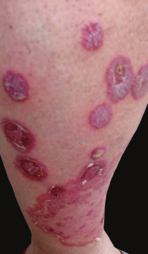
to increase. Therefore, it is essential to consider pets as          5. Nakamura Y, Kano R, Nakamura E, Saito K, Watanabe S,
                                                                        Hasegawa A. Case Report. First report on human ringworm
a potential source of infections and to treat them appro-               caused by Arthroderma benhamiae in Japan transmitted from
priately together with their owners in order to avoid re-               a rabbit. Mycoses. 2002;45:129-131. doi:10.1046/j.1439-
currences, interpersonal and inter-animal transmission.                 0507.2002.00732.x.
                                                                     6. Heidemann S, Monod M, Graser Y. Signature polymorphisms
                                                                        in the internal transcribed spacer region relevant for the dif-
RESPONSABILIDADES ÉTICAS                                                ferentiation of zoophilic and anthropophilic strains of Tricho-
                                                                        phyton interdigitale and other species of T. mentagrophytes
CONFLITOS DE INTERESSE: Os autores declaram a inex-                     sensu lato. Br J Dermatol. 2010;162:282-95. doi:10.1111/
                                                                        j.1365-2133.2009.09494.x.
istência de conflitos de interesse na realização do pre-
                                                                     7. Kawasaki M. Verification of a taxonomy of dermatophytes
sente trabalho.
                                                                        based on mating results and phylogenetic analyses. Med My-
                                                                        col. 2011;52:291-95. doi:10.3314/mmj.52.291.
FONTES DE FINANCIAMENTO: Não existiram fontes ex-
ternas de financiamento para a realização deste artigo.              8. Symoens F, Jousson O, Packeu A, Fratti M, Staib P, Mignon B,
                                                                        et al. The dermatophyte species Arthroderma benhamiae: in-
CONFIDENCIALIDADE DOS DADOS: Os autores declar-                         traspecies variability and mating behaviour. J Med Microbiol.
                                                                        2013;62:377-85. doi:10.1099/jmm.0.053223-0.
am ter seguido os protocolos da sua instituição acerca
                                                                     9. Contet-Audonneau N, Leyer C. Emergence of a dermato-
da publicação dos dados de doentes.
                                                                        phyte contracted from guinea pig and close to Trichophyton
                                                                        mentagrophytes var. erinacei: T. mentagrophytes var. por-
CONSENTIMENTO: Consentimento do doente para
                                                                        cellae. J Med Mycol. 2010;20:321-5. doi:10.1016/j.myc-
publicação obtido.                                                      med.2010.08.001.

PROVENIÊNCIA E REVISÃO POR PARES: Não comissio-                      10. Khettar L, Contet-Audonneau N. Cochon d’Inde et der-
                                                                         matophytose. Ann Dermatol Venereol. 2012;139:631-5.
nado; revisão externa por pares.                                         doi:10.1016/j.annder.2012.05.007.
                                                                     11. Hiruma J, Kano R, Harada K, Monod M, Hiruma M, Hasega-
                                                                         wa A, et al. Occurrence of Arthroderma benhamiae genotype
ETHICAL DISCLOSURES                                                      in Japan. Mycopathologia. 2015;179:219-23. doi:10.1007/
                                                                         s11046-014-9839-0.
CONFLICTS OF INTEREST: The authors have no conflicts
of interest to declare.                                              12. Monod M, Fratti M, Mignon B, Baudraz-Rosselet F. Dermato-
                                                                         phytes transmis par les animaux domestiques. Rev Med Suisse.
FINANCING SUPPORT: This work has not received any                        2014;10:749-53.

contribution, grant or scholarship.                                  13. Nenoff P, Uhrlaß S, Kruger C, Erhard M, Hipler UC, Seyfarth
                                                                         F, et al. Trichophyton species of Arthroderma benhamiae - a
CONFIDENTIALITY OF DATA: The authors declare that                        new infectious agent in dermatology. J Dtsch Dermatol Ges.
                                                                         2014;12:571-81. doi:10.1111/ddg.12390.
they have followed the protocols of their work center on
the publication of data from patients.                               14. Elewski BE, Hughey LC, Sobera JO, Hay R. Fungal diseases
                                                                         (Cap. 77). In: Bolognia JL, Jorizzo JL, Rapini LP, editors. Der-
PATIENT CONSENT: Consent for publication was ob-                         matology. 3rd ed. London: Elsevier; 2012. pag: 1251-84.

tained.
PROVENANCE AND PEER REVIEW: Not commissioned;
externally peer reviewed.

REFERENCES
1. de Hoog GS, Dukik K, Monod M, Packeu A, Stubbe D, Hen-
   drickx M, et al. Toward a novel multilocus phylogenetic taxon-
   omy for the dermatophytes. Mycopathologia. 2017;182:5-31.
   doi:10.1007/s11046-016-0073-9.
2. Sabou M, Denis J, Boulanger N, Forouzanfar F, Glatz I, Lipsker
   D, et al. Molecular Identification of Trichophyton Benhamiae in
   Strasbourg, France: A 9-year Retrospective Study. Med Mycol.
   2018;56:723-34. doi:10.1093/mmy/myx100.
3. Mochizuki T, Kawasaki M, Ishizaki H, Kano R, Hasegawa A,
   Tosaki H, et al. Molecular epidemiology of Arthroderma ben-
   hamiae an emerging pathogen of dermatophytoses in Japan,
   by polymorphisms of the non-transcribed spacer region of the
   ribosomal DNA. J Dermatol Sci. 2001;27:14-20. doi:10.1016/
   S0923-1811(01)00101-3.
4. El-Heis S, Borman AM, Szekely A, Godfrey KM. Tinea Corporis
   Caused by Arthroderma Benhamiae in a Child. Clin Exp Der-
   matol. 2016;41:955-7. doi:10.1111/ced.12966.

You can also read