Liposarcoma: A Rare Case Report with Review of Literature
←
→
Page content transcription
If your browser does not render page correctly, please read the page content below
Case Report                                                                                                              DOI: 10.17354/cr/2015/84

Liposarcoma: A Rare Case Report with Review of
Literature
Sneha Chandra1, M Parvathi Devi2, Apoorva Gupta1, S V Ravindra3
1
  Post-graduate Student, Department of Oral Medicine & Radiology, Teerthanker Mahaveer Dental College, Bagadpur, Moradabad, Uttar Pradesh, India,
2
  Professor and Head, Department of Oral Medicine & Radiology, Teerthanker Mahaveer Dental College, Bagadpur, Moradabad, Uttar Pradesh, India,
3
  Reader, Department of Oral Medicine & Radiology, Teerthanker Mahaveer Dental College, Bagadpur, Moradabad, Uttar Pradesh, India

     Liposarcoma is one of the most common malignant mesenchymal neoplasms, comprising approximately 15% of all soft-tissue sarcomas.
     First described by “Virchow” in 1857, it has been extensively reported in the literature, although its incidence remains exceedingly rare in the
     head and neck region with an annual incidence estimated to be 2.5/1 million inhabitants in population-based studies. It is one of the most
     common malignant mesenchymal neoplasms, comprising approximately 15% of all soft-tissue sarcomas. It is a heterogeneous disease with
     distinct sub-entities presenting with differential clinical behavior. The purpose of this article is to report an additional case of liposarcoma of
     the buccal vestibule and to review the literature.

     Keywords: Head and neck neoplasm, Liposarcoma, Mesenchymal neoplasms, Soft tissue sarcoma

                           INTRODUCTION                                          addressed and that its incidence is quite rare, representing
                                                                                 approximately 4-5% of liposarcomas. Most cases originated
Soft tissue sarcomas (STS) are a group of heterogenous                           in the neck (28%) followed by the head (scalp and face, 26%),
tumors that have their origin primarily in the embryonic                         larynx (20%), pharynx (18%), and mouth (8%).3 Liposarcoma
mesoderm. They can range from relatively slow growth,                            is a malignant mesenchymal neoplasm of the adipose tissue,
causing little destructive growth, to being locally aggressive,                  described first by Rudolf Virchow approximately 140 years
regionally destructive, and having a great potential for                         ago. It exhibits a predilection for the trunk and the lower
systemic metastases (Greene et al. 2002; Pelliteri et al., 2003).                extremities and is rare in the head and neck area. Adults
The approximate incidence for this kind of neoplasia is                          between the ages of 40 and 60 years are the primary targets.
3-4.5/100,000 (Zahm et al. 1997).1 The two most common                           Overall, males are affected slightly more than females.4
sarcomas in adults are malignant fibrous histiocytoma and                        Liposarcoma is a heterogeneous disease with distinct sub-
liposarcoma. Liposarcoma is the second most common                               entities presenting with different clinical behavior. Most
type of soft-tissue sarcoma, accounting for 10-35% of                            of them have been found located in the cheek, but others
these lesions.2 The World Health Organization describes                          have been reported in the floor of the mouth, soft palate,
three forms of liposarcoma that have unique clinical                             mandible, lip, and gingiva.5 Here we are reporting a case of
settings and behaviors: Atypical lipomatous tumor/well-                          liposarcoma arising in the right anterior to posterior region
differentiated liposarcoma (ALT/WDL), myxoid/round                               of the mandible in a 45-year-old female patient.
cell liposarcoma, and pleomorphic liposarcoma. ALT/WDL
comprises 40‑45% of all liposarcomas, whereas myxoid                                                       CASE REPORT
and pleomorphic liposarcomas represent 35-40% and 5%,
respectively. In their 1995 literature review, Golledge et al.                   A 45-year-old-woman visited to the Department of Oral
found that liposarcoma of the head and neck was poorly                           Medicine and Radiology, Teerthanker Mahaveer Dental
                                                                                 College and Research Centre with a chief complaint of
                          Access this article online                             swelling in the mandibular region since 2 years. She gave
                                                                                 the history of extraction 2 years ago due to a decayed
                                Month of Submission       :   03-2015            tooth in that region. The swelling didn’t subside even
                                Month of Peer Review      :   04-2015            after extraction of the tooth. The swelling gradually
                                Month of Acceptance       :   04-2015            increased in size to attain the present size. Now, she
                                Month of Publishing       :   05-2015
                                                                                 complains pain, and difficulty in eating and speaking
         www.ijsscr.com
                                                                                 since 6 months. Medical and family histories of the patient

    Corresponding Author:
    Dr. Apoorva Gupta, Department of Oral Medicine & Radiology, Teerthanker Mahaveer Dental College, Bagadpur, Delhi Road, Moradabad,
    Uttar Pradesh, India. E-mail: gupta_apoorva@live.com

    22                                                                                    IJSS Case Reports & Reviews | May 2015 | Vol 1 | Issue 12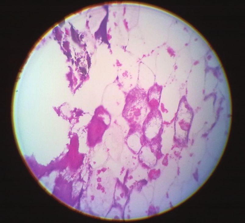
Chandra, et al.: Liposarcoma: A Rare Case Report
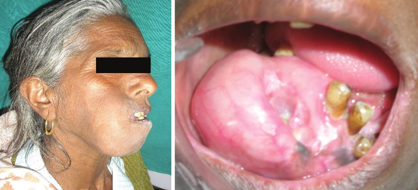
were non-contributory. On clinical examination, a 4 cm × liposarcoma. Patient was referred to the oncology center for
5 cm, non-tender, lobulated swelling was found in the the management of the tumor. Unfortunately, the patient
right half of the mandible. The swelling extends medially lost for the follow-up.
from the symphysis region, distally to 1 cm anterior to the
ramus of the mandible, superiorly from the nasolabial DISCUSSION
fold to lower border of the mandible crossing the midline.
(Figure 1a and b). Extensive induration around the lesion Liposarcoma is a soft tissue neoplasm with a rare entity.
was also noted which was soft in consistency. Lingually The guidelines for grading STS are mentioned in Table 1.5
displaced lower anterior and premolars were present Because of the rarity of STS most studies deal with the
clinically. Routine investigations, including complete entire group of STSs. The causes of STS are only poorly
blood counts and biochemical profile were within normal understood. The pathogenesis is based on various inherited
limits. Orthopantomogram shows involvement of the and environmental factors and also, on pre-existing
mandible on the right side with a diffuse radiolucency with conditions. Among environmental factors exposure to
radiopacity extending 36-48 region with an expansion of the ionizing irradiation including thorotrast, alkylating agents,
alveolar bone in the right posterior region of the mandible arsenical pesticides and medications, vinyl chloride,
(Figure 2). Computerized tomography scan reveals well immunosuppressive drugs, human immunodeficiency
defined irregular expansile osteolytic lesion involving right virus, herpes virus Type 8 and anabolic steroids are
ramus of mandible with internal residual bony separation described as risk factors. Inherited conditions associated
within causing an overlying superficial bulge. The lesion is with STSs are Li-Fraumeni syndrome, neurofibromatosis
causing cortical thinning with breach of buccal cortices of Type 1, Gardner’s syndrome, retinoblastoma, Werner’s
the mandible. There is resorption of the tooth at the site of syndrome, and nevoid basal cell carcinoma syndrome
the lesion with displaced anterior teeth (Figure 3a and b). (Gorlin’s syndrome). Pre-existing medical conditions
An incisional biopsy of the lesion was performed. Under ×40
magnification, the histopathological slide revealed mature
fat cells with relatively few, widely scattered lipoblasts
(Figure 4). All the clinical, radiographic, and histopathologic
features were suggestive of the well-differentiated type of
a b
Figure 3: (a) Three-dimensional (3D) computerized tomography (CT) head and
a b neck (coronal section), (b) 3D CT Head and neck (axial section)
Figure 1: (a) Extraoral swelling, (b) intraoral displacement of tooth visible on
the right side of face
Figure 2: Orthopantomograph revealing diffuse radiopacity and radiolucency Figure 4: Photomicrograph of liposarcoma showing diffuse lipoblasts
IJSS Case Reports & Reviews | May 2015 | Vol 1 | Issue 12 23Chandra, et al.: Liposarcoma: A Rare Case Report
such as long-standing lymphedema (Stewart-Treves clinical settings and behavior: ALT/WDL, myxoid/round
syndrome) can also cause STS.6 Several cases with trauma cell liposarcoma, and pleomorphic liposarcoma. ALT/WDL
preceding the development of STS have been reported in comprises 40-45% of all liposarcomas, whereas myxoid
the literature, but there is no proof of a causal relationship. and pleomorphic liposarcomas represent 35-40% and 5%,
Liposarcomas may occur wherever adipose tissue is respectively.9 Liposarcoma can easily be misdiagnosed
present, although the cause is not clear. The head and clinically, and it’s relatively indolent course often results in
neck region is an unusual site, and intraoral liposarcomas a misdiagnosis of a cyst or benign soft tissue neoplasm, it is
are even rarer with male predilection.7 No association frequently mistaken for lipoma.6 Therefore, histopathology
with race or geography is known. The most common is required for an appropriate diagnosis. An overview of
affected sites are the submandibular area, cheek, tongue, LPS subtypes are given in Table 2.9
the floor of the mouth, and soft palate. The present case 1. Myxoid: In this variant of liposarcoma, histopathologically
has the affected site at the right mandibular alveolus in a proliferating lipoblasts are seen in varying stages of
female patient, which is a rare finding. Most cases present differentiation. The “signet ring cells” are characteristic
as a slowly growing, painless, nonulcerated submucosal of a myxoid type of liposarcoma. A delicate plexiform
mass. However, some lesions grow rapidly and become capillary pattern, and a myxoid matrix with
ulcerated early. The clinical impression seems to be lipoma hyaluronidase-sensitive mucopolysaccharides
or fibroma in the majority of the cases. Tumor size ranged 2. Well-differentiated: It simulates lipoma with scattering
from 0.6 to 8.0 cm. Liposarcoma is most likely to arise from of lipoblasts with one or more of the lipid droplets
deep-seated, well-vascularized structures rather than from in the cytoplasm. The irregularly shaped cells with
submucocutaneous fat.8 The World Health Organization hyperchromatic nuclei and increased variation in
describes three forms of liposarcoma that have unique lipocyte size and shape can be appreciated. Four
subdivisions are recognized, namely Lipoma like,
Table 1: Grading of soft tissue neoplasms5 sclerosing, inflammatory, dedifferentiated
Tumors that are definitionally high grade 3. Round cell: This is closely related to myxoid but
Ewing sarcoma/MPNET characterized by proliferation of small uniformly shaped
Rhabdomyosarcoma (except the spindle cell variant) rounded cells with vesicular nuclei; myxoid and vascular
Angiosarcoma
Pleomorphic liposarcoma components are obscured by the vast number of tumor
Soft tissue osteosarcoma cells
Mesenchymal chondrosarcoma 4. Pleomorphic: This represents a disordered growth
Desmoplastic small cell tumor
Extrarenal rhabdoid tumor pattern and cellular pleomorphism with prominent
Tumors that are definitionally low grade mitotic activity. Two subtypes are recognized: Type I
WDL/ALT with large acidophilic giant cell and Type II with lipid
Dermatofibrosarcoma protuberans
droplet giant cells.
(except the fibrosarcomatous variant)
Infantile fibrosarcoma
Angiomatoid MFH The anatomic distribution of head and neck liposarcoma
Tumors that are not readily gradable but often metastasize within10 to is schematically represented in Figure 5.10 In the pre-
20 years of follow‑up
Alveolar soft part sarcoma operative diagnosis of soft tissue tumors, different biopsy
Clear cell sarcoma techniques are used depending on the site, size, and
Epithelioid sarcoma depth of the lesion. Core needle biopsy or longitudinally
Synovial sarcoma
“Low‑grade” fibromyxoid sarcoma oriented incisional biopsy can be used for extremity
Tumors of varying behavior for which grading may be prognostically masses. The advantage of using fine needle aspiration
useful cytology (FNAC) is the small, risk of seeding sarcoma
Myxoid liposarcoma
cells to the surrounding tissue and allowing a reduced
Leiomyosarcoma
Malignant peripheral nerve sheath tumor surgical margin, leading to better preservation of function.
Myxofibrosarcoma (myxoid MFH) Fine needle aspiration is also useful for diagnosing intra-
Fibrosarcoma abdominal, retroperitoneal and mediastinal masses. The
Tumors of varying behavior for which grading parameters are notyet
established disadvantage of using FNAC is that the cellular samples
Hemangiopericytoma are small, and it can be difficult to ascertain the tumor
Synovial sarcoma grade and histological type. A 90% diagnostic accuracy of
Myxoid chondrosarcoma
Malignant giant cell tumor differentiating a benign lesion from a liposarcoma has been
Malignant granular cell tumor reported with fine needle aspiration cytology. Reverse
Malignant mesenchymoma transcription-polymerase chain reaction is used to detect
MFH: Malignant fibrous histiocytoma, MPNET: Malignant peripheral
neuroectodermal tumor, ALT/WDL: Atypical lipomatous tumor/well‑differentiated
the fusion gene transcripts (FUS-DDIT3), pathognomonic
liposarcoma for Musladin-Leuke syndrome.
24 IJSS Case Reports & Reviews | May 2015 | Vol 1 | Issue 12Chandra, et al.: Liposarcoma: A Rare Case Report
Table 2: An overview of LPS subtypes9
Subtype Pathology Molecular characteristics/ MRI/CT appearance
“actionable” targets
ALT/WDLPS Low grade, positive IHC for MDM2 and CDK4 amplifications Large encapsulated lipomatous mass (high signal
MDM2, CDK4, p16 intensity both in T1‑weighted and T2‑weighted
DDLPS High grade, positive IHC for MDM2 and CDK4 amplifications MRI) with thick internal septations; signal loss
MDM2, CDK4, p16 on fat‑saturated T1‑weighted images, and focal
nodules (>1 cm is suggestive of a DDLPS)
MLPS and Low grade (percentage of round FUS‑CHOP fusion gene, PI3K Pathognomonically, low signal intensity in T1‑weighted
RCLPS cells important for grading) mutations (w20%) and marked signal intensity in T2‑weighted MRI
PLPS High grade, pleomorphic, Complex structural Nonspecific soft tissue mass, often including areas of
cellular sarcoma rearrangements necrosis and hemorrhage
IHC: Immunohistochemistry, ALT/WDLPS: Atypical lipomatous tumor/well‑differentiated liposarcoma, DDLPS: Dedifferentiated liposarcoma, MLPS: Myxoid liposarcoma,
RCLPS: Round cell liposarcoma, MRI: Magnetic resonance imaging, PLPS: Pleomorphic liposarcoma, CT: Computerized tomography
to tumor size at the time of radiation and radiation dose.
Chemotherapy is used partly in a palliative setting and
partly as adjuvant therapy for high-grade STSs in different
protocols. Doxorubicin and ifosfamide are the most
commonly used chemotherapeutics in the treatment of STSs.
Different adverse prognostic factors for local recurrence
and tumor related death have been identified for the
whole group of STS and specifically for liposarcoma. High
malignancy grade and positive microscopic margins have
been identified as obvious adverse prognostic factors for
recurrence of liposarcoma. Tumor related factors, such as
site, size and depth, presence of tumor necrosis, histological
subtype and recurrence, as well as treatment-related
Figure 5: Anatomic distribution of head and neck liposarcoma10
factors, such as non-wide resection, have been identified
as unfavourable prognostic factors for tumor related death.
Prognostic factors such as vascular invasion and infiltrative
Surgery is the most important treatment modality. Wide
growth pattern recognized as risk factors for metastases in
local excision with clear margins is important for local
STSs. They were not found to be independent predictors for
tumor control. The Scandinavian Sarcoma Group follow
clinical outcome in a study on liposarcomas.6
the definition of surgical margins introduced by Enneking
in 1980. Since 2006, there has been a modification in the
classifications of margins that are widely followed. They are:
CONCLUSION
1. “Positive margin” means that a gross tumor or
In summary, the rarity of liposarcoma makes it difficult to
microscopic tumor is left at the margin, which is
diagnose the disease based on the clinical characteristics,
reported.
and differential diagnosis of this tumor is difficult as WDLs
2. “Negative margin” means that there is no microscopic
are usually present in the retroperitoneum. Therefore,
tumor at the margin. The extent of the margin is
differentiations are important because there is little to
reported. More than 20 mm of normal tissue around
no malignant potential in lipomatosis tumors. A well-
the tumor, or fascia completely surrounding the tumor
is classified as “wide margin.” functioning collaboration between the radiologist and
cytopathologist is essential to diagnose STSs. Surgery is the
Radiotherapy in STS is an established method for main line of treatment, and close follow-up after surgery is
elimination of the microscopic tumor. mandatory due to the high rate of recurrence.
In 1998 Yang et al. in a prospective randomized trial, REFERENCES
assessed that adjuvant post-operative radiation therapy
1. Gonzalez RG, Molina RB, Maldonado OT, Burciaga RG,
in combination with conservative surgery improves local
Gastelúm MG. In: Derbel F, editor. Head and Neck Soft Tissue
control for STSs of both low and high grade in patients with Sarcoma, Soft Tissue Tumors. Rijeka: InTech; 2011.
microscopic negative, marginal or minimal microscopic 2. Davis EC, Ballo MT, Luna MA, Kumar S, Patel R, Roberts DB, et al.
positive surgical margins. Local tumor control was related Liposarcoma of the head and neck: The University of Texas M. D.
IJSS Case Reports & Reviews | May 2015 | Vol 1 | Issue 12 25Chandra, et al.: Liposarcoma: A Rare Case Report
Anderson Cancer Center experience. Head Neck 2009;31:28-36. 8. Shafer WG, Hine MK, Levy BM. Shafer’s Textbook of Oral
3. McElderry J, McKenney JK, Stack BC. High-grade liposarcoma Pathology. 7th ed. Philadelphia: Elsevier; 2012. p. 371-4.
metastatic to the gingival mucosa: C ase report and literature 9. Henze J, Bauer S. Liposarcomas. Hematol Oncol Clin North Am
review. Am J Otolaryngol 2008;29:130-4. 2013;27:939-55.
4. Gagari E, Kabani S, Gallagher GT. Intraoral liposarcoma: Case 10. McCulloch TM, Makielski KH, McNutt MA. Head and neck
report and review of the literature. Oral Surg Oral Med Oral Pathol liposarcoma. A histopathologic reevaluation of reported cases.
Oral Radiol Endod 2000;89:66-72. Arch Otolaryngol Head Neck Surg 1992;118:1045-9.
5. Nakahara H, Shirasuna K, Terada K. Liposarcoma of the floor of
the mouth: A case report. J Oral Maxillofac Surg 1994;52:1322-4.
6. Engström K, Bergh P, Gustafson P, Hultborn R, Johansson H, How to cite this article: Chandra S, Devi MP, Gupta A, Ravindra SV.
Liposarcoma: A Rare Case Report with Review of Literature. IJSS Case
Löfvenberg R, Bauer HC. Liposarcoma. Cancer. 12008;13(7), 1649-16. Reports & Reviews 2015;1(12):22-26.
7. Krishna MG, Acharatlal BH, Hirjibhai MP, Gopal U, Wadhwa MK.
Liposarcoma of the floor of the mouth: A case report. Turkish J
Source of Support: Nil, Conflict of Interest: None declared.
Cancer 2002;32:69-74.
26 IJSS Case Reports & Reviews | May 2015 | Vol 1 | Issue 12You can also read