Case Report Serous Borderline Tumor in Transgender Female-to-Male Individuals: A Case Report of Androgen Receptor-Positive Ovarian Cancer
←
→
Page content transcription
If your browser does not render page correctly, please read the page content below
Hindawi
Case Reports in Radiology
Volume 2021, Article ID 8861692, 6 pages
https://doi.org/10.1155/2021/8861692

Case Report
Serous Borderline Tumor in Transgender Female-to-Male
Individuals: A Case Report of Androgen Receptor-Positive
Ovarian Cancer

          Cristina Ferreira ,1 João Fraga,2 Célia Antunes,1 Manuela Gonçalo,1 and Paulo Donato1
          1
              Department of Radiology, University Centre Hospitals of Coimbra (CHUC), 3000-075 Coimbra, Portugal
          2
              Department of Pathology, University Centre Hospitals of Coimbra (CHUC), 3000-075 Coimbra, Portugal

          Correspondence should be addressed to Cristina Ferreira; cris.rcf@gmail.com

          Received 21 May 2020; Accepted 10 May 2021; Published 7 June 2021

          Academic Editor: Daniel P. Link

          Copyright © 2021 Cristina Ferreira et al. This is an open access article distributed under the Creative Commons Attribution
          License, which permits unrestricted use, distribution, and reproduction in any medium, provided the original work is
          properly cited.

          Ovarian cancer is the most fatal gynecologic malignancy. The incidence of ovarian cancer among female-to-male transsexuals
          receiving treatment with testosterone is unknown, and few cases have been reported in the literature. We report a recent case in
          our institution, a 23-year-old female-to-male transsexual patient who received testosterone supplementation. The patient
          underwent a pelvic magnetic resonance imaging to study an ovarian complex cyst that revealed the presence of a bilateral
          ovarian tumor with imaging features of borderline serous tumor. These masses were surgically removed and the pathology
          report confirmed the diagnosis associated with noninvasive peritoneal implants and the presence of numerous androgen
          receptors in the tumor cells. Although there is still insufficient data to validate a direct correlation between hormonotherapy and
          ovarian cancer in these patients, this case may reinforce previous reports on this association and highlights the relevance of
          radiological follow-up and bilateral salpingo-oophorectomy as part of gender reassignment surgery.

1. Introduction                                                            SBOT is an intermediate grade of neoplasm between
                                                                       benign and malignant serous ovarian tumors [2]. Patients
It has been established that the acquisition of secondary              frequently refer abdominal distension or pain and serum
sex characteristics of the other gender, based on sex ste-             CA-125 increases in half of patients [3, 4]. The more estab-
roids treatment, is fundamental for sex reassignment in                lished treatment is bilateral salpingo-oophorectomy, hyster-
transsexuals. However, an unresolved question is whether,              ectomy, and omentectomy [5, 6]. Compared to malignant
in the long term, the administration of cross-sex hormones             tumors, the prognosis of SBOT is far better, as the disease is
is safe [1].                                                           usually confined to the ovary, having a >95% 10-year survival
    Furthermore, it is believed to exist an underreporting             rate [7]. Nevertheless, 30% of SBOT are associated with peri-
of complications of cross-sex hormone therapy. This may                toneal implants that can be noninvasive or invasive [8]. In the
occur because although the initial treatment is mainly                 last case, survival rates of these patients fall considerably.
administrated in specialized centers, complications on                 Adjuvant chemotherapy can be offered to them, but there is
the long term are often seen in general practice, and they             no firm evidence of any survival benefits [9].
are then only occasionally reported in the scientific liter-                Currently, there is not enough evidence to support any
ature [1].                                                             recommendation to continue or stop the hormonal treat-
    We present a case of a female-to-male (FTM) transgen-              ment. However, its duration probably increases the risk of
der patient who developed serous borderline ovarian tumor              development of hormone-related malignancies, especially in
(SBOT) while taking testosterone injections.                           the case of long-term exposure.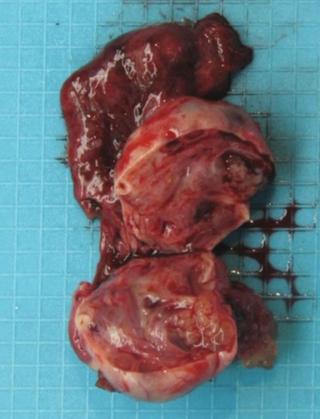
2                                                                                                                Case Reports in Radiology

                                            (a)                                                 (b)

                                            (c)                                                 (d)

Figure 1: Pelvic MRI. T2-weighted axial image (a). Dynamic study (b). T2-weighted coronal image (c, d). There is a complex cystic lesion of
the left ovary, with some solid papillary projections, which avidly enhance after contrast. Involving both ovaries there is another mixed mass,
with the same characteristics of the previously described but arising from the right ovary. Both ovaries have multiple follicles, the right with
polycystic appearance. Pedunculated leiomyoma (∗ ) of the right broad ligament. Small volume of peritoneal effusion in pelvic situation (1.5 T
MRI machine).

   Despite this, transsexuals are often reluctant in stopping             to our department of radiology in order to perform a pelvic
hormone administration for fear that the secondary sex                    magnetic resonance imaging (MRI) study.
characteristics of the acquired sex will diminish [1].                         MRI images were acquired using a 1.5 T MRI machine.
                                                                          Imaging parameters are provided in the figure legends
2. Case Report                                                            (shown in Figures 1 and 2). The exam revealed a complex
                                                                          cystic lesion arising from the left ovarian and measuring
We report the case of a 23-year-old FTM transgender who                   4:0 × 4:3 cm that displayed some solid intermediate to
had been taking testosterone since 2016 in order to gender                hypointense papillary projections, which avidly enhanced
reassignment. This patient suffered from asthma, treated                   after contrast, but without diffusion restriction. There
with budesonide/formoterol fumarate dihydrate and cetiri-                 was another mixed mass, involving both ovaries, with the
zine, without any other personal or family medical history.               same characteristics of the previously described, but aris-
Moreover, this patient had never been pregnant and had                    ing from the right ovary, being predominantly exophytic
not proceeded with any surgery.                                           and measuring 10 × 5:8 cm. Both ovaries had also multiple
    On the ultrasound routine exam dated of June 2019, a                  follicles, the right with polycystic appearance. The diagno-
complex cyst lesion was detected in the left ovary, with intra-           sis of bilateral serous borderline tumor has been suggested
cystic solid vegetation, measuring 3.0 cm. The patient was                in the radiology report—papillary cystic variant in the left
asymptomatic and did not stop the supplementation. Pelvic                 ovary and papillary superficial variant in the right ovary.
clinical examination was unremarkable. Blood tests showed                 The uterus was anteverted, with normal size and regular
high serum levels of CA-125 of 133 U/ml (institutional upper              contours. A hypointense nodular lesion was identified at
limit of normal, 27 U/ml). Thereby, the patient was presented             T1 and T2 sequences, next to the right uterine horn,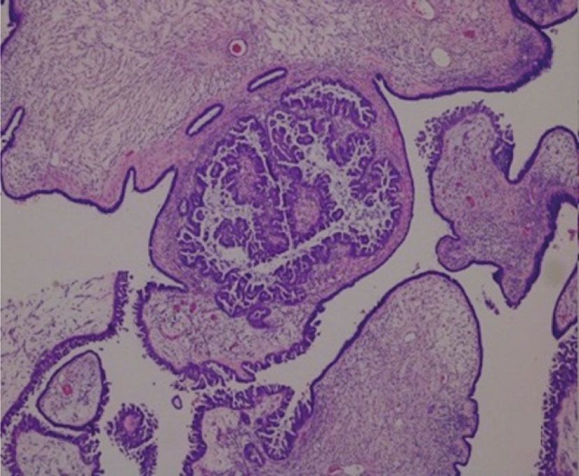
Case Reports in Radiology 3
(a) (b)
Figure 2: Axial diffusion-weighted (b1000) image (a) and diffusion coefficient map (b) show that papillary projections are nonrestricted on
diffusion-weighted imaging.
(a) (b)
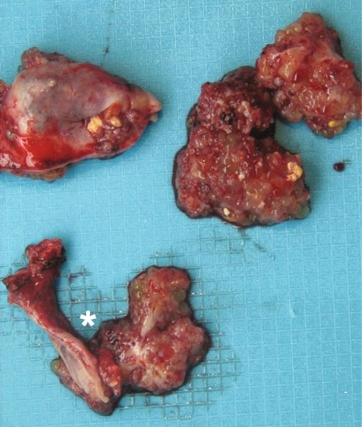
Figure 3: Macroscopic examination. Left ovary 6:5 × 3:5 × 1:5 cm, on the surface with friable nodular pink lesion, 4 × 5 × 1:4 cm. After
opening, the ovary is replaced by a cystic formation, with translucent and filant content, with abundant lobulated nodules adhering to
the inner surface, the largest with 1.5 cm in diameter, equally pink and friable. The cystic wall is thin and elastic (a). The contralateral
annex (∗ ) shows a 6 cm horn and a 3 × 2:5 × 2 cm ovary, with a mostly pink and smooth surface, with an exophytic and lobulated
lesion, with 2 × 1:5 cm. It is also identified a cystic formation with 1 cm, with a vegetation with 0.9 cm, which encompasses most of
the cystic lesion (b).
measuring 1.2 cm, suggesting pedunculated leiomyoma of that occupied the entire sac fundus of Douglas and was
the right broad ligament. The endometrium was fine and adherent to the anterior face to the bladder peritoneum.
regular. There was a small volume of peritoneal effusion The right ovary had normal volume, presenting numerous
in pelvic situation, and there was no evidence of suspected lesions on its surface. Between the two ovaries, the lesion
pelvic lymph nodes (shown in Figures 1 and 2). measured about 4.5 cm (shown in Figure 3). There was evi-
In view of the imaging findings, the patient underwent dence of another adhesion between sigmoid colon and the
surgery for resection of this mass, in October 2019. At the tumor. Isolated nodules were also noted on the bladder fold
time of the laparotomy, left ovary was enlarged due to a com- and on serosal surface of the sigmoid colon, suggesting
plex cystic formation, measuring about 6:5 × 3:5 × 1:5 cm, implants. Peritoneal serous effusion was also seen. Thus, a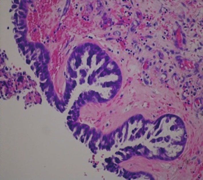
4                                                                                                              Case Reports in Radiology

                                             (a)                                                  (b)

Figure 4: Pathological examination. Neoplasm with exophytic growth on the ovarian surface and, internally, in the macroscopically observed
cystic areas. The lesion is mainly composed of papillae with large fibrovascular axes, with progressive and hierarchical branching, often with
edema, hyaline sclerosis, and stromal calcifications. Tumor cells have eosinophilic cytoplasm and oval, hyperchromatic nuclei with mild
atypia, with few mitotic figures being identified. There is no destructive invasion of stroma (H&E, 40x (a) and 100x (b)).

                                       (a)                                                        (b)

Figure 5: Autoimplants of the left ovary surface with dimensions ranging from 4 to 11 mm of larger axis, morphologically nodular, and
consisting of abundant desmoplastic stroma, which involves small aggregates of neoplastic cells (H&E, 40x (a) and 100x (b)).

full surgical staging procedure included total hysterectomy,             then, few cases of hormone-dependent tumors have been
bilateral salpingo-oophorectomy, and multiple biopsies of                reported in hormonally treated FTM transsexuals [1]. Only
the peritoneum. There were no other visible lesions in the               4 cases of ovarian cancer in FTM transsexuals were reported
remaining abdominal and pelvic cavities.                                 in the scientific literature [10].
     The pathology report confirmed a bilateral borderline                    This case report describes a new case of a FTM transsex-
ovarian serous tumor with noninvasive desmoplastics                      ual patient who was under testosterone supplementation and
implants (shown in Figures 4 and 5), and the immunohisto-                who developed an ovarian cancer, namely, a serous border-
chemical study revealed neoplastic cell positivity for WT1,              line ovarian tumor.
RE (80%), RP (80%), p53 wild-type labeling (faint 60%),                      Furthermore, this case report strengthens the role of
p16 (70%), and Ki-67 30% and AR (androgen receptors)                     radiology in the management of these patients. Indeed, ultra-
diffuse positivity (shown in Figure 6).                                   sound exam made it possible to detect the ovarian lesion.
     After that, the patient was referred to the oncology                Additionally, MRI study helped the recognition of this
department, for further management, and the hormone                      ovarian tumor and its characterization, suggesting a serous
therapy was temporarily suspended until revaluation.                     borderline tumor, as well as the papillary cystic and papillary
                                                                         superficial subtypes. In fact, an abundance of intermediate to
3. Discussion                                                            hyperintense papillary projections (edematous papillae) with
                                                                         hypointense internal branching (fibrous internal architecture
Cross-sex hormone administration is relatively recent in                 of the papillary projections) and ovarian stroma preservation
medicine. Actually, its history started in the 1970s, and since          with a hypointense ovarian capsular margin, on T2-weighted
Case Reports in Radiology                                                                                                                 5

                      (a)                                           (b)                                            (c)

                      (d)                                           (e)                                            (f)

                                                                    (g)

Figure 6: Immunohistochemistry study. Neoplastic cell positivity for WT1 (a), RE (80%) (b), RP (80%) (c), p53 wild-type labeling (faint 60%)
(d), p16 (70%) (e), Ki-67 30% (f), and AR showing diffuse positivity (g) (100x).

imaging, are features strongly suggestive of this tumor [11].                 The final diagnosis was confirmed by the anatomopatho-
When the high signal papillary projections are on the sur-                logical examination. In immunohistochemistry, WT1 is a
face of the ovary without any cystic component, it suggests               marker of tissues derived from the middle mesoderm inner
the diagnosis of the papillary superficial subtype. In the                 layer, combining positivity for estrogen receptors (ER) and
papillary cystic subtype, the papillary projections also occur            progesterone receptors (RP), proving to be a neoplasm
within the cystic component [11]. Papillary projections are               derived from the adnexal structures, so excluding the diag-
inconspicuous on T1WI. This tumor may restrict on                         nosis of endometrioid carcinoma. High-grade serous carci-
diffusion-weighted imaging, but, due to its variability, this              noma typically shows a p53 mutation with positivity in
feature is not mandatory to stablish the diagnosis. Never-                100% of cells, as well as positivity to p16 and cell prolifer-
theless, the enhancement after contrast is always present                 ation index (ki-67) around 100%, which was not observed
[11]. Moreover, the distinction between SBOT from benign                  in this clinical case report, thus reinforcing the diagnosis.
and malignant tumors, in MRI exam, can be helped by the                   Low-grade serous carcinoma can be immunohistochemi-
evaluation of the abundance and size of the papillary pro-                cally identical to the borderline serous tumor, but needs
jections, with a direct correlation between the number                    to have invasion criterion, namely, invasive implants.
and the dimension of these projections and the degree of                  Finally, the tumor reported in this case showed abundant
malignant suspicion [11].                                                 expression of androgen receptors, which also reinforces
6                                                                                                         Case Reports in Radiology

the diagnosis. It should be highlighted that androgen recep-       Conflicts of Interest
tors are present in almost all epithelial ovarian cancers.
Their function has been proposed to be associated with a           The authors declare that they have no conflicts of interest.
growth stimulatory effect of testosterone and a possibly
larger role in tumor progression.
    From the above mentioned, we may consider a possible           References
relationship between the testosterone supplementation and
                                                                    [1] A. Mueller and L. Gooren, “Hormone-related tumors in trans-
the development of the ovarian tumor in this patient, like
                                                                        sexuals receiving treatment with cross-sex hormones,” Euro-
few cases previously reported [12].                                     pean Journal of Endocrinology, vol. 159, no. 3, pp. 197–202,
    Studies have stated that short- and medium-term treat-              2008.
ment of testosterone in FTM transsexuals is safe, but long-         [2] G. Acs, “Serous and mucinous borderline (low malignant
term safety has been difficult to guarantee [13].                         potential) tumors of the ovary,” American Journal of Clinical
    Despite, in the current literature, there are no established        Pathology, vol. 123, article S1e45, 2005.
special guidelines regarding the follow-up imaging recom-           [3] M. Morotti, M. V. Menada, D. J. Gillott, P. L. Venturini, and
mendations for these patients neither defined dates for                  S. Ferrero, “The preoperative diagnosis of borderline ovarian
surgical procedures. These patients are, actually, advised to           tumors: a review of current literature,” Archives of Gynecology
adhere to the routine cancer screening protocols as in non-             and Obstetrics, vol. 285, no. 4, pp. 1103–1112, 2012.
transgender individuals, depending on the anatomic situa-           [4] P. P. Koonings, K. Campbell, D. R. J. Mishell, and D. A.
tion, according to birth-assigned sex [13].                             Grimes, “Relative frequency of primary ovarian neoplasms: a
    Regarding to the surgical procedures to prevent gyneco-             10-year review,” Obstetrics and Gynecology, vol. 74, article
logical cancer, it remains a controversial issue, because the           921e6, 1989.
oncological risk in FTM transsexuals, after the initiation of       [5] V. W. Chen, B. Ruiz, J. L. Killeen et al., “Pathology and classi-
testosterone therapy, is still inconclusive and lacks power             fication of ovarian tumors,” Cancer, vol. 97, no. S10, pp. 2631–
[13]. Some authors recommended salpingo-oophorectomy                    2642, 2003.
as the best treatment in FTM transsexuals, when they are fit         [6] I. Cadron, K. Leunen, T. van Gorp, F. Amant, P. Neven, and
for surgical sex reassignment, usually taking place 18–24               I. Vergote, “Management of borderline ovarian neoplasms,”
months after starting the testosterone administration [1].              Journal of Clinical Oncology, vol. 25, no. 20, pp. 2928–2937,
Other authors do not support this recommendation, claim-                2007.
ing the insufficient evidence linking exogenous androgens             [7] M. E. Sherman, P. J. Mink, R. Curtis et al., “Survival among
and ovarian malignancy [12].                                            women with borderline ovarian tumors and ovarian carci-
    On the other hand, there is also controversy on maintain-           noma: a population-based analysis,” Cancer, vol. 100, no. 5,
ing hormonal treatment in these patients. Hage et al. sug-              pp. 1045–1052, 2004.
gested that hormonal therapy should be stopped after the            [8] J. D. Seidman and R. J. Kurman, “Ovarian serous borderline
diagnosis of an androgen receptor-positive ovarian cancer               tumors: a critical review of the literature with emphasis on
[13]. However, Defreyne et al. argue that testosterone therapy          prognostic indicators,” Human Pathology, vol. 31, no. 5,
                                                                        pp. 539–557, 2000.
should be uninterrupted lifelong after oophorectomy in
order to maintain the secondary sex characteristics and to          [9] D. Fischerova, M. Zikan, P. Dundr, and D. Cibula, “Diagnosis,
avoid hypogonadism symptoms [13].                                       treatment, and follow-up of borderline ovarian tumors,” The
                                                                        Oncologist, vol. 17, no. 12, pp. 1515–1533, 2012.
    Lastly, it is important to remember that transgender indi-
viduals commonly report avoidance of medical care for being        [10] R. N. Gorton and L. Erickson-Schroth, “Hormonal and surgi-
                                                                        cal treatment options for transgender men (female-to-male),”
afraid of discrimination [14]. On the other hand, there is a
                                                                        Psychiatric Clinics of North America, vol. 40, no. 1, pp. 79–
lack of well-informed and culturally competent health pro-              97, 2017.
fessionals in relation to these issues [15].
                                                                   [11] J. Naqvi, E. Nagaraju, and S. Ahmad, “MRI appearances of
    Also, taking into account all these controversies, by
                                                                        pure epithelial papillary serous borderline ovarian tumours,”
reporting this case, we reinforce the importance of the role            Clinical Radiology, vol. 70, no. 4, pp. 424–432, 2015.
of imaging during the follow-up of these patients, during
                                                                   [12] S. Don, “Ovarian Cancer Associated with Testosterone Sup-
hormonal therapy or while waiting for surgery, allowing the             plementation in a Female-to-Male Transsexual Patient,” Gyne-
early detection and diagnosis of tumors, whose greater risk             cologic and Obstetric Investigation, vol. 62, no. 4, pp. 226–228,
cannot be excluded.                                                     2006.
                                                                   [13] J. Defreyne and G. T’Sjoen, “Transmasculine hormone ther-
                                                                        apy,” Endocrinology and Metabolism Clinics of North America,
4. Conclusion                                                           vol. 48, no. 2, pp. 357–375, 2019.
                                                                   [14] A. S. Cheung, K. Wynne, J. Erasmus, S. Murray, and J. D.
This case report emphasizes that, despite the few described             Zajac, “Position statement on the hormonal management of
cases of ovarian tumors in transgender patients under testos-           adult transgender and gender diverse individuals,” MJA,
terone therapy, we cannot underestimate its occurrence.                 vol. 211, no. 3, pp. 127–133, 2019.
Actually, this case was only detected by imaging studies,          [15] J. Lapinski, T. Covas, J. M. Perkins et al., “Best Practices in
namely, with MRI, thus, reinforcing the importance of the               Transgender Health: A Clinician's Guide,” Primary Care;
role of imaging in monitoring these patients.                           Clinics in Office Practice, vol. 45, no. 4, pp. 687–703, 2018.
You can also read