Activating HRAS Mutation in Agminated Spitz Nevi Arising in a Nevus Spilus
←
→
Page content transcription
If your browser does not render page correctly, please read the page content below
Research

                 Case Report/Case Series

                 Activating HRAS Mutation in Agminated Spitz Nevi
                 Arising in a Nevus Spilus
                 Kavita Y. Sarin, MD, PhD; Bryan K. Sun, MD, PhD; Charles D. Bangs; Athena Cherry, MD; Susan M. Swetter, MD;
                 Jinah Kim, MD, PhD; Paul A. Khavari, MD, PhD

                                                                                                                                    Supplemental content at
                    IMPORTANCE Spitz nevi are benign melanocytic proliferations that can sometimes be                               jamadermatology.com
                    clinically and histopathologically difficult to distinguish from melanoma. Agminated Spitz nevi
                    have been reported to arise spontaneously, in association with an underlying nevus spilus, or
                    after radiation or chemotherapy. However, to our knowledge, the genetic mechanism for this
                    eruption has not been described.

                    OBSERVATIONS We report a case of agminated Spitz nevi arising in a nevus spilus and use
                    exome sequencing to identify a clonal activating point mutation in HRAS (GenBank 3265)
                    (c.37G→C) in the Spitz nevi and underlying nevus spilus. We also identify a secondary copy                  Author Affiliations: Department of
                    number increase involving HRAS on chromosome 11p, which occurs during the development                       Dermatology, Stanford University
                                                                                                                                School of Medicine, Stanford,
                    of the Spitz nevi.                                                                                          California (Sarin, Sun, Swetter, Kim,
                                                                                                                                Khavari); Department of Pathology,
                    CONCLUSIONS AND RELEVANCE Our results reveal an activating HRAS mutation in a nevus                         Stanford University School of
                                                                                                                                Medicine, Stanford, California (Bangs,
                    spilus that predisposes to the formation of Spitz nevi. In addition, we demonstrate a copy
                                                                                                                                Cherry); Dermatology Service, VA
                    number increase in HRAS as a “second hit” during the formation of agminated Spitz nevi,                     Palo Alto Health Care System, Palo
                    which suggests that both multiple Spitz nevi and solitary Spitz nevi may arise through similar              Alto, California (Swetter, Khavari).
                    molecular pathways. In addition, we describe a unique investigative approach for the                        Corresponding Author: Kavita Y.
                    discovery of genetic alterations in Spitz nevi.                                                             Sarin, MD, PhD, Department of
                                                                                                                                Dermatology, Stanford University
                                                                                                                                School of Medicine, 450 Broadway
                    JAMA Dermatol. 2013;149(9):1077-1080. doi:10.1001/jamadermatol.2013.4745                                    St, Pavilion B, Fourth Floor, MC 5338,
                    Published online July 24, 2013.                                                                             Redwood City, CA 94063 (ksarin
                                                                                                                                @stanford.edu)

                 S
                         pitz nevi are benign melanocytic neoplasms composed                   bor the same mutations as solitary Spitz nevi or arise from an
                         of epithelioid or spindle cell melanocytes. While Spitz               alternate pathway. These lesions represent a compelling ap-
                         nevi have distinct histologic criteria for diagnosis, a sub-          proach to studying Spitz nevi since they may potentially arise
                 set of Spitz nevi can be clinically and histopathologically dif-              from an early mutation, producing a clone of melanocytes pre-
                 ficult to distinguish from malignant melanoma, leading to con-                disposed to developing into Spitz nevi. Herein, we applied
                 troversy regarding the nature of these lesions.1,2 Some Spitz                 exome sequencing to identify genetic changes in agminated
                 nevi harbor activating mutations in HRAS (GenBank 3265) and                   Spitz nevi arising in a nevus spilus and demonstrate a com-
                 BRAF (GenBank 673), serine-threonine kinases in the mitogen-                  mon mosaic mutation among them.
                 activated protein kinase pathway that play a critical role in epi-
                 dermal development, homeostasis, and tumor progression.3-5
                 In addition, approximately 20% of Spitz nevi, predominantly
                 those harboring HRAS mutations, have an increased copy num-
                                                                                               Report of a Case
                 ber of chromosomal locus 11p, where HRAS resides.3,6 These                    A 25-year-old man presented to the Stanford Pigmented Le-
                 HRAS mutations can be a favorable prognostic biomarker since                  sion and Melanoma Clinic with a 4-year history of pink pap-
                 HRAS is rarely mutated in melanoma.6,7                                        ules emanating in a large congenital pigmented tan patch on
                     Spitz nevi usually present as solitary skin tumors but can                his left lower back. Clinical examination revealed a more than
                 occur in multiple patterns, having agminated, dermatomal, and                 20-cm tan patch speckled with 1- to 2-mm hyperpigmented
                 disseminated forms.8-10 Agminated Spitz nevi occur rarely, with               macules, characteristic of a nevus spilus, and containing fif-
                 fewer than 50 cases reported in the literature. They have been                teen to twenty 4- to 6-mm pink papules, characteristic of Spitz
                 reported to arise spontaneously, in association with an under-                nevi (Figure 1A and B). The patient was otherwise healthy, with
                 lying nevus spilus, and after radiation or chemotherapy.10-12                 no personal or family history of malignant melanoma. Histo-
                 Despite the clinical and histopathologic resemblance to soli-                 pathologic specimens of 2 pink papules revealed symmetric,
                 tary Spitz nevi, the genetic alterations in these lesions re-                 well-demarcated melanocytic proliferations consisting of
                 main unknown. It is unclear if these agminated lesions har-                   spindle cell melanocytes with large vesicular nuclei splayed

                 jamadermatology.com                                                                       JAMA Dermatology September 2013 Volume 149, Number 9          1077

Downloaded From: http://jamanetwork.com/ on 05/18/2015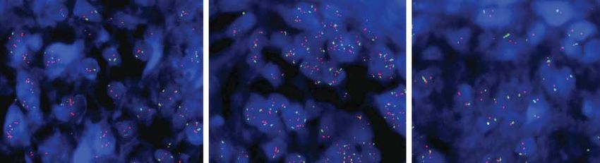
Research Case Report/Case Series                                                                                          Agminated Spitz Nevi in a Nevus Spilus

               Figure 1. Clinical and Histopathologic Features of the Agminated Spitz Nevi Arising in a Nevus Spilus

                  A                                                     B

                  C                                                                          D

               A and B, Photograph of a large tan patch on the left lower back with 1- to 2-mm   Spitz nevi (hematoxylin-eosin, original magnification ×10). D, Melanocytes with
               hyperpigmented macules and 4- to 6-mm pink papules. C, Pink papule showing        amphophilic cytoplasm in the dermis (hematoxylin-eosin, original magnification
               plump melanocytes splayed through desmoplastic collagen, consistent with          ×20).

               through the dermis, consistent with intradermal Spitz nevi                        tected (eMethods in the Supplement). Sanger sequencing con-
               (Figure 1C and D). To identify underlying genetic alterations,                    firmed the presence of the HRAS mutation in both Spitz le-
               we obtained specimens from 2 additional pink papules, with                        sions. We performed Sanger sequencing on DNA derived from
               histopathologic features also confirming the diagnosis of Spitz                   2 additional formalin-fixed, paraffin-embedded Spitz nevi ob-
               nevi. Our study complied with the Declaration of Helsinki and                     tained from the same patient that also demonstrated the HRAS
               was approved by the institutional review board at Stanford Uni-                   point mutation (Figure 2B). Therefore, all 4 Spitz nevi ob-
               versity School of Medicine. Genomic DNA was isolated from                         tained from our patient harbored the same single-nucleotide
               these 2 lesional samples along with the adjacent normal skin,                     variation.
               1 cm outside the boundaries of the nevus spilus, and sub-                              To evaluate for copy number changes, we used SeqGene-
               jected to exome sequencing (eMethods and eTable in the                            CNV on the exome sequencing data.13 This algorithm detects
               Supplement).                                                                      regions with abnormal copy number changes using circular bi-
                                                                                                 nary segmentation. This revealed a copy number increase in
                                                                                                 chromosome 11p in both Spitz nevi compared with the nor-
                                                                                                 mal skin control (Figure 2C). We then performed fluorescent
               Results                                                                           in situ hybridization using an HRAS probe that confirmed am-
               Comparison of recurrent variants from the exome sequenc-                          plification of HRAS in the melanocytes from 2 Spitz nevi
               ing identified an HRAS point mutation (c.37G→C, p.Gly13Arg)                       (Figure 2D) and polysomy in the melanocytes from a third Spitz
               in both Spitz nevi that was absent in the adjacent normal skin                    nevus (Figure 2E). No HRAS amplification was detected in ad-
               (Figure 2A). No other recurrent somatic mutations were de-                        jacent fibroblasts or epidermal keratinocytes (Figure 2F).

        1078   JAMA Dermatology September 2013 Volume 149, Number 9                                                                                      jamadermatology.com

Downloaded From: http://jamanetwork.com/ on 05/18/2015
Agminated Spitz Nevi in a Nevus Spilus Case Report/Case Series Research
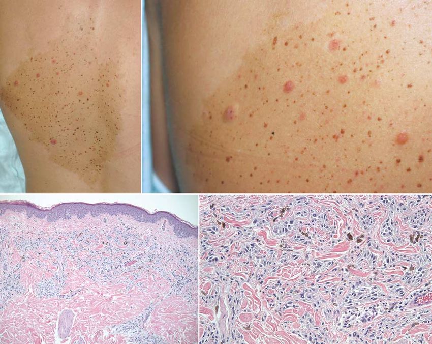
Figure 2. Activating HRAS Mutations and Amplification of Chromosome 11p in Agminated Spitz Nevi
A B
G
C G G C G T G T
C
Lesion HRAS
Spitz Spitz nevus 1 c.37 G>C (p. Gly13Arg)
nevus
Spitz nevus 2 c.37 G>C (p. Gly13Arg)
Spitz nevus 3 c.37 G>C (p. Gly13Arg)
Spitz nevus 4 c.37 G>C (p. Gly13Arg)
Normal Normal skin control WT
skin
C
Chromosome 1 2 3 4 5 6 7 8 9 10 11 12 13 14 15 16 17 18 19 20 21 22 X Y
Adjacent skin
Spitz 1
Spitz 2
D E F
HRAS HRAS HRAS
Centromere 11 Centromere 11 Centromere 11
G H
G
C G G C G T G T
C
WT
Spitz
nevus
Hit1 (HRAS G13R)
Spilus
Hit2 (amplified HRAS G13R)
Normal
skin
A, Sanger sequencing of a representative Spitz nevus and adjacent unaffected signals, indicating HRAS amplification (arrows). E, Dual-colored FISH showing a
skin demonstrates a c.37G→C, p.Gly13Arg mutation specific to the lesional focus of melanocytes with polysomy demonstrated by increased HRAS (red)
tissue. B, Table of HRAS mutations showing the HRAS mutation is present in all 4 and centromeric (green) signals in the nucleus (arrows). F, Dual-color FISH
Spitz nevi but undetectable in the adjacent normal skin. C, Chromosomal showing epidermal keratinocytes and papillary dermal fibroblasts with
amplifications predicted by SeqGene CNV and displayed with an Integrative equivalent red and green signals. G, Sanger sequencing of AciI1-digested DNA
Genomics Viewer (http://www.broadinstitute.org/igv/). Both Spitz nevi have a from a Spitz nevus, nevus spilus, and the adjacent normal skin demonstrating
predicted amplification (red bars) over chromosome 11p. D, Dual-color the HRAS mutation in the nevus spilus and Spitz nevus but not in the normal
fluorescent in situ hybridization (FISH) with HRAS probe (red signals) and a skin. H, Diagram of 2-hit model of a nevus spilus, with the first hit leading to the
reference centromeric probe for chromosome 11 (green signals) showing a focus macular portion of the nevus spilus and the second hit leading to the formation
of melanocytes with increased red signal significantly above reference green of Spitz nevi. WT indicates wild type.
jamadermatology.com JAMA Dermatology September 2013 Volume 149, Number 9 1079
Downloaded From: http://jamanetwork.com/ on 05/18/2015Research Case Report/Case Series Agminated Spitz Nevi in a Nevus Spilus
Spitz nevi are heterogeneous melanocytic tumors, with less nevi. Mosaic HRAS mutations were recently recognized in the
than 20% of these lesions harboring HRAS activating muta- nevi sebacei and nevi spili in patients with phacomatosis
tions and even fewer containing the HRAS point mutation.3 pigmentokeratotica.14 This report extends this finding by dem-
Thus, it would be highly improbable for all Spitz nevi ob- onstrating an HRAS mutation in a sporadic nevus spilus. In-
tained from our patient to develop identical mutations if they terestingly, the HRAS point mutation, in particular, has been
represented independent lesions. We hypothesized that these detected in a variety of benign skin neoplasms, including epi-
Spitz nevi arose in an agminated fashion from a common dermal and sebaceous nevi, Spitz nevi, and nevi spili, provid-
postzygotic clone of melanocytes, likely demarcated by the ne- ing a unique example of genetic pleiotropy within the ecto-
vus spilus. To improve our sensitivity to detect this mutation dermal lineage.15
in the nevus spilus, we performed polymerase chain reaction Our data indicate that multiple Spitz nevi may have a simi-
amplification of the genomic DNA followed by enzymatic di- lar pathogenesis to that of solitary Spitz nevi since a subset of
gestion with Aci1, which digests the wild-type sequence but solitary Spitz nevi also harbors activating mutations in HRAS
not the mutant sequence (eMethods and eFigures 1 and 2 in and copy number increases in chromosome 11p.3 However,
the Supplement). Subsequent Sanger sequencing reproduc- similar to solitary Spitz nevi, other genetic alterations also may
ibly detected the HRAS point mutation in the nevus spilus and play a role in the pathogenesis of multiple Spitz nevi. Gantner
Spitz nevi but not in the adjacent normal skin (Figure 2G). This et al9 recently demonstrated the absence of HRAS-activating
implicates the HRAS point mutation as the initiating muta- mutations in a patient with eruptive Spitz nevi, suggesting that
tion predisposing melanocytes to develop into Spitz nevi. In alternate genetic alterations may be responsible for the le-
this model, a “second hit” may be required for the formation sions in this patient. It is tempting to speculate that many cases
of Spitz nevi (Figure 2H). Our data support HRAS amplifica- of multiple Spitz nevi may result from an early clonal muta-
tion as a secondary change because its mechanism was not tion, as demonstrated in our patient.
identical in all Spitz nevi, with 1 nevus demonstrating poly- Recently, significant progress has been made in under-
somy of chromosome 11. standing the genetic alterations in cutaneous tumors, in part
due to the advances in sequencing technology. Many of these
technologies rely on a large number of samples to determine
recurrent mutations. This approach may be difficult in soli-
Discussion tary Spitz nevi since the lesions are uncommon and possess
Multiple Spitz nevi can occur rarely in agminated and dissemi- heterogeneous mutations. Identifying clonal mutations in pa-
nated forms, but the genetic alterations that lead to these oc- tients with multiple Spitz nevi presents a promising ap-
currences are unknown. To our knowledge, this is the first re- proach to distinguish genetic alterations in all Spitz nevi. In-
port demonstrating mosaicism in agminated Spitz nevi and sight into these genetic changes is critical to improve our ability
identifying an activating HRAS mutation in agminated Spitz to diagnose and manage these controversial lesions.
ARTICLE INFORMATION 2. Mones JM, Ackerman AB. “Atypical” Spitz’s 9. Gantner S, Wiesner T, Cerroni L, et al. Absence of
Accepted for Publication: April 2, 2013. nevus, “malignant” Spitz’s nevus, and BRAF and HRAS mutations in eruptive Spitz naevi.
“metastasizing” Spitz’s nevus: a critique in historical Br J Dermatol. 2011;164(4):873-877.
Published Online: July 24, 2013. perspective of three concepts flawed fatally. Am J
doi:10.1001/jamadermatol.2013.4745. 10. Boone SL, Busam KJ, Marghoob AA, et al. Two
Dermatopathol. 2004;26(4):310-333. cases of multiple Spitz nevi: correlating clinical,
Author Contributions: Drs Sarin, Swetter, Kim, and 3. Bastian BC, LeBoit PE, Pinkel D. Mutations and histologic, and fluorescence in situ hybridization
Khavari had full access to all the data in the study copy number increase of HRAS in Spitz nevi with findings. Arch Dermatol. 2011;147(2):227-231.
and take responsibility for the integrity of the data distinctive histopathological features. Am J Pathol.
and the accuracy of the data analysis. 11. Aloi F, Tomasini C, Pippione M. Agminated Spitz
2000;157(3):967-972. nevi occurring within a congenital speckled
Study concept and design: Sarin, Kim.
Acquisition of data: Sarin, Sun, Bangs, Cherry, 4. Fullen DR, Poynter JN, Lowe L, et al. BRAF and lentiginous nevus. Am J Dermatopathol.
Swetter, Kim. NRAS mutations in spitzoid melanocytic lesions. 1995;17(6):594-598.
Analysis and interpretation of data: Sarin, Sun, Mod Pathol. 2006;19(10):1324-1332. 12. Berk DR, Lane AT. Acquired bilateral agminated
Bangs, Cherry, Kim, Khavari. 5. Wiesner T, Murali R, Fried I, et al. A distinct Spitz nevi in a child with Langerhans cell
Drafting of the manuscript: Sarin, Bangs, Swetter, subset of atypical Spitz tumors is characterized by histiocytosis. Pediatr Dermatol. 2010;27(3):
Kim. BRAF mutation and loss of BAP1 expression. Am J 282-284.
Critical revision of the manuscript for important Surg Pathol. 2012;36(6):818-830. 13. Deng X. SeqGene: a comprehensive software
intellectual content: Sarin, Sun, Cherry, Swetter, 6. Bastian BC, Wesselmann U, Pinkel D, Leboit PE. solution for mining exome- and transcriptome-
Kim, Khavari. Molecular cytogenetic analysis of Spitz nevi shows sequencing data. BMC Bioinformatics. 2011;12:267.
Statistical analysis: Sarin. clear differences to melanoma. J Invest Dermatol. doi:10.1186/1471-2105-12-267.
Administrative, technical, and material support: Sun, 1999;113(6):1065-1069.
Bangs, Cherry, Kim, Khavari. 14. Groesser L, Herschberger E, Sagrera A, et al.
Study supervision: Kim, Khavari. 7. van Dijk MC, Bernsen MR, Ruiter DJ. Analysis of Phacomatosis pigmentokeratotica is caused by a
mutations in B-RAF, N-RAS, and H-RAS genes in the postzygotic HRAS mutation in a multipotent
Conflict of Interest Disclosures: None reported. differential diagnosis of Spitz nevus and spitzoid progenitor cell [published online January 21, 2013].
melanoma. Am J Surg Pathol. 2005;29(9):1145-1151. J Invest Dermatol. doi:10.1038/jid.2013.24.
REFERENCES
8. Harris K, Florell SR, Papenfuss J, et al. Melanoma 15. Hafner C, Groesser L. Mosaic RASopathies. Cell
1. Da Forno PD, Fletcher A, Pringle JH, Saldanha GS. mimic: a case of multiple pagetoid Spitz nevi. Arch Cycle. 2013;12(1):43-50.
Understanding spitzoid tumours: new insights from Dermatol. 2012;148(3):370-374.
molecular pathology. Br J Dermatol.
2008;158(1):4-14.
1080 JAMA Dermatology September 2013 Volume 149, Number 9 jamadermatology.com
Downloaded From: http://jamanetwork.com/ on 05/18/2015Agminated Spitz Nevi in a Nevus Spilus Case Report/Case Series Research
NOTABLE NOTES
Euphorbia peplus: 18th-Century Insights on a 21st-Century Therapy
Navya S. Nambudiri, MBBS; Vinod E. Nambudiri, MD, MBA
In 2012, the US Food and Drug Administration approved a new thera-
Figure. Eighteenth-Century Monograph Describing the Dermatologic
peutic agent, ingenol mebutate, for the topical treatment of actinic kera-
Use of Euphorbia peplus, the Plant From Which Ingenol Mebutate Is
toses. Ingenol mebutate is a diterpene ester with the chemical formula Derived
C25H34O6 and is extracted from the sap of the plant species Euphorbia
peplus, also known as the petty spurge. Euphorbia peplus extract has been
used for centuries as a topical agent for the treatment of a variety of skin
conditions in traditional medicine systems from around the world.
Euphorbia peplus was first taxonomically categorized in the West-
ern scientific community by Carl Linnaeus in the 1750s and presented
in a thesis defended by his student Johannes Wiman at Uppsala Univer-
sity in Sweden.1 Linnaeus described a variety of medicinal uses for the
genus of Euphorbia plants as topical treatments and systemic agents for
gastrointestinal tract purging. Members of this genus were known to
cause skin irritation on contact with the plant’s sap. The genus was named
after the ancient Greek physician Euphorbus, who in the first century
AD documented the laxative properties of the spurges.
A monograph published in London, England, circa 1770 highlights
specific insights into several plants, including E peplus.2 The manu-
script (Figure), published in both Latin and English, likely represents one
of the earliest documentations of the dermatologic applications after Lin-
naean classification. The monograph authors describe “the milky fluid
which it abounds with, is by some applied to Warts, which it is said to
destroy.”2 The other members of the Euphorbia genus, particularly Eu-
phorbia helioscopia, or sun spurge, were also recognized to have sap with
similar properties in the monograph.
A later selection from the same monograph discusses the sun spurge
or “wart-wort” species in greater detail, including its toxicity. “My friend
Mr William Wavell lately informed me of a case which fell under his no-
tice in the Isle of Wight, where from the application of the juice of this
Spurge [E helioscopia] to some Warts near the eye of a little girl, the whole
face became inflamed to a very great degree,” noted the author of the
monograph.2
Consistent with these case reports from more than 2 centuries ear-
lier, most patients enrolled in clinical trials demonstrating the efficacy
of ingenol mebutate for actinic keratoses developed clinically signifi-
cant erythema at the site of application.3 It is also notable that a lower
concentration of the drug is approved for treatment of the face and that Reproduced with permission from the Bodleian Libraries, University of Oxford,
the most common adverse effects of ingenol mebutate in the aforemen- Oxford, England.
tioned clinical trials were pruritus, irritation, and pain—echoing the cau-
tionary case described in the monograph. As future work unfolds exam-
ining additional applications for topical ingenol mebutate, looking back 1. Linnaeus CV, Wiman J. Euphorbia ejusque historia naturalis et medica.
into the past may help uncover other natural remedies awaiting our re- Uppsala, Sweden, 1752. Cambridge, MA: Harvard University Library.
http://books.google.com/books?id=xCm6tgAACAAJ. Accessed April 10, 2013.
discovery.
2. Euphorbia peplus. Small garden spurge. London, circa 1770. Eighteenth
Author Affiliations: Cochin Medical College, Kochi, Kerala, India (N. S. Century Collections Online. Oxford, England: JJ Horticulture Folder, Bodleian
Nambudiri); Department of Dermatology, Harvard Medical School, Boston, Libraries, University of Oxford. http://find.galegroup.com.ezp-prod1.hul
Massachusetts (V. E. Nambudiri). .harvard.edu/ecco/infomark.do?&source=gale&prodId=ECCO&
Corresponding Author: Vinod E. Nambudiri, MD, MBA, Department of userGroupName=camb55135&tabID=T001&docId=CW109002308&type=
Dermatology, Harvard Medical School, 55 Fruit St, Bartlett Hall, Sixth Floor, multipage&contentSet=ECCOArticles&version=1.0&docLevel=FASCIMILE.
Boston, MA 02114 (vnambudiri@partners.org). Accessed April 10, 2013.
Additional Contributions: We thank the JJ Horticulture Folder, The Bodleian 3. Lebwohl M, Swanson N, Anderson LL, Melgaard A, Xu Z, Berman B. Ingenol
Libraries, The University of Oxford, for image permissions. mebutate gel for actinic keratosis. N Engl J Med. 2012;366(11):1010-1019.
jamadermatology.com JAMA Dermatology September 2013 Volume 149, Number 9 1081
Downloaded From: http://jamanetwork.com/ on 05/18/2015You can also read