A HUGE GIANT CELL TUMOUR OF THE EXTENSOR TENDON SHEATH OF THE FOOT: A CASE REPORT AND LITERATURE REVIEW
←
→
Page content transcription
If your browser does not render page correctly, please read the page content below
Case report                                 East African Orthopaedic Journal

A HUGE GIANT CELL TUMOUR OF THE EXTENSOR TENDON SHEATH OF THE
FOOT: A CASE REPORT AND LITERATURE REVIEW

A. Younus, FC Orthopedics (SA), T. Joseph, MBChB (SA), Helen Joseph Hospital, Johannesburg, South
Africa and A. Kelly, FC Neurosurgery (SA), Dr George Mukhari Academic Hospital, Pretoria, South Africa

Correspondence to: Dr Adrian Kelly, P.O. Box Medunsa, Pretoria, South Africa. Email: adriankelly1000@
yahoo.co.uk

ABSTRACT

   Giant cell tumours have a predilection for the hand, where after ganglion cysts, they are the most
   frequent tumour type. Only 3-5% of giant cell tumours occur in the foot, and even here they tend
   to occur in the forefoot, with hindfoot giant cell tumours being a rarity. While relatively common
   overall, their exact nature, as to whether they are truly neoplastic or simply inflammatory, is a
   subject of significant controversy. They are benign slow growing lesions, best treated with gross
   total excision under magnification. Despite their subcutaneous nature, they can become markedly
   infiltrative, and unless completely excised exhibit recurrence rates of between 14% and 44%. Factors
   predictive of recurrence include pressure erosion on X-ray imaging, interphalangeal joint location,
   concomitant degenerative joint disease, and incomplete excision. We present an adult female patient
   who presented to our unit with a giant cell tumour extending over the dorsum and medial side of
   her foot of an alarming size. While the lesion was largely asymptomatic, she was offered surgery for
   functional reasons. Through the creation of a local flap over the tumour, and using magnification, we
   were able to achieve a gross total excision. According to our review of the PubMed literature, this is
   the largest giant cell tumour described in this location, and as such our case report adds value to the
   world-body of orthopaedic knowledge on the subject.

   Key words: Giant cell tumour, Extensor tendons foot

INTRODUCTION                                               neoplastic, while others remain steadfast and
                                                           regard them as non-neoplastic, polyclonal, and
While giant cell tumours are the second most               inflammatory (8,9). Evidence for this controversy
tumour in the hand after ganglion cysts, their             comes from karyotype analysis which has
occurrence in the foot is relatively rare. The relative    identified aneuploidy DNA, abnormal mitosis, and
distribution between these two sites is in fact so         genetic aberrations in giant cell tumours indicative
marked, that while several studies report that             of neoplasia, while polymerase chain reaction
85% of cases occur in the fingers, only 3-5% of            assays of the X-linked human androgen receptor
giant cell tumours occur in the foot and ankle (1-         gene note polyclonal proliferation indicating
3). Another study supports the rarity of giant cell        only a reactive neoplastic hyperplastic process
tumours in the foot, and notes that while 85% of           (10). Further evidence for the latter is proposed in
these tumours occur in the hand, outside of the            another study which notes a history of antecedent
hand the knee is the second most common site               trauma, and a subsequently likely inflammatory
accounting for 12% of cases, with their occurrence         origin, to be present in approximately 50% of
in the foot being a relative rarity (4). Another           cases (11). Whether neoplastic or not, giant cell
epidemiological clustering occurs in individuals           tumours are now agreed to be a distinct pathology
in their 3rd -5th decade, and regarding gender             from pigmented villonodular synovitis. The most
distribution displays a female predominance                important distinguishing fact is that while giant
(5,6). Giant cell tumours were first described by          cell tumours may rarely secondarily extend into
Jaffe in the early 1940’s, and in this early study         the intra-articular space, it is largely accepted that
they were proposed to be a variant of pigmented            they originate from the extra-articular tendon
villonodular synovitis, and thus classified as non-        sheath and synovium. Pigmented villonodular
neoplastic (7). Ongoing controversy in more recent         synovitis on the other hand arises exclusively from
studies persists, with some proposing them to be           the intra-articular synovium, and hence primarily

    56                                                                                  Volume 15 No. 1, March 2021
East African Orthopaedic Journal

    involves the intra-articular chondral and osseous                                        Figure 1
    structures, almost invariable destroying the                  Pre-operative patient photograph showing the large
    involved joint (1,12). Giant cell tumours are benign,           soft tissue mass on the antero-lateral aspect of the
    slow-growing, soft tissue tumours of uncertain                  left foot. The alarming size is appreciated. The long
    origin comprising 1.6% of all soft tissue tumours                history provided alluded to the fact that the mass
    (13). They are largely non-infiltrative, and any bone                   was in all likelihood benign in anture
    erosion seen is most commonly only from a local
    Caspressure effect. Despite these features being
    present in the majority of cases, bone invasion
    has been reported to occur in approximately 5%
    of cases (14), as has malignant transformation
    (15). Important differential diagnoses for soft
    tissue tumours of the foot include lipoma, synovial
    cyst, pigmented villonodular synovitis, fibroma of
    tendon sheath, synovial sarcoma, undifferentiated
    pleomophic sarcoma, leiomyosarcoma, and
    rheumatoid nodule (13,16,17). We present a
    middle-aged female patient who presented to
    our unit with an 8-year history of a progressive,
    painless, soft tissue mass on the dorsum of the left
    foot, of alarming size, that on histopathological
    analysis proved to be a diffuse giant cell tumour.
    According to our review of the Pub Med literature    Due to the extensive differential diagnosis of a mass of this size, a fine needle
    this is the largest giant cell tumour of the foot aspiration
                                                      ever       biopsy wasDue   to the which
                                                                              performed   extensive     differential
                                                                                                 reported the mass todiagnosis
                                                                                                                      be a diffuse giant cell
    reported, and as such adds value to the world-    tumour. Weof     a massthisoffortunate
                                                                   considered         this size,   a fine had
                                                                                             as malignancy  needle   aspiration
                                                                                                               been excluded, recognized a
    body of knowledge on the subject.                             biopsy was performed which reported the mass
                                                                  to be a diffuse giant cell tumour. We considered
    CASE REPORT                                                   this fortunate as malignancy had been excluded,
                                                                  recognized a complete surgical excision to be her
    A 54-year old female patient presented to our unit            best chance of cure, and would primarily consider
    with progressively increasing in size, painless, mass         re-operation in case of recurrence. As part of pre-
    on the dorsum of the left foot of 8-years duration.           operative planning X-rays of her left foot were
    What had brought her to the hospital now was                  performed which excluded bony involvement by
    purely the fact that she was having difficulty                no osteolysis being noted (Figure 2).
    wearing shoes. She denied having any difficulty or
    pain ambulating. In terms of her medical history she                                     Figure 2
    was known with hypertension that was controlled                  Pre-operative X-ray of her left foot showed disuse
    medically, and she had recently been tested and                  osteopenia however no overt osteolytic focus was
    found to be HIV negative. On examination she was                                            noted
    a healthy-looking adult female with no stigmata
    of immunosuppression. General examination
    was non-contributory, including no popliteal or
    inguinal lymphadenopathy. Examination of her
    left leg revealed a normal range of ankle and
    toe movement on passive testing, however on
    active testing she had a decreased range of toe
    flexion. Examination of the mass revealed a 14cm
    by 11cm multinodular soft tissue mass over the
    anteromedial aspect of the left foot. The mass
    had discreet edges, was fixed to the underlying
    tissues, of firm consistency, with no overlying skin
    changes. The mass did not trans-illuminate and
    was non-pulsatile (Figure 1).

                                                                A pre-operative MRI confirmed the multilobulated nature of the mass,

Volume 15 No. 1, March 2021                                                                                                     57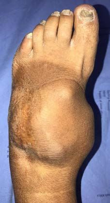
East African Orthopaedic Journal
A pre-operative MRI confirmed the multi- Using magnification, the tumour was dissected
lobulated nature of the mass, demonstrated its free from the extensor tendon mass, with special
insinuation between the extensor tendons of the attention afforded to the tumour edges to ensure
left foot, and demonstrated the circumferential a gross total resection had been achieved. The
nature of the mass which extended from the tumour was noted to have most likely arisen from
dorsum of the left foot over and onto the medial the synovial sheath of extensor hallucis longus
side (Figure 3). tendon, insinuated between the extensor tendons
over the dorsum of the foot, and infiltrated beneath
Figure 3 the tibialis anterior tendon on the medial aspect of
Pre-operative MRI T2W sagittal and axial views the foot (Figure 5).
showing the hyperintense multi-lobulated nature
of the mass extending over the dorsum and medial Figure 5
aspect of the left foot. The largely subcutaneous Intra-operative patient photograph showing the
nature of the lesion was regarded as favorable and medial aspect of the tumour where it had infiltrated
we planned for a gross total resection beneath the tibialis anterior tendon (blue arrow)
She was taken to the operating room and the tourniquet was insufflated without
She was taken to the operating room and
the tourniquet was insufflated without prior
exsanguination to allow for visualization of the
vasculature. An 8cm longitudinal skin incision
was made over the dorsum of the left foot, and a
second 10cm lazy-S skin incision, stemming from
the first, was made directed inferior medially over
the tumour mass, towards the medial aspect of A meticulous dissection was performed until a
the foot (Figure 4). The great saphenous vein gross total resection, confirmed by magnification,
was identified, ligated, transected, and its ends had been achieved. A necessary part of the
A meticulous dissection was performed until a gross total resection, confirmed by
cauterized. operation was to resect part of the extensor
tendon sheath of the extensor hallucis longus
Figure 4 muscle (Figure 6).
Intra-operative patient photograph of the skin
incision performed to maximize access to the Figure 6
tumour mass. The flap created (red arrow), assisted Intra-operative patient photograph showing the
with this purpose, and was reflected inferiorally. The tumour bed after gross total resection of the tumour.
white fleshy nature of the tumour, directly beneath The exposed tendons of the extensor hallucis longus
the skin, was immediately visible muscle (yellow arrow), and the tibialis anterior
muscle (blue arrow), can be seen
Using magnification, the tumour was dissected free from the extensor tendon mass
58 Volume 15 No. 1, March 2021
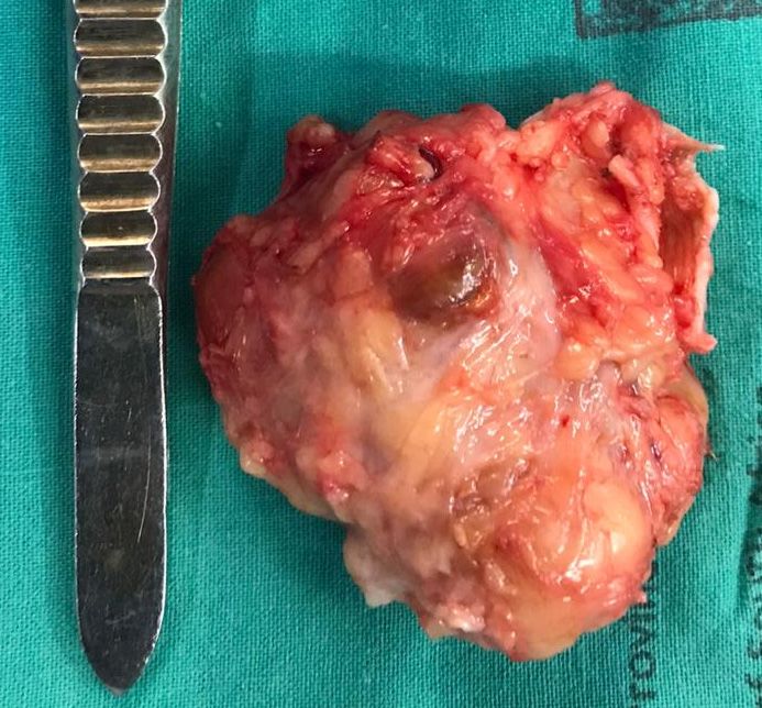
The fleshy nature of the main tumour mass was sent for histopathological analysis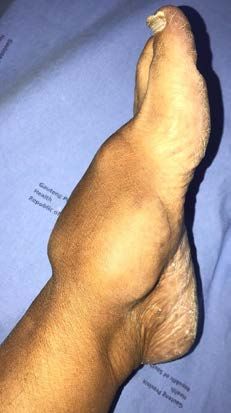
East African Orthopaedic Journal

               The fleshy nature of the main tumour mass was           for re-consideration depending on the size of the
            sent for histopathological analysis (Figure 7), the        recurrence and findings of the surgery.
            wound irrigated with hydrogen peroxide, washed
            with saline, and closed in layers. The skin was
            closed with interrupted nylon mattress sutures.            DISCUSSION

                                   Figure 7                     Several classification systems exist by which to
              Intra-operative photograph of the main tumour     classify giant cell tumours. The first is a clinical-
                                                                radiological classification, proposed by Al Qattan,
               mass on a sterile drape. The typical solid, fleshy
              nature, of a giant cell tumour can be appreciated which divides giant cell tumours into three
                                                                types. Type 1 is a single tumour that is round
                                                                and multilobulated, such as that in our case.
                                                                Type 2 refers to two or more distinct separate
                                                                tumours. Type 3 refers to recurrent satellite lesions
                                                                after an incomplete primary excision (17). The
                                                                histopathological classification, proposed by the
                                                                World Health Organization (WHO), classifies giant
                                                                cell tumours as fibriohistiocytic tumours. Here a
                                                                localized nodular type, characterized by a relatively
                                                                hypocellular nature with numerous giant cells, is
                                                                differentiated from a diffuse type characterized
                                                                by hyper cellularity with numerous polygonal
                                                                mononuclear cells and less frequent giant cells (18).
                                                                Giant cell tumours of the extensor tendon mass, of
                                                                whatever histopathological type, are noted to be
                                                                found in the subcutaneous tissue plane arising from
                                                                the tendon sheath, with extensions that infiltrate
                                                                over and under the surrounding structures. The
                                                                neurovascular bundle is often found displaced
               The patient was discharged on the 3rd post-      by the tumour mass, and hence the importance
           operative day, fully ambulant, for out-patient       of magnification is proposed to make separation
ent was discharged on the 3rd post-operative day, fully ambulant,
                                                                possiblefor  out-
                                                                          (19). Gross total excision is recognized by
           follow-up. At her 2-week follow-up appointment
           the histopathological analysis became available.     several studies to offer the best chance of cure (19-
           According to this report the specimen was            21). Despite this aim, recurrence rates are reported
           mostly comprised of nests of large mononuclear       to be between 14% and 44% (21,22). Factors
           polygonal cells with an eosinophilic cytoplasm,      predictive of local recurrence include pressure
           contained within a collagenous hyalinized stroma.    erosion on X-ray imaging, interphalangeal joint
           Osteoclast-like multinucleated giant cells were      location, concomitant degenerative joint disease,
           randomly distributed within the tumour matrix,       and incomplete excision (22,23). One prospective
           as were scattered foam cells. A lymphocytic          study, that gave an adjunctive total dose of 20G of
           infiltrate was further noted. Hemosiderin deposits   radiotherapy, in 2G divided doses, to 14 patients
           were also seen. Vascular invasion, necrosis, and     with incomplete excision, mitotic figures, or bone
           mitotic figures were not seen. Regarding staining    involvement, noted no recurrences. This study
           both the mononuclear and multinucleated cells        proposes a role for adjunctive radiotherapy in the
           stained positive with CD-68, however only the        management of this specific post-operative patient
           multinucleated cells demonstrated membrane           subset (21). Regarding the pre-operative diagnosis
           positivity for CD-45. The entirety of the tumour     by fine needle aspiration, as was performed in
           was negative for S-100 and desmin. The conclusion    our patient, this is supported by one study as an
           of the histopathological report was that the lesion  important tool for pre-operative surgical planning
           was a giant cell tumour- diffuse type. At her 6-week to prevent recurrence (24). Another study, albeit
           out-patient appointment the patient was noted to     only a case report, supports our practice and
           be wearing closed shoes, and on examination her      similarly used hydrogen peroxide lavage of the
           wound had healed well. She was booked for 1-year     tumour bed post removal of a giant cell tumour
           out-patient review. In the event of recurrence, we   from the ankle of a 58-year old adult male. In this
           planned to consider re-operation as our first line   study no recurrence had occurred at 2-year follow-
           intervention and reserved adjunctive radiotherapy    up (25).

        Volume 15 No. 1, March 2021                                                                              59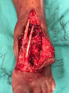
East African Orthopaedic Journal

CONCLUSION                                                      nodular tenosynovitis): clinicopathological
                                                                features of 71 cases. J Clin Pathol. 2001;
Giant cell tumours are relatively rare on the                   54:404-407.
dorsum of the foot compared to their incidence            7.    Jaffe, H., Lichtenstein, L. and Sutro, C.J.
on the hand. When they are present, they most                   Pigmented villonodular synovitis, bursitis, and
commonly affect the forefoot and their presence                 tenosynovitis. Arch Pathol. 1941; 31:731-765.
over the hindfoot is a rarity. One study that             8.    Brandal, P., Bjerkehagen, B. and Heim, S.
considered 14 giant cell tumours of the foot, noted             Molecular cytogenetic characterization of
11/14 (79%) to involve the forefoot, and only 3/14              tenosynovial giant cell tumors neoplasia.
(21%) involved the hindfoot (26). While benign in               Neoplasia (New York, NY). 2004; 6(5):578–583.
nature, their propensity to recur makes gross total       9.    Rukavina, I. and Caleta, D. Giant-cell tumour of
excision under magnification the treatment of                   the tendon sheath: a review. OA Orthopaedics.
choice. Recurrence is similarly best treated with re-           2014; 2(2)11.
operation. Our case report provides an account of         10.   Vogrincic, G.S., O’Connell, J.X. and Gilks,
a giant cell tumour of the extensor tendon sheath               C.B. Giant cell tumor of tendon sheath is a
of the foot of alarming size. Our study hopes to                polyclonal cellular proliferation. Hum Pathol.
assist foot and ankle orthopaedic surgeons who                  1997; 28: 815-819.
will in all probability need to manage this specific      11.   Dartoy, C., Fenoll, B., Leroy, J.P., Dubrana, F.,
tumour at some point in their careers.                          Nen, D.L. and Behannin, B. Giant cell tumor of
                                                                the tendon sheath of the long extensor muscle
Funding: This research did not receive any specific             of the thumb in a 7-year-old child. Ann Chir
grant from funding agencies in the public,                      Main Memb Super. 1994; 13(3):198-201.
commercial, or not-for-profit sectors.                    12.   Reilly, K.E., Stern, P.J. and Dale, J.A. Recurrent
                                                                giant cell tumors of the tendon sheath. J Hand
Declaration of interest: None of the authors have               Surg Am. 1999; 24: 1298-1302.
any financial nor personal relationships with other       13.   Lo, E. and Ketterer, D. Giant cell tumor of tendon
                                                                sheath in the toe. JAPMA. 2000; 90(5):270-272.
people, or organizations, that could inappropriately
                                                          14.   Walsh, E.F., Mechrefe, A., Akelman, E. and
influence (bias) their work, all within 3 years of the
                                                                Schiller, A.L. Giant cell tumor of tendon sheath.
beginning the work submitted.
                                                                Am J Orthop (Belle Mead NJ). 2005; 34(3):116–
                                                                121.
REFERENCES                                                15.   Bertoni, F., Unni, K., Beabout, J. and Sim, F.
                                                                Malignant giant cell tumor of the tendon
1. Phalen, G.S., McCormack, L.G. and Gazale, W.J.               sheaths and joints (malignant pigmented
   Giant-cell tumor of tendon sheath (benign                    villonodular synovitis). Am J Surg Pathol. 1997;
   synovioma) in the hand: evaluation of 56 cases.              21(2):153-163.
   Clin Orthop. 1959; 15:140–151.                         16.   Goni, V., Gopinathan, N.R., Radotra, B.D.,
2. Zhang, Y., Huang, J., Ma, X., Wang, X., Zhang,               Viswanathan, V.K., Logithasan, R.K. and Balaji,
   C. and Chen, L. Giant cell tumor of the tendon               S. Giant cell tumour of peroneus brevis
   sheath in the foot and ankle: Case series and                tendon sheath - a case report and review of
   review of the literature. J Foot Ankle Surg. 2013;           literature. BMJ Case Rep. 2012; 13. doi: 10.1136/
   52: 24-27.                                                   bcr.01.2012.5703.
3. Qin, Y., Zhang, B. and Song-Cen, L.V. A rare           17.   Al-Qattan, M. Giant cell tumors of tendon sheath:
   localized giant cell tumor of the tendon sheath              classification and recurrence rate. J Hand Surg.
   in hip joint: A case report and review of the                2001; 26B:72–75.
   literature. Biomed Res. 2017; 28:6510-13.              18.   Fletcher, C.D., Krishnan, K., Unni, K.K. and
4. Ghnaimat, M., Alodat, M., Aljazazi, M., Al-Zaben,            Mertens, F. World Health Organization
   R. and Alshwabkah, J. Giant cell tumor of                    classification of tumors, pathology, and
   tendon sheath in the knee. Electron Physician.               genetics of tumors of soft tissue and bone.
   2016; 8(8):2807-09. doi:10.19082/2809.                       Lyon, France: IARC Press; 2002:112–114.
5. Adams, E.L., Yoder, E.M. and Kasdan, M.L. Giant        19.   Kanta, K.S., Manava, A.K., Abhinavb, R.K.,
   cell tumor of the tendon sheath: experience                  Sinhaa, V.K. and Sharma, A. Giant cell tumour
   with 65 cases. Eplasty. 2012; 12: e50.                       of tendon sheath and synovial membrane: A
6. Monaghan, H., Salter, D.M. and Al-Nafussi, A.                review of 26 cases. J Clin Orthop Trauma. 2017;
   Giant cell tumour of tendon sheath (localized                8S:S96-S99.

    60                                                                                   Volume 15 No. 1, March 2021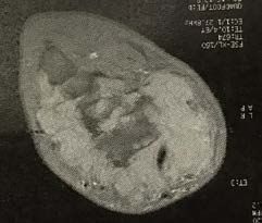
East African Orthopaedic Journal

    20. Liu, T. Endoscopic resection of giant cell tumor       24. Ng, V.Y., Thomas, K., Crist, M., et al. Fine needle
        of the extensor tendon of the foot. Arthroscopy            aspiration for clinical triage of extremity
        Tech. 2017; 6(2):e303-e309.                                soft tissue masses. Clin Orthop Rel Res. 2010;
    21. Kotwal, P.P., Gupta, V. and Malhotra, R. Giant-cell        468:1120-28.
        tumour of the tendon sheath. Is radiotherapy           25. Chen, Y., Yu, X.C., Xu, S.F. and Wang, B. Giant cell
        indicated to prevent recurrence after surgery?             tumor of the tendon sheath originating from
        J Bone Joint Surg Br. 2000; 82:571-573.                    the ankle capsule: A case report and literature
    22. Reilly, K.E., Stern, P.J. and Dale, J.A. Recurrent         review. Oncology Letters. 2016; 11:3461-64.
                                                               26. Gibbons, C.L., Khwaja, H.A., Cole, A.S., Cooke,
        giant cell tumors of the tendon sheath. J Hand
                                                                   P.H. and Athanasou, N.A. Giant-cell tumour of
        Surg Am. 1999; 24: 1298-1302.
                                                                   the tendon sheath in the foot and ankle. J Bone
    23. Grover, R., Grobbelaar, A.O., Richman, P.I., et al.
                                                                   Joint Surg. 2002; 84-B:1000-3.
        Measurement of invasive potential provides
        an accurate prognostic marker for giant cell
        tumor of tendon sheath. J Hand Surg. 1998;
        23B:728-731.

Volume 15 No. 1, March 2021                                                                                     61
You can also read