Cell Function and Viability in the Spontaneously Diabetic GK Rat
←
→
Page content transcription
If your browser does not render page correctly, please read the page content below
-Cell Function and Viability in the Spontaneously
Diabetic GK Rat
Information From the GK/Par Colony
B. Portha, M.-H. Giroix, P. Serradas, M.-N. Gangnerau, J. Movassat, F. Rajas, D. Bailbe, C. Plachot,
G. Mithieux, and J.-C. Marie
The GK rat model of type 2 diabetes is especially con- dicted and thus it is possible to dissect the pathogenic mech-
venient to dissect the pathogenic mechanism necessary anism necessary for the emergence of overt diabetes. The
for the emergence of overt diabetes because all adult Goto-Kakizaki Wistar rat (GK rat) is especially useful
rats obtained in our department (GK/Par colony) to because all adult animals of both sexes exhibit type 2 dia-
date have stable basal mild hyperglycemia and because betes. This spontaneous diabetes model was produced by
overt diabetes is preceded by a period of normo-
glycemia, ranging from birth to weaning. The purpose of
selective breeding (with glucose intolerance as a selection
this article is to sum up the information so far available index) repeated over many generations, starting from a non-
related to the biology of the -cell in the GK/Par rat. In diabetic Wistar rat colony. The characteristics of GK ani-
terms of -cell function, there is no major intrinsic mals bred in our colony in Paris (GK/Par) for more than 10
secretory defect in the prediabetic GK/Par -cell, and years (1) are very stable and remain close to those of the ani-
the lack of -cell reactivity to glucose (which reflects mals in the original Japanese colony (2): all of the rats have
multiple intracellular abnormalities), as seen during a basal mild hyperglycemia and impaired glucose tolerance.
the adult period when the GK/Par rats are overtly dia- Males and females are similarly affected, and their diabetic
betic, represents an acquired defect (perhaps gluco- state is stable over 72 weeks of follow-up (3). In adult GK rats,
toxicity). In terms of -cell population, the earliest plasma insulin release in vivo in response to intravenous
alteration so far detected in the GK/Par rat targets the
size of the -cell population. Several convergent data
glucose is abolished (1,3). In vitro studies of insulin release
suggest that the permanently reduced -cell mass in with the isolated perfused pancreas (1) or with perifused
the GK/Par rat reflects a limitation of -cell neogene- islets (4) indicate that both early and late phases of glucose-
sis during early fetal life, and it is conceivable that induced insulin release are markedly affected in the adult GK
some genes among the set involved in GK diabetes rat. Concerning insulin action in adult GK rats, we have
belong to the subset of genes controlling early -cell reported decreased insulin sensitivity in the liver, in parallel
development. Diabetes 50 (Suppl. 1):S89–S93, 2001 with moderate insulin resistance in extrahepatic tissues,
i.e., muscle and adipose tissue (5,6). In our colony (GK/Par),
hyperglycemia is preceded by a period of normoglycemia,
ranging from birth to weaning (7). Therefore, during this
T ype 2 diabetes develops as a consequence of
interplay among -cell dysfunction, peripheral
insulin resistance, and elevated hepatic glucose
production. However, it is not known which is
the primary abnormality and which are abnormalities sec-
ondary to elevated plasma glucose, so-called glucose toxic-
ity. To delineate the primary abnormalities, it is desirable to
period, young GK rats can be considered to be prediabetic.
DECREASED -CELL NUMBER AND MULTIPLE -CELL
FUNCTIONAL DEFECTS IN THE ADULT GK/Par RAT
WITH OVERT DIABETES
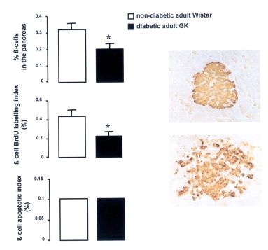
In the adult GK rat, total pancreatic -cell mass is decreased
(by 60%) in the range of the decrease in pancreatic insulin
analyze individuals destined to become diabetic before the stores (1,7,8) (Fig. 1). This alteration of the -cell population
development of the disease. The advantage of using an ani- cannot be ascribed to increased -cell apoptosis but is
mal model is that the development of diabetes can be pre- related, at least partly, to significantly decreased -cell repli-
cation (Fig. 1). Moreover, the adult GK pancreas exhibits
From the Laboratoire de Physiopathologie de la Nutrition (B.P., M.-H.G., P.S., two different populations of islets: large islets, which are dis-
M.-N.G., J.M., D.B., C.P., J.-C.M.), Université D. Diderot, Paris; and the rupted by connective tissue (7) and display heterogeneity in
Institut National de la Santé et de la Recherche Médicale (F.R., G.M.), Fac-
ulté Laënnec, Lyon, France. the staining of the -cells, and small islets, with heavily
Address correspondence and reprint requests to Prof. B. Portha, Labo- stained -cells and normal architecture (Fig. 1).
ratoire de Physiopathologie de la Nutrition, CNRS ESA 7059, Université D. The islets of adult GK/Par rats, at least after collagenase
Diderot/Paris 7, 2 Place Jussieu, Tour 33, 75251 Paris Cedex 05, France.
E-mail: portha@paris7.jussieu.fr.
digestion, show decreased -cells and low insulin content
Received for publication 21 May 2000 and accepted in revised form compared with control islets. The islet DNA content was
28 August 2000. decreased to a similar extent; this is consistent with our mor-
This article is based on a presentation at a symposium. The symposium phometric data (Fig. 2), which indicate that there is no major
and the publication of this article were made possible by an unrestricted
educational grant from Les Laboratoires Servier. change in the relative contribution of -cells to total
GLP, glucagon-like protein; SERCA, calcium-ATPase. endocrine cells in the GK islets. In addition, in GK islets,
DIABETES, VOL. 50, SUPPLEMENT 1, FEBRUARY 2001 S89-CELL FUNCTION AND VIABILITY IN GK RAT
in the pancreas
% -cells
-cell BrdU labeling
index (%)
-cell apoptotic
index (%)
FIG. 1. Left panel: Percentage of -cells in total pancreas, -cell mitotic index (BrdU labeling index), and -cell apoptotic index in 4-month-
old male GK/Par rats and control Wistar rats. Values are expressed as means ± SE. *P < 0.001 compared with Wistar rats. Right panel:
Immunoperoxidase staining for insulin in pancreas of 4-month-old male GK/Par rats (original magnification 125). Adult GK/Par pancreas
exhibits two different populations of islets: large islets, which are disrupted by connective tissue and display heterogeneity in the staining of
-cells (bottom), and small islets with heavily stained -cells and normal architecture (top).
insulin content, when expressed relative to DNA, remains exhibit a specific decrease in the activities of flavin adenine
lower than in control islets, which supports degranulation in dinucleotide–dependent glycerophosphate dehydrogenase
the -cells of diabetic animals. (4,10) and branched-chain ketoacid dehydrogenase (12).
The notion that in the GK -cell the lesion responsible for Although this certainly may contribute to lower oxidation
loss of glucose-induced insulin secretion is mostly upstream rates, it does not exclude other mechanisms. Indeed, we
of the effector system is supported by data indicating that GK found that the -cells of adult GK rats had a significantly
islets are duly responsive to nonnutrient secretagogues, such smaller mitochondrial volume than control -cells (13). No
as sulfonylureas or a combination of barium and theophylline major deletion or restriction fragment polymorphism could
(9). We have reported that glucose transport and glucose be detected in mtDNA from adult GK islets (13); however, they
phosphorylating activity are not modified in the GK -cells contained markedly less mtDNA than did control islets. The
(4,10). Consistent with these conclusions is the present obser- lower islet mtDNA was paralleled by decreased content of
vation that the expression of glucose transporter GLUT2 (by some islet mt mRNAs, such as cytochrome b (13). In accor-
reverse transcriptase–polymerase chain reaction) is normal in dance with this, insufficient increase of ATP generation in
GK islets (Fig. 3). It is also noteworthy that in GK (as well as response to high glucose was shown by our group (4)
Wistar) rat islets, we were unable to detect expression of glu- (Fig. 4). Finally, the impaired insulin response to glucose
cose-6-phosphatase (whereas in GK rat liver, glucose-6-phos- may be attributed to impaired elevation of intracellular Ca2+
phatase was easily detected) (Fig. 3). This is an interesting concentration, which seems to be a consequence of the fail-
point because in another GK colony (11), it has been reported ure by glucose to augment L-type Ca2+ channel activity
that islet glucose-6-phosphatase activity was increased, as because of insufficient plasma membrane depolarization,
was cycling between glucose and glucose-6-phosphate (11). reflecting impaired closure of ATP-sensitive K+ channels
We have shown that impaired glucose-induced insulin (Fig. 4). We have recently obtained data supporting the view
release in GK islets is associated with perturbation of multi- that abnormal Ca2+ handling by the endoplasmic reticulum
ple mitochondrial functions. More specifically, we reported may also participate in defective Ca2+ signaling; we investi-
that aerobic, but not anaerobic, glycolysis is impaired in GK gated the cytosolic calcium response to high glucose in sin-
islets (4,9,10), and we showed that mitochondria of GK islets gle perifused GK islets, as measured by dual-wave spec-
S90 DIABETES, VOL. 50, SUPPLEMENT 1, FEBRUARY 2001B. PORTHA AND ASSOCIATES
FIG. 2. Characteristics of collagenase-isolated islets from 4-month-old male GK/Par rats and control Wistar rats. Data are means ± SE. The num-
ber of islet preparations is between 3 (morphometric studies) and 18 (biochemical determinations). In each experiment, DNA and insulin con-
tent values were obtained from two to five groups of 20 islets each, and the percentages of -cells and non- endocrine cells (, , and PP)
were estimated in four to five islets. **P < 0.05, ***P < 0.001 compared with related value in control Wistar group.
trophotometry using fura-2 (14). The most prominent differ-
ence is detected in the first 5 min following high glucose,
because the GK islet lacks the initial reduction of cytosolic cal-
cium. This initial reduction is thapsigargin-sensitive in the nor-
mal islet, suggesting that the sequestration of calcium by
endoplasmic reticulum, attributed to activation of calcium-
ATPases (SERCAs), is impaired in the GK -cell (Fig. 4).
Such a conclusion is consistent with the report that SERCA3
gene expression is downregulated in GK islets (15). Alterna-
tively, impaired calcium sequestration in the GK -cell can also
be accounted for by insufficient cytosolic ATP generation in
response to high glucose (Fig. 4).
FIG. 4. Model for defective glucose-induced insulin release in the
GK/Par -cell. Impaired insulin response to glucose may be attributed
to impaired elevation of intracellular Ca2+ concentration, which seems
to be a consequence of the failure by glucose to augment L-type Ca2+
channel activity, in its turn due to insufficient plasma membrane
depolarization, reflecting impaired closure of the ATP-sensitive
K+ channels; this is the result of insufficient cytosolic ATP generation
by glucose. Abnormal Ca2+ handling by the endoplasmic reticulum
(ER) in response to high glucose may also participate in the defective
FIG. 3. Detection of glucose-6-phosphatase (Glc6Pase) and GLUT2 Ca2+ signaling: the sequestration of calcium by ER during high-glucose
mRNAs in islet and liver from 4-month-old male GK/Par rats and con- exposure (attributed to activation of the SERCAs) is impaired in the
trol Wistar (W) rats. Total RNAs were purified from freshly isolated GK rat -cell. Impaired calcium sequestration can also be accounted
islets and a frozen liver sample from GK and Wistar rats, using the Qia- for by insufficient cytosolic ATP generation in response to high glu-
gen purification procedure. Glucose-6-phosphatase cDNA (1,095 bp, cose: in GK islets, glucose fails to increase inositol triphosphate (IP3)
exons 1–5) was amplified from 1 µg total RNA in 50 µl amplification accumulation. This is linked to an anomaly in targeting the phospho-
medium as previously described (25), and 20 µl was analyzed on 1.5% rylation of phosphoinositides: the activity of phosphatidylinositol
agarose gel. GLUT2 cDNA (808 bp, exons 6–11) was amplified from the kinase, the first of the two phosphorylating activities responsible for
same RNA samples and analyzed accordingly. Digestion products of generating phosphatidylinositol biphosphate, is reduced. Moreover,
phage- by HindIII/EcoRI were analyzed as size markers (DNA frag- deficient calcium handling and ATP supply in response to glucose
ments appearing were 2,027, 1,904, 1,584, 1,330, 983, 832, and 564 bp probably also contribute to abnormal activation of phosphatidylinos-
long, as shown on the left of the figure from top to bottom). itol kinases and phospholipase C.
DIABETES, VOL. 50, SUPPLEMENT 1, FEBRUARY 2001 S91-CELL FUNCTION AND VIABILITY IN GK RAT
(µg/mg pancreas)
total -cell mass
pancreas weight
(mg/pancreas)
-cell mass
(mg)
index of -cells (%)
apoptotic index
BrdU labeling
of -cells (%)
FIG. 5. Pancreas weight, total -cell mass, -cell BrdU labeling index, and -cell apoptotic index of 7-day-old GK/Par and Wistar (W) rat neonates.
Values are expressed as means ± SE; the number of observations is four in each group. ***P < 0.001 compared with Wistar rats.
Finally, we investigated phosphoinositides (16) and cAMP
metabolism (17) in GK islets. Whereas carbachol was able to
promote normal inositol generation in GK islets, high glucose
failed to increase inositol phosphate accumulation (16). The
inability of glucose to stimulate inositol phosphate production
is not related to defective phospholipase C activity per se
(total activity in islet homogenates is normal). Rather, it is
linked to abnormal targeting of the phosphorylation of phos-
phoinositides; the activity of phosphatidylinositol kinase, ·
which is the first of the two phosphorylating activities
responsible for the generation of phosphatidylinositol
biphosphate, is clearly reduced (16). Moreover, deficient cal-
cium handling and ATP supply in response to glucose prob-
ably also contribute to abnormal activation of phosphatidyl-
inositol kinases and phospholipase C.
Concerning cAMP, it is remarkable that its intracellular
content is already very high in GK -cells at low glucose (17).
This is related to increased expression of the cyclase isoforms
2, 3, and 7, and of the Golf form of Gs proteins (18). Fur-
thermore, cAMP is not further enhanced at increasing glucose
concentrations (at variance with the situation in normal
-cells) (17). This suggests that there exists a block in the
steps linking glucose metabolism to activation of adenylate
cyclase in the GK -cell. This contrasts strikingly with the
capacity of the GK -cell to respond to glucagon-like protein
(GLP)-1 such that it is able to restore the secretory compe-
tence to glucose with a clear biphasic response (17). This
proves that the glucose-incompetence of the GK -cell is not
irreversible and emphasizes the usefulness of GLP-1 as a
therapeutic agent in type 2 diabetes.
FIG. 6. In vitro insulin release by freshly isolated islets from 1-week-
old GK/Par rats in response to nutrient secretagogues. Data are
-CELL POPULATION AND -CELL FUNCTION IN THE expressed as absolute values (top) or as percentage of basal value
GK/Par RAT DURING THE PREDIABETIC PERIOD determined at 2.8 mmol/l glucose in each group of islets (bottom).
Extensive follow-up of the animals after delivery has Each bar represents mean ± SE for 32–42 batches of islets obtained
from six distinct islet preparations. ***P < 0.001 compared with the
revealed that GK/Par pups become overtly hyperglycemic respective values obtained at 2.8 mmol/l glucose or 10 mmol/l leucine.
between 3 and 4 weeks of age. In GK neonates, total -cell Experiments with leucine or glutamine were carried out in the
mass is clearly decreased compared with that in Wistar rats absence of glucose.
S92 DIABETES, VOL. 50, SUPPLEMENT 1, FEBRUARY 2001B. PORTHA AND ASSOCIATES
(Fig. 5). Such -cell growth retardation cannot be ascribed to 9. Giroix MH, Vesco L, Portha B: Functional and metabolic perturbations in iso-
decreased -cell replication or to increased apoptosis lated pancreatic islets from the GK rat, a genetic model of non-insulin depen-
dent diabetes. Endocrinology 132:815–822, 1993
(Fig. 5). We therefore postulate that the recruitment of new 10. Giroix MH, Sener A, Portha B, Malaisse WJ: Preferential alteration of oxyda-
-cells from the precursor pool is defective in the young GK tive relative to total glycolysis in islets of rats with inherited or acquired non-
rat; other data from our group (19–23) suggest that the per- insulin dependent diabetes. Diabetologia 36:305–309, 1993
manently reduced -cell mass in the GK model reflects a lim- 11. Östenson CG, Abdel-Halim SM, Andersson A, Efendic S: Studies on the patho-
itation of -cell neogenesis during early fetal life. genesis of NIDDM in the GK (Goto-Kakisaki) rat. In Lessons from Animal Dia-
betes VI. Shafrir E, Ed. Boston, Birkhäuser, 1996, p. 301–317
At the same age, under in vitro static incubation conditions, 12. Giroix MH, Saulnier C, Portha B: Decreased pancreatic islet response to
GK rat islets release less insulin at basal glucose; however, L-leucine in the spontaneously diabetic GK rat: enzymatic, metabolic and
they amplify their secretory response to high glucose, secretory data. Diabetologia 42:965–977, 1999
leucine, or leucine plus glutamine to the same extent as Wis- 13. Serradas P, Giroix MH, Saulnier C, Gangnerau MN, Borg H, Welsh M, Portha
B, Welsh N: Mitochondrial DNA content is specifically decreased in adult, but
tar rat islets (Fig. 6). Therefore, there does not seem to exist not fetal, pancreatic islets of the GK rat, a genetic model of non-insulin depen-
a major intrinsic secretory defect in the prediabetic GK rat dent diabetes. Endocrinology 136:5623–5631, 1995
-cell. Consequently, the lack of -cell reactivity to glucose, 14. Marie JC, Bailbé D, Portha B: Defective glucose-dependent cytosolic Ca2+ han-
as seen during the adult period, when the GK rats are overtly dling in islets of GK and nSTZ rat models of NIDDM (Abstract). Diabetes
diabetic, represents an acquired defect (glucotoxicity?). 48:A334, 1999
15. Varadi A, Molnar E, Ostenson CG, Ashcroft SJH: Isoforms of endoplasmic retic-
In conclusion, the earliest alteration so far detected in the ulum Ca2+-ATPase are differentially expressed in normal and diabetic islets of
GK/Par rat targets the size of the -cell population. It is con- Langerhans. Biochem J 319:521–527, 1996
ceivable that some genes among those involved in GK/Par dia- 16. Giroix MH, Morin L, Wolf B, Portha B: Defective islet phosphoinositide metab-
betes (24) belong to the subset of genes controlling early olism and insulin secretion in the GK rat model of type 2 diabetes (Abstract).
-cell development. Diabetologia. In press
17. Dachicourt N, Serradas D, Bailbé D, Portha B: Abnormal cAMP content and
insulin release in islets of spontaneously diabetic GK rats (Abstract). Dia-
betologia 40:A114, 1997
REFERENCES 18. Frayon S, Pessah M, Giroix MH, Mercan D, Boissard C, Malaisse WJ, Portha
1. Portha B, Serradas P, Bailbé D, Suzuki K, Goto Y, Giroix M-H: -cell insensi- B, Garel JM: Golf identification by RT-PCR in purified normal pancreatic B
tivity to glucose in the GK rat, a spontaneous non-obese model for type II dia- cells and in islets from rat models of non-insulin-dependent diabetes. Biochem
betes. Diabetes 41:486–491, 1991 Biophys Res Commun 254:269–272, 1999
2. Goto Y, Kakisaki M, Masaki N: Spontaneous diabetes produced by selective 19. Serradas P, Gangnerau MN, Giroix MH, Saulnier C, Portha B: Impaired pan-
breeding of normal Wistar rats. Proc Jpn Acad Ser B 51:80–85, 1975 creatic -cell function in the fetal GK rat: impact of diabetic inheritance. J Clin
3. Berthelier C, Kergoat M, Portha B: Lack of deterioration of insulin action with Invest 101:899–904, 1998
aging in the GK rat: a contrasted pattern as compared to non-diabetic rat. 20. Movassat J, Portha B: Beta cell growth in the neonatal Goto-Kakisaki rat and
Metabolism 46:890–896, 1997 regeneration after treatment with streptozotocin at birth. Diabetologia 42:1098–
4. Giroix MH, Sener A, Bailbé D, Leclercq-Meyer V, Portha B, Malaisse WJ: 1106, 1999
Metabolic, ionic and secretory response to D-glucose in islets from rats with 21. Plachot C, Movassat J, Saulnier C, Portha B: -Cell regeneration after pan-
acquired or inherited non-insulin-dependent diabetes. Biochem Med Metab createctomy in the adult GK rat, a genetic model of MIDDM (Abstract). Dia-
Biol 50:301–321, 1993 betes 48:A244, 1999
5. Bisbis S, Bailbé D, Tormo M-A, Picarel-Blanchot F, Derouet M, Simon J, 22. Portha B, Movassat J, Plachot C, Saulnier C: Anomalies de la croissance et du
Portha B: Insulin resistance in the GK rat: decreased number but normal potentiel régénératif des cellules beta pancréatiques chez le rat GK, modèle de
kinase of insulin receptor in the liver. Am J Physiol 265:E807–E813, 1993 DNID. Flammarion Mèdecine-Sciences, Journées de Diabétologie, 25–38, 1999
6. Picarel-Blanchot F, Berthelier C, Bailbé D, Portha B: Impaired insulin secre- 23. Miralles F, Portha B: Early development of -cells is impaired in the GK rat
tion and excessive hepatic glucose production are both early events in the dia- model of NIDDM. Diabetes 50 (Suppl. 1):S84–S88, 2001
betic GK rat. Am J Physiol 271: E755–E762, 1996 24. Gauguier D, Froguel P, Parent V, Bernard C, Bihoreau MT, Portha B, James MR,
7. Movassat J, Saulnier C, Portha B: -Cell mass depletion precedes the onset Pénicaud L, Lathrop M, Ktorza A: Chromosomal mapping of genetic loci asso-
of hyperglycaemia in the GK rat, a genetic model of NIDDM. Diabet Metab ciated with non-insulin dependent diabetes in the GK rat. Nat Genet 12:38–43,
21:365–370, 1995 1996
8. Movassat J, Saulnier C, Serradas P, Portha B: Impaired development of pan- 25. Rajas F, Bruni N, Montano S, Zitoun N, Mithieux G: The glucose-6-phos-
creatic -cell mass is a primary event during the progression to diabetes in the phatase gene is expressed in human and rat small intestine: regulation of
GK rat. Diabetologia 40:916–925, 1997 expression in fasted and diabetic rats. Gastroenterology 117:132–139, 1999
DIABETES, VOL. 50, SUPPLEMENT 1, FEBRUARY 2001 S93You can also read