Characteristics of normal human retinal pigment epithelium cells with extremes of autofluorescence or intracellular granule count
←
→
Page content transcription
If your browser does not render page correctly, please read the page content below
Original Article
                                                                                                                                      Page 1 of 9

Characteristics of normal human retinal pigment epithelium cells
with extremes of autofluorescence or intracellular granule count
Katharina Bermond1,2, Andreas Berlin1, Ioana-Sandra Tarau1, Christina Wobbe1, Rainer Heintzmann3,4,
Christine A. Curcio5, Kenneth R. Sloan5, Thomas Ach6
1
    Department of Ophthalmology, University Hospital Würzburg, Würzburg, Germany; 2Department of Ophthalmology, Ludwigshafen Hospital,
Ludwigshafen, Germany; 3Leibniz Institute of Photonic Technology, Jena, Germany; 4Institute of Physical Chemistry and Abbe Center of Photonics,
Friedrich-Schiller University Jena, Jena, Germany; 5Department of Ophthalmology, University of Alabama at Birmingham, Birmingham, AL, USA;
6
Department of Ophthalmology, University Hospital Bonn, Bonn, Germany
Contributions: (I) Conception and design: K Bermond, KR Sloan, T Ach; (II) Administrative support: K Bermond, T Ach; (III) Provision of study
materials or patients: CA Curcio, R Heintzmann, KR Sloan, T Ach; (IV) Collection and assembly of data: K Bermond, A Berlin, C Wobbe, IS Tarau,
CA Curcio, R Heintzmann, T Ach; (V) Data analysis and interpretation: K Bermond, A Berlin, CA Curcio, T Ach; (VI) Manuscript writing: All
authors; (VII) Final approval of manuscript: All authors.
Correspondence to: Thomas Ach, MD, FEBO. Department of Ophthalmology, University Hospital Bonn, Ernst Abbe Straße 2, Bonn, 53127,
Germany. Email: thomas.ach@ukbonn.de.

                  Background: Cells of the retinal pigment epithelium (RPE) accumulate different kinds of granules
                  (lipofuscin, melanolipofuscin, melanosomes) within their cell bodies, with lipofuscin and melanolipofuscin
                  being autofluorescent after blue light excitation. High amounts of lipofuscin granules within the RPE have
                  been associated with the development of RPE cell death and age-related macular degeneration (AMD);
                  however, this has not been confirmed in histology so far. Here, based on our previous dataset of RPE granule
                  characteristics, we report the characteristics of RPE cells from human donor eyes that show either high or
                  low numbers of intracellular granules or high or low autofluorescence (AF) intensities.
                  Methods: RPE flatmounts of fifteen human donors were examined using high-resolution structured
                  illumination microscopy (HR-SIM) and laser scanning microscopy (LSM). Autofluorescent granules were
                  analyzed regarding AF phenotype and absolute number of granules. In addition, total AF intensity per cell
                  and granule density (number of granules per cell area) were determined. For the final analysis, RPE cells with
                  total granule number below 5th or above the 95th percentile, or a total AF intensity ± 1.5 standard deviations
                  above or below the mean were included, and compared to the average RPE cell at the same location. Data
                  are presented as mean ± standard deviation.
                  Results: Within 420 RPE cells examined, 42 cells were further analyzed due to extremes regarding total granule
                  numbers. In addition, 20 RPE cells had AF 1.5 standard deviations below, 28 RPE cells above the mean local AF
                  intensity. Melanolipofuscin granules predominate in RPE cells with low granule content and low AF intensity.
                  RPE cells with high granule content have nearly twice (1.8 times) as many granules as an average RPE cell.
                  Conclusions: In normal eyes, outliers regarding autofluorescent granule load and AF intensity signals
                  are rare among RPE cells, suggesting that granule deposition and subsequent AF follows intrinsic control
                  mechanisms at a cellular level. The AF of a cell is related to the composition of intracellular granule types.
                  Ongoing studies using AMD donor eyes will examine possible disease related changes in granule distribution
                  and further put lipofuscin´s role in aging and AMD further into perspective.

                  Keywords: Retinal pigment epithelium (RPE); granules; autofluorescence (AF); lipofuscin; melanolipofuscin;
                  melanosomes

                  Received: 04 August 2020. Accepted: 20 October 2020; Published: 15 March 2021.
                  doi: 10.21037/aes-2021-01
                  View this article at: http://dx.doi.org/10.21037/aes-2021-01

© Annals of Eye Science. All rights reserved.                                         Ann Eye Sci 2021;6:3 | http://dx.doi.org/10.21037/aes-2021-01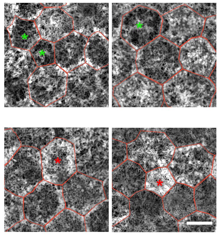
Page 2 of 9                                                                                           Annals of Eye Science, 2021

Introduction                                                     hypothesized that RPE cells at the upper and lower end in
                                                                 granule load or AF will not markedly differ from an average
The retinal pigment epithelium (RPE) cell layer is
                                                                 RPE cell. Based on our previous data on characterization
embedded between the photoreceptors and the Bruch
                                                                 and number of intracellular granules within RPE cells,
membrane. It is a monolayer of polygonal cells that play
                                                                 we analyzed those RPE cells with high/low granule load
important roles in maintaining outer retinal health, protein
                                                                 or high/low total AF regarding their retinal location, cell
and oxygen transport, and in the phagocytosis of shed
                                                                 size, intracellular granule density, and relation to age. We
photoreceptor outer segments. RPE cells incorporate
                                                                 present the following article in accordance with the MDAR
different kinds of pigmented and non-pigmented
                                                                 reporting checklist (available at http://dx.doi.org/10.21037/
organelles of imaging significance (granules): lipofuscin
                                                                 aes-2021-01).
(L), melanolipofuscin (ML), melanosomes (M), and
mitochondria (MC) (1). Some of them, L and ML, are
autofluorescent after excitation with blue light (2,3).          Methods
   Blue light fundus autofluorescence (FAF) is widely used
in routine clinical diagnostics and disease monitoring (4,5);    The study was conducted in accordance with the
however, the histological basis of FAF and age-related as        Declaration of Helsinki (as revised in 2013) and the
well as disease-related autofluorescence (AF) have been          Harmonized Tripartite Guideline for Good Clinical Practice
investigated only recently (6,7). We added information on        from the International Conference on Harmonization.
histological RPE AF, showing that RPE AF depends on              The use of human tissue was approved by the Institutional
retinal position in relation to the fovea and age. Despite the   Review Boards of the University of Alabama at Birmingham
significant increase in AF, RPE cells maintain a continuous      and the University of Würzburg (208/15). Due to its
monolayer, though individual RPE cell geometry                   retrospective character, individual consent was waived.
might change (i.e., cell shape, number of neighbors). In
degenerative diseases like age-related macular degeneration      Tissue preparation and imaging
(AMD), RPE cells overall tend to lose AF due to loss of
autofluorescent granules, although there are intensely           Tissue collection, preparation, and granule counts
hyperautofluorescent regions within cells, due to granule        within RPE cells is based on 15 RPE/BrM flat mounts
aggregation. This diminution of AF in AMD has been               (15 Caucasian donors; eight ≤51 years, seven >80 years), as
shown both in ex vivo histology (8) and in vivo quantitative     previously reported (11,13).
FAF (9,10).                                                          In brief, globes were collected from the Advancing Sight
   Recently, we further classified >195,000 intracellular        Network (formerly the Alabama Eye Bank; Birmingham,
RPE granules (L, ML, M) within 450 RPE cells based               AL, USA), and inspected under a dissection microscope to
on their autofluorescent properties using high-resolution        exclude macular or retinal pathologies. The neuroretina
structured illumination microscopy (HR-SIM) (11). A              and choroid were removed in a multistep preparation and
major finding was that the major contributor to AF at the        imaging process to ensure preservation of the exact foveal
fovea is the relative high ML load within foveal RPE cells,      position (13). RPE/BrM flat mounts were imaged using
while L dominate RPE cells at the perifovea. RPE cells can       a laser scanning confocal fluorescence microscope (LSM)
incorporate hundreds of intracellular granules, while still      and a structured illumination microscope (SIM). Excitation
maintaining geometric precision, suggesting that they are        wavelength was set at 488 nm for both microscopes and
healthy and highly functional.                                   AF emission recorded from 490 to 695 nm in 24 channels
   Previous reports suggested that high AF and massive           (LSM), or between 510 and 750 nm (SIM), respectively.
load of L granules within RPE cells is a precursor of            The scans were conducted from apical RPE (first granules
AMD or AMD related changes (12). However, subsequent             in focus) to basal RPE (last granules out of focus),
histological studies of human eyes, both in normal aging         representing the cushion of granules, in 390 nm (LSM) or
and in AMD (8,13,14) eyes provided no evidence for this          110 nm steps (SIM), respectively. Identical areas were
theory of L-instigated RPE cell death.                           imaged at three predefined locations: fovea, superior
   Here, to further support the idea that granule load           perifovea, and near-periphery at the superior edge of the
within RPE cells is well-regulated and organized we              flat mount (11,13).

© Annals of Eye Science. All rights reserved.                           Ann Eye Sci 2021;6:3 | http://dx.doi.org/10.21037/aes-2021-01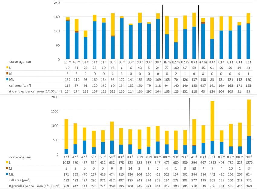
Annals of Eye Science, 2021 Page 3 of 9
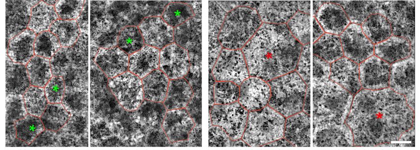
82 y/o male 83 y/o female 83 y/o female 90 y/o female
perifovea fovea near periphery near periphery
Figure 1 Retinal pigment epithelium (RPE) cells with low/high granule load. The colored asterisks mark the cells with low (green) and high
(red) amounts of granules. Individual RPE cells are red-rimmed. Structured Illumination Microscopy (summation of the whole image stack).
Scale bar: 10 µm.
Cell selection and granule classification density) was calculated.
Using custom FIJI plugins (https://fiji.sc) (15), at each
location, 10 adjacent cells were selected from reconstructed Analysis of RPE cell bodies with high/ low granule content
SIM images, and on LSM images the identical cells were or high/low AF-intensity
marked (10 cells/location for each of the 15 donors = 450
To identify cells with remarkably high or low granule
cells), as previously described. Three foveas (donor ages 36,
content, total number of granules was calculated for each
82, and 88 years) were excluded from the current analysis of the 420 cells. Cells with a granule content below the 5th
since RPE cell boundaries were not clearly detectable, or above the 95th percentile were selected for this study.
resulting in 420 RPE cells for final analysis. Absolute numbers of M, L and ML as well as mean AF-
Within SIM images, all detectable granules within the intensity per cell and cell area are reported. Also, for each
RPE cell bodies were manually tagged and classified on the cell, the (M+ML)/L-ratio was calculated.
basis of granule morphology. Nine different phenotypes To identify and characterize cells with high or low AF-
were identified based on the AF pattern and structural intensity, mean AF per cell ± standard deviation for each
properties [for details see Figure 1 in Bermond et al. (11)] location were calculated and only cells at one specific
using a custom written FIJI plugin. For each tagged granule, location within one donor were compared since our
the classification code and its x, y, z coordinates within the LSM imaging setting does not qualify for quantitative AF
stack were registered. In the current study, we distinguish measurements between locations and donors. Thus, cells
only among the classic three intracellular RPE granule with an AF-intensity 1.5 standard deviations higher or
classes: Lipofuscin granules (L, nearly spherical granules lower than the mean AF of the ten cells at a specific location
with homogenous hyper-AF), Melanolipofuscin granules were selected for further analysis. For each cell, mean AF-
(ML, round or spindle-shaped granules with iso- to hypo- intensity per cell, cell area and absolute numbers of M, L
AF core and hyper-AF coating) and Melanosomes (M, and ML per cell were reported. Also, for each of these cells,
hypo-AF round or spindle shaped granules without hyper- the (M+ML)/L-ratio was calculated, meaning that melanin
AF coating). Using LSM images from the identical cells, containing granules predominate when >1, and pure L
total AF per cell (AF intensity expressed as a planimetric granules predominate whenPage 4 of 9 Annals of Eye Science, 2021
Statistical analysis Characteristics of RPE cells with low or high AF-intensity
Data collection, organization and analysis was performed At each of the analyzed 42 locations, RPE cells with an
using SPSS statistic (IBM SPSS 25.0, IBM corporation, AF-intensity 1.5 standard deviations lower or higher than
Armonk, NY, USA) and Microsoft office software packages the location’s mean AF value were defined as cells with
(Microsoft Corporation, Redmond, WA, USA). Categorical remarkably low or high AF-intensity, respectively. Twenty
variables are presented as numbers and percentages, cells with low AF-intensity and twenty-eight cells with high
continuous variables are expressed as means ± standard AF-intensity were identified (Table 1).
deviation (STD). RPE cells with low AF-intensity had about 300 granules
in total (316.5±127.9), while RPE cells with high AF-
intensity contained about 500 granules (499.6±359.3).
Results
RPE cells with low AF-intensity were found at all three
Data on total number of granules, cell area, number of locations (5 fovea, 6 perifovea, 9 near periphery) and were
granules per cell area, and (M + ML)/L-ratio in an average distributed equally between the two age groups (10 ≤51
RPE cell related to age and the specific retinal location is years group; 10 >80 years group). They were characterized
reported in the Table 1 (11). For the current study, granule by a high (M+ML)/L-ratio at all three locations (Table 1).
data of 420 RPE cells from 15 RPE/BrM flat mounts Foveal and perifoveal RPE cells with low AF-intensity were
without retinal pathologies at three predefined locations (11) smaller than the average foveal and perifoveal RPE cell
were included (total of 42 locations). while cell area at near periphery was comparable with the
average RPE cell at this location.
RPE cells with high AF-intensity were found at all three
Characteristics of RPE cells with low or high granule load
locations (8 fovea, 9 perifovea, 11 near periphery) and in
RPE cells with low granule content (below the 5 th both age groups (13 ≤51 years group; 15 >80 years group).
percentile; total of 21 cells) were predominantly found They were characterized by a low (M+ML)/L-ratio and a
within the >80 years group (14 of 21 cells) and at the fovea cell area about the same size as an average RPE-cell (Table 1,
(11 fovea, 4 perifovea, 6 near periphery). Eight of these 21 Figure 3).
cells are from the same donor (83 y/o female). RPE cells
with low granule load are characterized by a high (M +
Discussion
ML)/L-ratio, a small cell area, and a low granule density, as
compared to normal RPE cells at the respective locations Cells of the RPE can host hundreds of granules within
(Table 1, Figure 1). their cell body, including granules of AF relevance (L,
RPE cells with high granule content (above the 95 th MLF, M) (11,16) and mitochondria (17). L and ML show
percentile; total of 21 cells) were predominantly found AF properties after blue light excitation and are the major
within the >80 years group (14 of 21 cells), and at the contributor to fundus AF (3). Elevated FAF has been
perifovea. Interestingly, none of these fully packed cells linked to excessive L load of RPE cells and to subsequent
were found at the fovea (0 fovea, 14 perifovea, 7 near development of AMD and RPE atrophy (18), though
periphery). Six of the 21 cells are from the same donor supporting histological evidence from well-characterized
(88 y/o male). RPE cells with high granule content are human eyes was lacking.
characterized by a low (M+ML)/L-ratio and a large cell area The results presented here confirm our previous data
(Table 1, Figure 1). that L load seems to be regulated at a cellular level, at least
Figure 2 displays the characteristics of those cells in normal aging eyes, and to be related to topography of
with high or low granule load. Those RPE cells with a the overlying photoreceptors. While there is an increase
high number of intracellular granules (>95 th percentile) in granule load with age, RPE cells that have massive L
contained about two times more granules compared to overload or significant increased granule density have not
an average RPE cell, and about six times more granules been found. Even those cells with the highest amount of
than an RPE cell below the 5th percentile, at the respective L granules had only twice as many granules in their cell
locations. body as the average RPE cell at the respective location. The
© Annals of Eye Science. All rights reserved. Ann Eye Sci 2021;6:3 | http://dx.doi.org/10.21037/aes-2021-01Annals of Eye Science, 2021 Page 5 of 9
Table 1 Characteristics of RPE cells with extremes of granule load and autofluorescence (AF)
RPE-cells with
Location Group Characteristic Low granule High granule All RPE-cells
Low AF-intensity High AF-intensity
content content
Fovea ≤51 years number of cells n=5 n=0 n=2 n=6 n=70
group
area [µm²] 112.0±18.5 n/a 116.0±26.9 143.0±42.6 162.3±34.4
(M+ML)/L-ratio 7.2 ±5.7 n/a 14.3±7.5 2.8±1.9 5.9±5.4
number of granules per 148.8 ±19.3 n/a 231.0±66.9 177.7±65.9 191.4±39.4
cell area [×/100 µm²]
>80 years number of cells n=6 n=0 n=3 n=2 n=50
group
area [µm²] 112.2±35.3 n/a 117.0±18.5 181.0±36.8 172.3±57.8
(M+ML)/L-ratio 15.8 ±13.4 n/a 10.3±11.9 2.1±1.4 4.9±6.6
number of granules per 177.2 ±77.4 n/a 143.3±20.6 201.3±89.3 204.7±53.1
cell area [×/100 µm²]
Perifovea ≤51 years number of cells n=1 n=6 n=3 n=2 n=80
group
area [µm²] 94.3 403.3±62.5 157.7±20.7 250.0±2.8 224.7±74.3
(M+ML)/L-ratio 1.4 0.6±0.3 1.8±0.3 0.2±0.1 1.0±0.8
number of granules per 193.1 248.3±26.2 224.0±3.9 303.1±21.8 236.2±36.0
cell area [×/100 µm²]
>80 years number of cells n=3 n=8 n=3 n=7 n=70
group
area [µm²] 144.1±7.6 317.9±73.8 143.7±11.0 204.1±47.7 215.9±65.6
(M+ML)/L-ratio 1.8 ±0.9 0.5±0.2 1.6±1.1 0.4±0.2 0.7±0.4
number of granules per 127.7 ±4.3 283.8±45.7 178.8±87.1 247.5±45.5 230.4±52.6
cell area [×/100 µm²]
Near ≤51 years number of cells n=1 n=1 n=5 n=5 n=80
periphery group
area [µm²] 437.3 576.5 194.8 ±48.3 182.2±56.6 214.0±63.4
(M+ML)/L-ratio 10.5 0.4 2.5±1.8 0.8±0.4 1.6±1.4
number of granules per 39.6 210.1 221.3±57.2 217.9±62.0 194.0±36.4
cell area [×/100 µm²]
>80 years number of cells n=5 n=6 n=4 n=6 n=70
group
area [µm²] 168.3±19.2 365.5±237.3 200.3±61.8 276.2±217.8 2151±89.7
(M+ML)/L-ratio 3.7±3.7 0.5±0.3 4.0±4.2 0.6±0.3 1.5±1.6
number of granules per 106.1±12.3 405.1±114.3 175.8±79.0 280.5±163.4 228.6±87.4
cell area [×/100 µm²]
L, lipofuscin; ML, melanolipofuscin; M, melanosomes. (M+ML)/L-ratio, melanin containing granules predominate when >1, and pure L
granules predominate whenPage 6 of 9 Annals of Eye Science, 2021
A Fovea Perifovea Near periphery
Number of granules per cell
Low granule load
Perifovea Near periphery
B
Number of granules per cell
High granule load
Figure 2 Characteristics of cells with low (A) high (B) granule load. F, female; m, male; L, lipofuscin; ML, melanolipofuscin; M,
melanosomes. The Y-axis in A is only ~10% the height of the Y-axis in B.
Noticeable differences, however, were observed in AF intensities (above or below 1.5 standard deviations
granule density between RPE cells with low and high of the mean AF at the distinct location), as compared to
granule content. Cells with a high number of granules are the adjacent cells. This finding confirms that in healthy
more densely packed, despite the fact that they already aging, AF signal arises equally from the different cells in a
have a larger area and volume. These tight packed RPE given location. However, regional differences are visible,
cells were predominantly located at the perifovea, probably as described ex vivo (13,21) and in vivo (22,23), related to
resulting from intense rod photoreceptor-RPE cell the proportion of L and ML granules and photoreceptor
interactions during the visual cycle and daily disk shedding topography (24). Whether these findings can be confirmed
with the subsequent deposition of lipofuscin granules (19). in AMD eyes is basis of ongoing studies.
On the contrary, RPE cells with low granule content show Massive lipofuscin load has been intensively discussed
predominately melanolipofuscin granules, a feature of foveal as a major contributor to the development of AMD (12).
cells, as recently shown (11). Future studies could focus on However, Rudolf et al. and others showed that the hyper-
the contribution of the different photoreceptor systems autofluorescent signal in the junctional zone of geographic
(cones versus rods) to lipofuscin accumulation within the atrophy in late-stage non-neovascular AMD could be
RPE cells and whether lipofuscin originating from cone explained by the phenomena of stacked RPE cells, i.e.,
outer segment tips differs from that of rod outer segment ectopic RPE cells that left the monolayer and migrated
tips. Imaging mass spectrometric studies of photoreceptors toward the inner retina, rather than individual RPE cells
and RPE cells might further help to clarify this (20). with massive granule load (14,25). Granule density in RPE
Only a few RPE cells showed increased/decreased cells with high AF in our study did not exceed the mean
© Annals of Eye Science. All rights reserved. Ann Eye Sci 2021;6:3 | http://dx.doi.org/10.21037/aes-2021-01Annals of Eye Science, 2021 Page 7 of 9
90 y/o female, fovea 16 y/o male, near periphery
88 y/o male, near periphery 49 y/o male, fovea
Figure 3 Retinal pigment epithelium (RPE) cells with low/high autofluorescence (AF) intensity. The colored asterisks mark the cells with low
(green) and high (red) total autofluorescence intensity. Individual RPE cells are red-rimmed. Structured Illumination Microscopy (summation
of the whole image stack). Scale bar: 10 µm.
AF of an average RPE cell by more than a factor of 1.8, predominate in the fovea, with mostly cone photoreceptors,
supporting the conclusions of Rudolf et al. (14). and L granules predominate in the perifovea, with mostly
Limitations of our study include the analysis of a small rod photoreceptors. These findings highlight the potential
number of RPE cells per location. It cannot be excluded of RPE cells to regulate intracellular granule accumulation
that results might differ in an analysis of larger sample sizes. as part of their normal physiology. Variations in diseases like
The use of tissue of normal donor eyes does not allow any AMD are currently examined.
conclusions on RPE granule distribution in diseased eyes
and its possible relationship to AMD; however, studies
Acknowledgments
examining AMD tissues are currently being conducted in
our lab. Funding: Dr. Werner Jackstädt Foundation (TA), NIH/NEI
Strengths of our analysis include RPE measurements at 1R01EY027948 (TA, RH, CAC), 1R01EY06109 (CAC).
exactly predefined locations (13), especially in relation to
the fovea and the overlying photoreceptor distribution.
Footnote
Granule distribution data revealed that RPE cells with
high granule content had only twice as many granules Provenance and Peer Review: This article was commissioned
within the cell body as an average RPE cells. RPE cells by the Guest Editor (R Theodore Smith) for the series
with massive granule loads were not detectable. Also, “Retinal Imaging and Diagnostics” published in Annals of
intracellular granule type proportion seems to follow Eye Science. The article was sent for external peer review
overlying photoreceptor distribution in that ML granules organized by the Guest Editor and the editorial office.
© Annals of Eye Science. All rights reserved. Ann Eye Sci 2021;6:3 | http://dx.doi.org/10.21037/aes-2021-01Page 8 of 9 Annals of Eye Science, 2021
Reporting Checklist: The authors have completed the MDAR Retinal Pigment Epithelium Organelles Significant for
reporting checklist. Available at http://dx.doi.org/10.21037/ Clinical Imaging. Invest Ophthalmol Vis Sci 2020;61:13.
aes-2021-01 2. Feeney-Burns L, Berman ER, Rothman H. Lipofuscin
of human retinal pigment epithelium. Am J Ophthalmol
Data Sharing Statement: Available at http://dx.doi. 1980;90:783-91.
org/10.21037/aes-2021-01 3. Delori FC, Goger DG, Dorey CK. Age-related
accumulation and spatial distribution of lipofuscin in
Conflicts of Interest: The authors have completed the RPE of normal subjects. Invest Ophthalmol Vis Sci
ICMJE uniform disclosure form (available at http://dx.doi. 2001;42:1855-66.
org/10.21037/aes-2021-01). The series “Retinal Imaging 4. Schmitz-Valckenberg S, Holz FG, Bird AC, et al. Fundus
and Diagnostics” was commissioned by the editorial office autofluorescence imaging: review and perspectives. Retina
without any funding or sponsorship. Dr. Heintzmann 2008;28:385-409.
reports grants from NIH/NEI 1R01EY027948, during 5. Delori FC, Dorey CK, Staurenghi G, et al. In vivo
the conduct of the study. Dr. Curcio reports grants fluorescence of the ocular fundus exhibits retinal pigment
from NEI/NIH 1R01EY06109, grants from NEI/NIH epithelium lipofuscin characteristics. Invest Ophthalmol
1R01EY027948, during the conduct of the study; grants Vis Sci 1995;36:718-29.
from Heidelberg Engineering, grants from Genentech/ 6. Feeney L. Lipofuscin and melanin of human retinal
Hoffman LaRoche, other from MacRegen Inc, outside the pigment epithelium. Fluorescence, enzyme cytochemical,
submitted work. Dr. Sloan reports other from MacRegen, and ultrastructural studies. Invest Ophthalmol Vis Sci
outside the submitted work. Dr. Ach reports grants from 1978;17:583-600.
NIH/NEI 1R01EY027948, grants from Dr Werner 7. Weiter JJ, Delori FC, Wing GL, et al. Retinal pigment
Jackstädt Foundation, other from MacRegen, during the epithelial lipofuscin and melanin and choroidal melanin in
conduct of the study. The authors have no other conflicts of human eyes. Invest Ophthalmol Vis Sci 1986;27:145-52.
interest to declare. 8. Ach T, Tolstik E, Messinger JD, et al. Lipofuscin
redistribution and loss accompanied by cytoskeletal
Ethical Statement: The authors are accountable for all aspects stress in retinal pigment epithelium of eyes with age-
of the work in ensuring that questions related to the accuracy related macular degeneration. Invest Ophthalmol Vis Sci
or integrity of any part of the work are appropriately 2015;56:3242-52.
investigated and resolved. The study was conducted in 9. Gliem M, Muller PL, Finger RP, et al. Quantitative
accordance with the Declaration of Helsinki (as revised in Fundus Autofluorescence in Early and Intermediate
2013). The study was approved by institutional review boards Age-Related Macular Degeneration. JAMA Ophthalmol
of the University of Alabama at Brimingham and University 2016;134:817-24.
of Würzburg (NO.: 208/15). Due to its retrospective 10. Orellana-Rios J, Yokoyama S, Agee JM, et al. Quantitative
character, informed consent was waived from all participants. Fundus Autofluorescence in Non-Neovascular Age-
Related Macular Degeneration. Ophthalmic Surg Lasers
Open Access Statement: This is an Open Access article Imaging Retina 2018;49:S34-42.
distributed in accordance with the Creative Commons 11. Bermond K, Wobbe C, Tarau IS, et al. Autofluorescent
Attribution-NonCommercial-NoDerivs 4.0 International Granules of the Human Retinal Pigment Epithelium:
License (CC BY-NC-ND 4.0), which permits the non- Phenotypes, Intracellular Distribution, and Age-Related
commercial replication and distribution of the article with Topography. Invest Ophthalmol Vis Sci 2020;61:35.
the strict proviso that no changes or edits are made and the 12. Dorey CK, Wu G, Ebenstein D, et al. Cell loss in the
original work is properly cited (including links to both the aging retina. Relationship to lipofuscin accumulation
formal publication through the relevant DOI and the license). and macular degeneration. Invest Ophthalmol Vis Sci
See: https://creativecommons.org/licenses/by-nc-nd/4.0/. 1989;30:1691-9.
13. Ach T, Huisingh C, McGwin G, Jr., et al. Quantitative
autofluorescence and cell density maps of the human
References
retinal pigment epithelium. Invest Ophthalmol Vis Sci
1. Pollreisz A, Neschi M, Sloan KR, et al. Atlas of Human 2014;55:4832-41.
© Annals of Eye Science. All rights reserved. Ann Eye Sci 2021;6:3 | http://dx.doi.org/10.21037/aes-2021-01Annals of Eye Science, 2021 Page 9 of 9
14. Rudolf M, Vogt SD, Curcio CA, et al. Histologic basis of Landscape of the Human Retina and Supporting Tissues
variations in retinal pigment epithelium autofluorescence Revealed by High-Resolution Imaging Mass Spectrometry.
in eyes with geographic atrophy. Ophthalmology J Am Soc Mass Spectrom 2020. [Epub of print]. doi:
2013;120:821-8. 10.1021/jasms.0c00119.
15. Schindelin J, Arganda-Carreras I, Frise E, et al. Fiji: an 21. Wing GL, Blanchard GC, Weiter JJ. The topography
open-source platform for biological-image analysis. Nat and age relationship of lipofuscin concentration in the
Methods 2012;9:676-82. retinal pigment epithelium. Invest Ophthalmol Vis Sci
16. Pollreisz A, Messinger JD, Sloan KR, et al. Visualizing 1978;17:601-7.
melanosomes, lipofuscin, and melanolipofuscin in human 22. von Rückmann A, Fitzke FW, Bird AC. Distribution
retinal pigment epithelium using serial block face scanning of fundus autofluorescence with a scanning laser
electron microscopy. Exp Eye Res 2018;166:131-9. ophthalmoscope. Br J Ophthalmol 1995;79:407-12.
17. Pollreisz A, Neschi M, Sloan KR, et al. Deciphering 23. Greenberg JP, Duncker T, Woods RL, et al. Quantitative
subcellular signal sources for optical coherence fundus autofluorescence in healthy eyes. Invest
tomography (OCT) and autofluorescence (AF) imaging Ophthalmol Vis Sci 2013;54:5684-93.
of the human retinal pigment epithelium (RPE). Invest 24. Curcio CA, Sloan KR, Kalina RE, et al. Human
Ophthalmol Vis Sci 2019;60:1574. photoreceptor topography. J Comp Neurol
18. Dorey C, Staurenghi G, Delori F. Lipofuscin in aged and 1990;292:497-523.
AMD eyes. Retinal degeneration. Boston, MA: Springer, 25. Zanzottera EC, Ach T, Huisingh C, et al. Visualizing
1993:3-14. Retinal Pigment Epithelium Phenotypes in the Transition
19. Crouch RK, Koutalos Y, Kono M, et al. A2E and to Geographic Atrophy in Age-Related Macular
Lipofuscin. Prog Mol Biol Transl Sci 2015;134:449-63. Degeneration. Retina 2016;36 Suppl 1:S12-25.
20. Anderson DMG, Messinger JD, Patterson NH, et al. Lipid
doi: 10.21037/aes-2021-01
Cite this article as: Bermond K, Berlin A, Tarau IS, Wobbe C,
Heintzmann R, Curcio CA, Sloan KR, Ach T. Characteristics of
normal human retinal pigment epithelium cells with extremes
of autofluorescence or intracellular granule count. Ann Eye Sci
2021;6:3.
© Annals of Eye Science. All rights reserved. Ann Eye Sci 2021;6:3 | http://dx.doi.org/10.21037/aes-2021-01You can also read