Innovative Methods Of Miscellaneous Skin Inclusions Removal And Post-Inflammatory Hyperpigmentation Correction
←
→
Page content transcription
If your browser does not render page correctly, please read the page content below
European Journal of Molecular & Clinical Medicine
                                                  ISSN 2515-8260          Volume 07, Issue 05, 2020

  Innovative Methods Of Miscellaneous Skin
  Inclusions Removal And Post-Inflammatory
         Hyperpigmentation Correction
                               Anastasiya V. Fridman-Sorokina

 Graduate student, Kemerovo State University, Kemerovo 650000 Russia, 6 Krasnaya Street

                                 e-mail: sibtatoo@gmail.com.

Abstract: Scarless tattoo extraction remains as a major problem in a cosmetology field
nowadays. At the moment laser removing method is a basic procedure for tattoo removal.
However, this technology is painful and leads to significant trauma of the skin cover. Thise
article presents a new combination of laser and chemical technique for skin inclusions
removal. The group of techniques belongs to the chemical industry and represents a
lightning and neutralizing composition and a method for removing a dye introduced under
the skin. The skin area with dye is exposed to a laser effect. After laser exposure an
oxidizing composition of 2-3 mm thickness is applied. Oxidizing composition: carboxylic
acids, fermented celandine oil extracts and propylene-glycol celandine extracts. The main
novelty of the method is presented by several chain processes: in applying of the oxidizing
composition on the skin surface after laser radiation, a significant role is played by
physicochemical characteristics changes of the skin. Without the skin surface damaging,
the percentage of chemical reactions dissociations aimed at removing the pigment is
raising which shows better efficiency comparing with classical laser method of dye
particles removing.
Keywords: tattoos, laser, oxidizing, hyperpigmentation, dye removal

   1. INTRODUCTION

        Despite significant advances in cosmetic surgery, scar less tattoo removing as well as
post inflammatory hyperpigmentation (PIH) correction remains as the leading problems of
modern cosmetology [1]. The chemical composition and features of extracted agents is quite
different: coal (carbon), bitumen, gunpowder, mechanical dirt, graphite particles. Most
frequent foreign pigment removal happens as a result of unsuccessful cosmetic procedures,
cosmetic tattooing, under specific working conditions (such as the coal industry, varnish
industry, metallurgy), occupations associated with increased danger (emergency situations),
as well as creative professions associated with a certain risk (stuntman). Foreign body
extraction is the elimination of dye large particles, as well as particles of potentially coloring
substances that enter the skin when injured (coal, ground) [2]. Carbon fractions have the
highest inertness and are difficult to extract from the skin [3].
        Tattoos removal growth. Procedure of intradermal multicolor pigment granules
insertion is called tattooing [4]. Also, tattoo formation can be the result of accidents and
trauma. Tattoos can broadly be divided into professional, amateur, cosmetic, traumatic, or
medical tattoos. Professional tattoos are applied with a tattoo machine into the deeper layer of
the dermis, and are applied to be permanent in nature [5]. The worldwide tattoo prevalence is
10-20% depending on the region, population of the country and the time of survey

                                                                                               214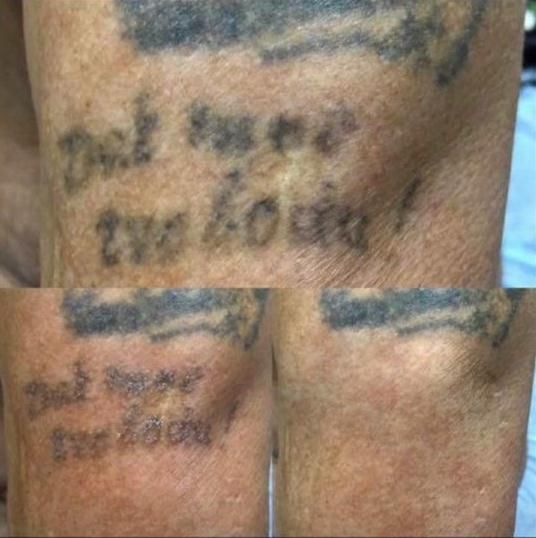
European Journal of Molecular & Clinical Medicine
                                                 ISSN 2515-8260         Volume 07, Issue 05, 2020
performing [6]. Unfortunately, there are still no strict and common legislation for the tattoo
safety regulation, therefore quantity of tattoos complication is tending to grew up [7].
        Due to the rising popularity of tattoos, demand for their removal has also increased
[8]. Unfortunately, the removal of tattoos is generally costlier and time consuming than
acquiring them [5]. Motivation for tattoo removal includes new jobs or careers, the need to
portray a certain image at work or in new social circles, and new, negative feelings towards
old tattoos [9]. Some patients with tattoo reactions have reduced quality of life and suffer
from itch [10].
        “Coal Tattooing”. Despite the fact deep coal mines are closing, there are still
negative effects of mining on physical health that need to be addressed. Mining exposes
workers to a variety of potentially harmful agents, including fuels, reagents, solvents,
detergents, chemicals, coal dust, silica dust, diesel particulate matter, asbestos, welding
fumes, poisonous plants and metal dust. These may be inhaled, ingested or absorbed through
the skin, eyes, mucous membranes or ears. Miners are often exposed to them for decades
before any adverse effects are noticed. In the past, they may not always have been adequately
instructed about the health risks involved and the safety precautions required [11].
        Coal tattooing, also known as „colliers‟ stripes‟ is resulted from scratches and small
injuries that healed without scarring, in which coal dust was deposited before healing was
complete. Commonly found on the face, forearms and hands, they presented as light greyish-
blue linear or angular markings, measuring up to 1 inch in length [12-13].
        Chronic (cumulative) irritant contact dermatitis used to be commonly seen in miners.
Coal dust is presented in miners working process, therefore unprotected by work-wear human
skin resorb some portion of the dust. Coal, is a black dye, freely moves in the air and through
the sweat glands enters the various layers of the dermis reaching the lymph nodes, with
further fibrous tissue overgrow and cosmetic defect causing. The work conditions, the mine‟s
geographical location, depth, temperature, humidity and ventilation, and the physical and
chemical properties of the extracted mineral can all have a role in its aetiology [14-15].
        Post inflammatory hyperpigmentation (PIH). Post inflammatory hyperpigmentation
(PIH), also known as post inflammatory melanosis, is a reactive hypermelanosis of the skin
that occurs as a sequela of cutaneous inflammation. Common causes of PIH include acne
vulgaris, eczematous dermatoses, and burn injury. PIH is a frustrating problem that can have
a strong psychologic toll on affected patients. The provoking inflammatory process that leads
to post inflammatory hyperpigmentation (PIH) can be endogenous or exogenous. Common
endogenous causes of PIH include acne vulgaris, atopic dermatitis, irritant contact dermatitis,
allergic contact dermatitis, psoriasis, and lichen planus. Accidental burns, nonionizing
radiation therapy, phototoxicity, chemical peels, and laser procedures are examples of
exogenous causes [16-17].
        Abnormal hyperpigmentation. Post‐inflammatory hyperpigmentation is one of the
most common and rather persistent in dark‐skinned people. The different skin conditions like
inflammatory dermatoses, trauma and medical interventions (such as laser therapy) are in
dark people often the etiology of remaining hyperpigmentation. Sunlight, some medication
and chemicals often worsen the spots. The dyschromia follows the pattern and distribution of
the original dermatoses, but its intensity is not necessarily related to the degree of previous
inflammation. Epidermal pigmentation is mostly brown and fades out in several months.
Dermal pigmentation has a grey‐brown color and is generally permanent for years.3
Treatment of post‐inflammatory hyperpigmentation is difficult. The primary goal of therapy
is treating the etiology. Most significant clinical improvement for the lesions is directly
correlated with different topical therapies such as depigmenting agents. Particularly important
is the combination of these therapies with the frequented use of sunscreens [18].

                                                                                             215
European Journal of Molecular & Clinical Medicine
ISSN 2515-8260 Volume 07, Issue 05, 2020
Skin inclusions elimination approaches. Through the years, many different methods
of tattoo removal have been explored. Older techniques involve removal of the outer skin
layers using mechanical (dermabrasion and salabrasion), chemical, or thermal (cryosurgery
and cauterization) methods. Progress in laser technology offers alternative treatments to
patients with cutaneous discolorations, including post inflammatory hyperpigmentation and
tattoos [19-20].
Lasers have revolutionized the way tattoos are treated and have become the gold
standard of treatment. To achieve optimal cosmetic outcome of treatment, lasers emitting
high energies and short pulses are required to adequately destroy tattoo ink [21]. Lasers based
on the principle of selective photothermolysis are now being used to remove black as well as
colorful tattoos with varying successes [22].
Among modern methods of ink removal are chemical subcutaneous, chemical
external, laser [23]. Currently, the only FDA approved tattoo removal devices are laser based;
FDA has not approved the topical or injectables for tattoo removal [24].
We are presenting a new combination of laser and chemical technique for skin
inclusions removal.
2. MATERIAL AND METHODS
Application of a lightening agent after laser radiation exposure on the target area (A
variant). Pigment particles in the skin are unevenly located, with different depths (vary from
the epidermis border and the dermis, to presence in the lymph nodes and subcutaneous fat).
These particles also have an uneven spot shaped (dashed) dot-like deposition, various
concentrations and relation to the skin relief (hypotrophic, hypertrophic type), different
fraction, density, and other physicochemical characteristics [25-26]. The skin area with dye is
exposed to a laser effect equal to one impulse of the untreated skin zone. For this technique
we use different types of lasers: Nd-YAG, QSwitched, Picosecond, Alexandrite, Erbium. The
power for each laser modification should be minimal, without increasing at further stages of
removal. The nozzle of the laser has be 532 nm, 1064 nm, depending on the source. After
laser exposure an oxidizing composition of 2-3 mm thickness is applied, the exposure time is
1 minute. Oxidizing composition: carboxylic acids, fermented celandine oil extracts and
propylene-glycol celandine extracts (Table 1). The maximum processing area should not
exceed 23 cm2. After 2-3 minutes, the area is wiped with a dry disk; the zone is cooled
according to the laser modification algorithm.
Table 1. Oxidizing composition components
The part of Active
Initial
the substance substance
A composition concentration of
Ingredient in the content in the pH
component the ingredient
finished finished
active substance
product, % product, %
32.2 % water
Oxidizing agent Glycolic acid 15-20 4.9-5.9 3.6
solution
28.9 % water
Oxidizing agent Lactic acid 15-20 3.1-4.1 3.6
solution
Propylene 15.0 % water
Entering agent 30-40 14.0 3.6
glycol solution
Celandine
Lightening agent 99.5 % 30-40 24.8-26.8 3.6
extract
Viscosity Glycerin 99.5 % 2-5 0.5-2.2 3.6
216European Journal of Molecular & Clinical Medicine
ISSN 2515-8260 Volume 07, Issue 05, 2020
stabilizing agent
Solvent Water 100 % 12-14 78.5-79.2 3.6
The lightening area is neutralized by a composition containing triethanolamine or
diethanolamine. A sterile dressing is applied then, and the regeneration process begins [28].
After the subject area is occurred by laser, a laser's acoustic wave produces
photomechanical fragmentation of the particle is formed. Herewith the particles of the
extracting pigment are decaying into small fractions over the extracted dye area, remaining
directly in the skin layers. The tissue at this moment remains visually intact, but its physical
and chemical condition changes. The temperature increasing leads to the hydrogen bonds
rupturing and raising of capillary pressure and microcirculation. The molecules structure of
the irradiated tissue is disturbed.
The rate of chemical and biological processes of such tissue increases with the
oxidizing agent participation deposited after laser exposure to the lightning area. Pigment
particles presented by oxides, organic and inorganic compounds, enter into various types of
reactions with an oxidizing composition. Oxidizing composition applying reduces the time of
dye particles complete removal and minimize the risk of color inversion.
The main novelty of our method is presented by several chain processes: in applying
of the oxidizing composition on the skin surface after laser radiation, a significant role is
played by physicochemical characteristics changes of the skin. Without the skin surface
damaging, the percentage of chemical reactions dissociations aimed at removing the pigment
is raising which shows better efficiency comparing with classical laser method of dye
particles removing. The various characteristics of foreign intradermal particles are not
fundamental, which is a significant advantage of the combined method.
Application of a lightening agent before laser radiation exposure on the target area
(B variant). A thin layer of the oxidizing composition is applied on the treated skin for 1
minute, and then the skin is exposed to laser radiation. The skin area is cooled according to
the algorithm established by the laser removal procedure, the area is neutralized, a sterile
dressing is applied, and then the regeneration process takes place.
The high lightning effect of the oxidizing agent is ensured by penetrating into the
tissue in non-invasive way without skin integrity violation by the laser radiation. The
lightning composition enters the upper dermal layers using an amplified laser pulse and
enters various skin chemical reactions simultaneously with acoustic effects of laser. Under
the laser radiation influence, the oxidizing moisture of the upper dermal skin layer
evaporates. Thus the lightning agent resorption percentage at the pigment location level is
raising. Due to these mechanisms, dye removal is greatly enhanced. Variant B requires
minimal power of lasers.
Advantages of B variant. Despite the fact some lasers enters deep skin layers up to 10
mm, our method ensures increasing tissue absorption coefficient of the radiation, allowing the
skin tissues avoiding laser beam damaging. Consequently, pathological processes of
excessive skin damage are reducing.
3. RESULTS AND DISCUSSION
The chemical and laser combined method of intradermic particles removal allows to
work at minimum laser power, achieving better results without the skin integrity violation,
avoiding burn-form skin complications and overheating of large skin areas, due to the
enlarged time of intradermic dye processing. The level of laser operation control is reduced,
the output result is increased, regardless of the dye type dye and color introduced, and the risk
of color gamut inversion is reduced. On the large processing areas while combined method
217European Journal of Molecular & Clinical Medicine
ISSN 2515-8260 Volume 07, Issue 05, 2020
using, the risk of general overheating and scar formation is reduced. The risk of skin damage
is reduced by decreasing the time of temperature exposure and the absence of the necessity
for increasing laser power. The better result of combined method using is also observed in
areas with high dye concretion, in "expanded" areas that are considered the most dangerous.
It becomes possible to study color tattoos with a single nozzle, absorbing more quantity of his
dye, without losing the result. For example, a nozzle with a 1064 nm wavelength without
using a nozzle 532 nm, or vice versa 532 nm nozzle, without using 1064 nm. The technology
can be applied for mixed tattoos, which could have simultaneously black, red, green, blue,
yellow, and other (Fig. 1).
Figure 1. Tattoo removal procedure with combination of chemical and laser methods
During the subcutaneous chemical method, the upper epidermis layer undergoes
mechanical destruction to ensure maximum resorption of the active substance by tissues
containing the previously introduced dye. The disadvantage of the chemical method is the
skin integrity destruction, the inability to predict the exact timing of rehabilitation, excessive
injuries, and absence of special tables to establish approximate dates of skin regeneration.
External chemical method of intradermic skin particles extraction provides high
concentrations of acids and alkalis applying, which can destroy the deeper layers of the skin.
The disadvantage of this method is the high percentage of chemical burns, a large
rehabilitation period, and uncontrolled destruction of the dermis and epidermis layers arising
from the aggressive effects of reagents on the skin. As a result, various pathological tissue
changes leading to various types of life disorders. The laser method does not destroy the skin
when used correctly, but the risk is increased with an unprofessional approach. For the
classical method of operation, combinations of nozzles with various wave parameters are
recommended. Another difficulties arising from the danger of re-applying the beam and
removing the “colors of risk” - beige, white, light, blue, all colors with an admixture of
titanium dioxide. There is also a risk of over large tissue areas overheating, the risk of burns,
the risk of color inversion, limitation of work on the skin with increased production of
melanin [28-31].
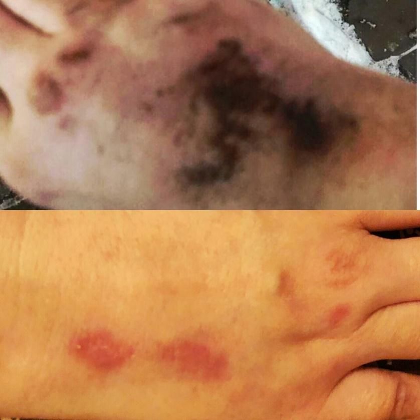
Complications of Laser Tattoo Removal. Laser removal method complications divided
into immediate (pain, blistering, urticarial reaction) and delayed (pigmentary changes,
paradoxical darkening, allergic reactions, surface changes). The most common complication
218European Journal of Molecular & Clinical Medicine
ISSN 2515-8260 Volume 07, Issue 05, 2020
is pigmentary changes, either hypopigmentation or hyperpigmentation. These occur 4-6
weeks after laser treatment and most of them are transient [32] (Fig. 2).
Figure 2. Example of hyperpigmentation correction with chemical and laser methods usage.
4. CONCLUSION
We discovered an optimal method to treat scars after tattooing and hyperpigmentation
correction was, which can be used to remove permanent makeup (tattoo) or tattoos. The
benefit of the introduction of these techniques is the reduction in the time required to remove
the dye, the reduction of skin trauma, the reduction of pain, and the increase in the healing
rate of the treated area.
5. REFERENCES
[ 1] Ozerskaya, О.S. (2007). Skin scars and their dermatocosmetological correction.
St.Peterburg: Iskustvo Rossii, 224 p. [in Russian].
[ 2] Sorokina, А.V. (2019). A modern look at the treatment of scars after tattooing. Scient.
and Pract. Conf.: Medicine. Sociology. Philosophy. Applied research, 6: 3-5. [in
Russian].
[ 3] Mathur, R.B. Singh, B.P., Pande, Sh. (2017). Carbon Nanomaterials Synthesis,
Structure, Properties and Applications, 1: 284. doi: 10.1201/9781315371849.
[ 4] Graudenz, K., Greve, B., Raulin, C. (2003). Diffused traumatic dirt and decorative
tattooing. Removal by Q-switched lasers. Hautarzt. 54(8): 756-759.
doi:10.1007/s00105-003-0493-6.
[ 5] Ho, S.G., Goh, C.L. (2015). Laser tattoo removal: a clinical update. Journal of
cutaneous and aesthetic surgery, 8(1): 9–15. doi: 10.4103/0974-2077.155066.
[ 6] Serup, J., Kluger, N., Bäumler, W. (2015). Tattooed Skin and Health. Curr Probl
Dermatol. Basel, Karger, 48: 6-20. doi: 10.1159/000369175.
219European Journal of Molecular & Clinical Medicine
ISSN 2515-8260 Volume 07, Issue 05, 2020
[ 7] Ortiz, A.E., Alster, T.S. (2012). Rising Concern over Cosmetic Tattoos. Dermatol Surg,
38: 424-429. doi:10.1111/j.1524-4725.2011.02202.x.
[ 8] Naga, L.I., Alster, T.S. (2017). Laser Tattoo Removal: An Update. Am J Clin Dermatol,
18: 59–65. doi: 10.1007/s40257-016-0227-z.
[ 9] Armstrong, M.L., Roberts, A.E., Koch, J.R., Saunders, J.C., Owen, D.C., Anderson,
R.R. (2008). Motivation for Contemporary Tattoo Removal: A Shift in Identity. Arch
Dermatol.144(7):879–884.
[ 10] Hutton, C.K., Serup, J. (2015), Patients with tattoo reactions have reduced quality of
life and suffer from itch. Skin Res Technol, 21: 101-107. doi:10.1111/srt.12164.
[ 11] Scott, D.F., Grayson, R.L. (2001). Selected Health Issues Mining [Electronic Resource].
Access: https://www.cdc.gov/niosh/mining/UserFiles/works/pdfs/shiim.pdf
(08.07.2020).
[ 12] Hodgson, G. (1955) Skin hazards of coal mining with particular reference to dermatitis.
British Journal of Dermatology, 67(12): 426-433.
[ 13] Bettley, F.R. (1940). Colliers‟ stripes: the coal-miners‟ dermatosis. British Journal of
Dermatology, 52(4): 129-130.
[ 14] Williamson, D.M. (1981). Skin hazards in mining. British Journal of Dermatology,
105(21): 41-44.
[ 15] Lawton, S., Miles, G. (2019). Occupational skin and lung disease in coalfield
communities. Nursing Times, 115: 7, 58-60. Issn Print: 0954-7762.
[ 16] Callender, V.D., St. Surin-Lord, S., Davis, E.C. et al. (2011). Postinflammatory
Hyperpigmentation. Am J Clin Dermatol 12: 87–99. doi: 10.2165/11536930-
000000000-00000.
[ 17] Davis, E.C., Callender, V.D. (2010). Postinflammatory hyperpigmentation: a review of
the epidemiology, clinical features, and treatment options in skin of color. The Journal
of clinical and aesthetic dermatology, 3(7), 20–31.
[ 18] Nieuweboer‐Krobotova, L. (2013), Hyperpigmentation: types, diagnostics and targeted
treatment options. Journal of the European Academy of Dermatology and Venereology,
27: 2-4. doi:10.1111/jdv.12048.
[ 19] Gómez, C., Martin, V., Sastre, R., Costela, Á., García-Moreno, I. (2009). In Vitro and
In Vivo Laser Treatments of Tattoos: High Efficiency and Low Fluences. Arch
Dermatol, 146(1): 39–45. doi:10.1001/archdermatol.2009.321.
[ 20] Kirby, W., Chen, C. L., Desai, A., Desai, T. (2013). Causes and recommendations for
unanticipated ink retention following tattoo removal treatment. The Journal of clinical
and aesthetic dermatology, 6(7), 27–31.
[ 21] Naga, L.I., Alster, T.S. (2017). Laser Tattoo Removal: An Update. Am J Clin Dermatol,
18, 59–65. doi: 10.1007/s40257-016-0227-z.
[ 22] Choudhary, S., Elsaie, M.L., Leiva, A. et al. (2010). Lasers for tattoo removal: a review.
Lasers Med Sci, 25: 619–627. doi: 10.1007/s10103-010-0800-2.
[ 23] Kazanddjieva, J., Tsankov, N. (2007). Tattoos: dermatological complications. Clin
Dermatol, 25: 375-382.
[ 24] Juhasz, M., Cohen, J.L. (2018). Treatment of Hypertrophic Scarring Attempted Caustic
Tattoo Removal. Skin Res Technol. 24(4):636-641. doi: 10.1111/srt.12578.
[ 25] Sorokina, A.V. Method for treating scar tissue and compositions for its implementation.
Patent 2686310, Russian Federation, MPK A61K31 / 19, A61K31 / 185, A61K31 / 164,
A61P17 / 02. №2018111195, Published 25.04. 2019.
[ 26] Sorokina, A.V. Method for combined chemical and laser removal of dye injected under
the skin. Invention patent 2019132117, Russian Federation, A61Q1 / 14, A61Q19 / 02,
A61N5 / 067, A61B18 / 20. №2019132117.04(063297), Published 21.07.2020.
220European Journal of Molecular & Clinical Medicine
ISSN 2515-8260 Volume 07, Issue 05, 2020
[ 27] Gurevich, K. G., A. L Urakov, L. I Bashirova, A. V Samorodov, P. P Purygin, V. A
Yermokhin, A. S Gilmutdinova, and N. A Bondareva. The hemostatic activity of bis (2-
aminoethan-1-sulfonate) calcium. Asian Journal of Pharmaceutical and Clinical
Research, Vol. 11, no. 11, Nov. 2018, pp. 452-5, doi:10.22159/ajpcr.2018.v11i11.29049
[ 28] Sorokina, A.V. Method for removing dye introduced under the skin and composition for
its implementation. Patent 2650630, Russian Federation, MPK A61K8 / 36, A61K8 /
41, A61Q19 / 02. №2017119180; Published 16.04.2018.
[ 29] Tredget, E.E., Shankowsky, H.A., Pannu, R. (2008). Transforming growth factor-beta
in thermally-injured patients with hypertrophic scars: effects of interferom alpha-b.
Plast. Reconstr.-Sung. 102(5): 1317-1328.
[ 30] Sorokina, А.V. (2019). Combination of laser and chemical removal of dye injected
under the skin. Int. Conf.: Refrigeration and biotechnology, Kemerovo, 97-100.
[ 31] Sorokina, A.V. (2019). Modern aspects of the treatment of scar tissue using the patent
"Method for the treatment of scar tissue and compositions for its implementation".
Medicine. Sociology. Philosophy. Applied Research, 5: 13-16.
[ 32] Khunger, N., Molpariya, A., Khunger, A. (2015). Complications of Tattoos and Tattoo
Removal: Stop and Think Before you ink. Journal of cutaneous and aesthetic surgery,
8(1), 30–36. doi: 10.4103/0974-2077.155072.
221You can also read