Optimizing Operating Points for High Performance Lesion Detection and Segmentation Using Lesion Size Reweighting
←
→
Page content transcription
If your browser does not render page correctly, please read the page content below
Medical Imaging with Deep Learning 2021

                                               Optimizing Operating Points for High Performance Lesion
                                               Detection and Segmentation Using Lesion Size Reweighting

                                           Brennan Nichyporuk1,2                                      brennan.nichyporuk@mail.mcgill.ca
                                           Justin Szeto1,2                                                      justin.szeto@mail.mcgill.ca
                                           Douglas L. Arnold3                                                    douglas.arnold@mcgill.ca
                                           Tal Arbel1,2                                                                 arbel@cim.mcgill.ca
                                           1
                                               Centre for Intelligent Machines, McGill University, Montreal, Canada
                                           2
                                               MILA (Quebec Artificial Intelligence Institute), McGill University, Montreal, Canada
arXiv:2107.12978v1 [eess.IV] 27 Jul 2021

                                           3
                                               Montreal Neurological Institute, McGill University, Montreal, Canada

                                                                                           Abstract
                                                 There are many clinical contexts which require accurate detection and segmentation of all
                                                 focal pathologies (e.g. lesions, tumours) in patient images. In cases where there are a mix of
                                                 small and large lesions, standard binary cross entropy loss will result in better segmentation
                                                 of large lesions at the expense of missing small ones. Adjusting the operating point to
                                                 accurately detect all lesions generally leads to oversegmentation of large lesions. In this
                                                 work, we propose a novel reweighing strategy to eliminate this performance gap, increasing
                                                 small pathology detection performance while maintaining segmentation accuracy. We show
                                                 that our reweighing strategy vastly outperforms competing strategies based on experiments
                                                 on a large scale, multi-scanner, multi-center dataset of Multiple Sclerosis patient images.
                                                 Keywords: multiple sclerosis, segmentation, detection, deep learning

                                           1. Introduction
                                           Many clinical contexts require accurate detection and segmentation of multiple lesions of
                                           varying sizes within a single patient image, either to diagnose or stage a disease or de-
                                           termine treatment efficacy (Doyle et al., 2017). Methods based on the UNet architecture
                                           (Ronneberger et al., 2015) use pixel-wise loss functions to learn the appropriate segmen-
                                           tation output given an input MRI and a target. Although voxel-wise loss functions have
                                           proven effective to train models to produce accurate segmentations as measured by voxel-
                                           wise metrics such as DICE, they suffer from an inherent bias towards larger lesions that
                                           contain more voxels. As a result, voxel-wise loss functions typically miss smaller lesions at
                                           operating points that are favorable to voxel-wise metrics such as DICE (Nair et al., 2020).
                                           Reducing the detection threshold to an operating point that is more suited for detection is
                                           a feasible workaround, but this comes at the cost of over-segmenting larger lesions. Given
                                           that the optimal operating point for detection and segmentation are different, simultane-
                                           ously achieving both objectives is not possible with standard loss functions. Recent research
                                           (Shirokikh et al., 2020) suggests that re-weighing the voxels of each lesion in a manner that
                                           is inversely proportional to that lesion’s size can be an effective way to improve small lesion
                                           detection performance. Although this approach directly deals with the size imbalance be-
                                           tween multiple lesions, it assigns equal weight to each lesion, which can be problematic in
                                           contexts such as cancer and Multiple Sclerosis, where lesions span a wide range of sizes (and
                                           can be quite small). In this work, we propose a novel weighing function that is much less

                                           © 2021 B. Nichyporuk, J. Szeto, D.L. Arnold & T. Arbel.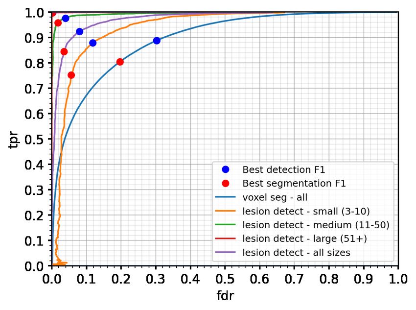
Nichyporuk Szeto Arnold Arbel
prone to the training instability caused by assigning a high weight to smaller lesions that
are typically much more uncertain. Our approach closes the detection/segmentation perfor-
mance gap, showing that with the right lesion reweighing strategy, high overall simultaneous
detection and segmentation accuracies are possible. Through large scale experiments on a
large propriety dataset of Multiple Sclerosis patient images, the proposed method outper-
forms the competing baseline and several other common loss functions.
2. Lesion Size Reweighting
We propose a lesion weighing function, where the objective is to have the optimal detection
and segmentation operating points converge by assigning more weight to small lesions than
would otherwise be assigned by binary cross entropy. Although small lesions can be weighed
more, they should still be assigned less weight than larger lesions, which are typically much
more certain. Our conjecture is that assigning too much weight to small lesions can produce
suboptimal results.
Formally, each lesion Lj is assigned a weight Wj that is a function of the number of
voxels |Lj | that comprise that lesion. In practice, weights must be assigned to individual
voxels rather than individual lesions, so we also define the voxel weight wj , related to Wj
W
via wj = |Ljj| .
− β1 (|Lj |−1) α − β1 (|Lj |−1)
Wj = |Lj | + αe wj = 1 + e (1)
|Lj |
where α and β are hyperparameters such that α ≤ β to ensure monotonicity in the weight
with respect to lesion size. Background (i.e. non lesions) voxels retain a weight of 1.
3. Experiments and Results
We train a UNet architecture (Ronneberger et al., 2015) to segment T2 lesions with bi-
nary cross entropy (BCE), weighted BCE (WBCE), focal loss (FL) (Lin et al., 2017), BCE
with the proposed lesion size reweighting (BCE+LSR), and BCE with inverse weighting
(BCE+IW) (Shirokikh et al., 2020). Hyperparameters common to all methods (augmenta-
tion, dropout, etc.) were first tuned for our baseline BCE model. We then freeze these hy-
perparameters for all subsequent experiments, modifying only the loss function and learning
rate. Hyperparameters for the proposed BCE+LSR loss function were tuned on a log2 scale
(α = 4 and β = 4 performed best in our experiments). Our dataset (train/validation/test),
contains 1350/175/175 MRI scans from 575/175/175 subjects obtained over the course of a
2 year clinical trial. The train split contains 1-3 scans per subject, each taken 1 year apart.
MRI sequences used include FLAIR, PDW, T2, T1, and gadolinium enhanced T1.
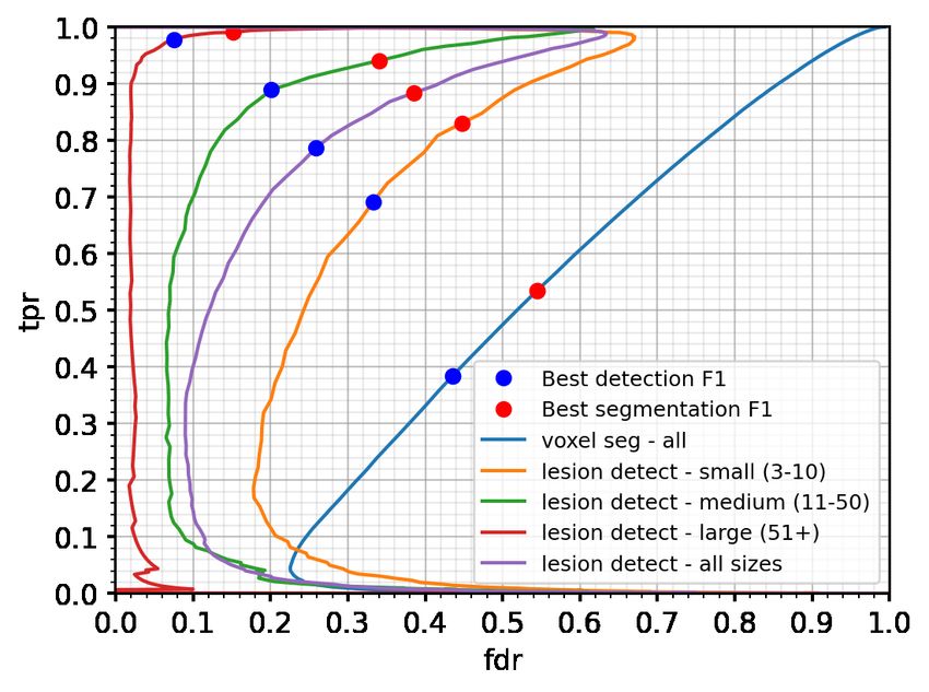
Figure 1 shows the TPR vs FDR curves and compares overall segmentation performance
with detection performance for small (3-10 voxels), medium (11-50 voxels), and large (51+
voxels) lesions for the proposed BCE+LSR, as compared to BCE and BCE+IW. In the
case of BCE+LSR, the optimal operating points for segmentation and detection (red and
blue dots) overlap and the method performs well on both tasks. This is in contrast to BCE,
for which the optimal operating points are comparatively far apart, and which shows a
degree of over-segmentation at the optimal detection operating points (and under-detection
2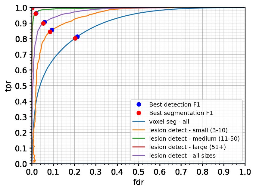
LSR for Optimal Lesion Detection and Segmentation

at the optimal segmentation operating point, particularly for small lesions). WBCE and FL
exhibited performance characteristics similar to BCE. For BCE+IW, the distance between
the optimal detection and segmentation operating points is even larger, and the method
significantly underperforms all others. Given the significant decrease in performance for
BCE+IW relative to both BCE and BCE+LSR, further analysis revealed that BCE+IW
applied substantial weight to extremely small lesions. Since the lesion weights computed by
BCE+IW ranged over several orders of magnitude, training was extremely unstable. On
the other hand, using the proposed BCE+LSR, the weights remain in a reasonable range,
upper bounded by 1 + |Lαj | . Since smaller lesions are considerably more uncertain, using a
weighting scheme with a reasonable upper bound prevented training instability.

       (a) BCE+LSR                       (b) BCE                    (c) BCE+IW

Figure 1: TPR vs FDR curves: voxel-level segmentation and lesion-level detection. The
          best detection F1 operating point (blue dot) is based on the lesion - all curve.
          The best segmentation F1 operating point (red dot) is based on the voxel - all
          curve. The closer the operating points the better. The operating points overlap
          for the proposed BCE+LSR (i.e. BCE+LSR achieves the highest simultaneous
          detection and segmentation F1).

Acknowledgments
This work was supported by an award from the International Progressive MS Alliance
(PA-1603- 08175) and by funding from the Canada CIFAR AI Chairs Program.

References
Andrew Doyle et al. Lesion detection, segmentation and prediction in multiple sclerosis
 clinical trials. In MICCAI Brainlesion Workshop, 2017.
Tsung-Yi Lin et al. Focal loss for dense object detection. In ICCV, 2017.
Tanya Nair et al. Exploring uncertainty measures in deep networks for multiple sclerosis
  lesion detection and segmentation. MedIA, 2020.
Olaf Ronneberger et al. U-net: Convolutional networks for biomedical image segmentation.
  In MICCAI, 2015.
Boris Shirokikh et al. Universal loss reweighting to balance lesion size inequality in 3d
  medical image segmentation. In MICCAI, 2020.

                                             3
You can also read