Melanoma Skin Cancer Classification Using Deep Learning
←
→
Page content transcription
If your browser does not render page correctly, please read the page content below
Medico-legal Update, July-September 2020, Vol.20, No. 3   351

   Melanoma Skin Cancer Classification Using Deep Learning
               Convolutional Neural Network

                            S. Mohan Kumar1, J. Ram Kumar2, K. Gopalakrishnan3
    1
     Research Scholar, Department of Mechanical Engineering, Indian Institute of Technology, Kanpur, India,
       2Professor, Department of Mechanical Engineering, Indian Institute of Technology, Kanpur, India,
                     3Dean- R&D, New Horizon College of Engineering, Bangalore, India

                                                      Abstract
    In the recent years skin cancer skin cancer is emerging as one of the most complex diseases in which
    diagnosis is very challenging. Melanoma is generally characterized by the uncontrolled growth of body
    cells which might be caused due to prolonged exposure to UV rays produced by sun. Skin cancer can be
    categorized as basal cell carcinoma, squamous cell carcinoma and melanoma among which melanoma is
    considered as the most difficult to detect and if detected on time, melanoma is curable. Computer vision
    and Image processing toolboxes plays a pivotal portion in the field of medical imaging and diagnosis and
    is widely used. This paper focuses on a computer aided tool for skin cancer detection (i.e. melanoma).
    Dermoscopic images are used as inputs to the CAD system which is subjected to further image processing
    in which segmentation, feature extraction and classification is done to finally to differentiate between normal
    and melanoma images.

    Keywords: Skin cancer, Computer Aided Diagnosis, Feature Extraction, Convolutional Neural Network

                    Introduction                             male and female) are Australia, New Zealand, Norway,
                                                             Denmark, Netherland, Sweden, Germany, Switzerland,
     Cancer is second is the ranked the second cause
                                                             etc.
of worldwide deaths. Cancer is mainly caused by the
uncontrolled growth and division of cells. A survey               Malignant melanoma is caused due to lesser
conducted by WHO shos that there are around 9.8              amount of derma tint which is mainly caused by ultra
million deaths caused by cancer in the year 2018. Cancer     violet (UV) rays from Sun (i.e.) pollution caused due
is considered the cause of 1 out of 6 deaths throughout      to reduction in ozonosphere and exorbitant exposure
the world. In developing and poor countries (i.e. less and   to sun. The excessive use of cosmetics, radiation and
middle level income countries) nearly 70% of deaths          pollution are major causes of skin cancer. Skin lesions
are caused due to cancer. The human skin is the largest      can be categorized as either malignant or begnin based
organ of the integumentary system and outer most             on various external characteristics as the nature of the
covering layer of the body. Immunity which present in        lesion, whether the lesion is moving and also the size
human skins plays a vital aspect or role in protecting our   and shape of the lesion.
human body opposing to pathogens.
                                                                  In paper [1], suggests the image segmentation process
     Skin cancer can be categorized as basal cell            is performed based on snake active counter and support
carcinoma, squamous cell carcinoma and melanoma              vector machine. It will help us to finding the parameters
among which melanoma is considered as the most               from SVM and Snake algorithm. To make the Snake
difficult to detect and if detected on time, melanoma        algorithm effective appropriate selecting of the initial
is curable. According to the WCRF (World Cancer              curve and snake parameters is done. The following
Research Fund) survey in 2018, melanoma affects both         shapes like rectangle, eclipse and curve are predicted by
men and women equally and also around 0.3 million of         using the initial curve. In order to decrease the level of
new cases were detected. The top countries which have        complexity, without any deterioration these shapes are
highest levels of melanoma-skin cancer in 2018 (both         chosen to keep the SVM implementation.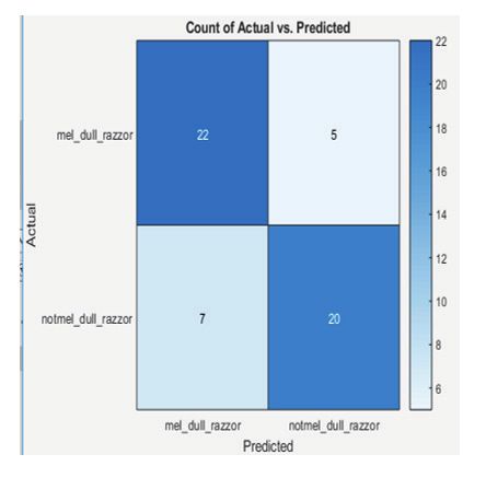
352 Medico-legal Update, July-September 2020, Vol.20, No. 3
In testing the dataset, the images are used for In paper [7] explains about the thresholding methods
template creation and also to determine the edges based and maximum entropy methods, and these features such
on accuracy. These testing results of snake algorithms as correlations, energy, and unsymmetrical features are
will show the finding of edge. To get the good results, obtained from gray level co-occurrence matric [8-10].
segmentation and classification of these algorithms is And finally, feed forward and artificial neural network
required. method is used for melanoma detection.
In paper [2] it describes about the detection the skin Proposed Algorithms:
cancer from captured images of the affected tumor to
determine the tumor is cancerous or normal. Diagnosis The algorithm that is being proposed for the
of melanoma at an early stage reduces the risk of death. diagnosis of skin cancer is explained here.
Computer aided techniques will help the dermatologist DB Image & Category Split/Count
to find out the skin cancer using image processing. In
this work, graph cut algorithm type is used to detect the Load the Pre Trained Network
melanoma from the images and also the features like
color, shape and geometry features are extracted from the Preprocess with CNN features
images using image processing. Based on the extracted Resize the Image & Visualize Weight
features, the images will be classified as malignant or
benign stage by support vector machine using radial Feature Layer &Train CNN Features by SVM
basis of kernel.
Predict the Category with Trained Label
The paper [3] tells about the usage of segmentation
of image based on lesion detection using deep learning Predicted Class & Accuracy finding
of pixel wise labeling scheme. The architectural network In our proposed scheme, melanoma classification
is used for testing the public data and the ISIC database is done through by using conventional neural network
images are used for training. These results provide good of deep learning technique. Here we are using the pre-
accuracy rate and perform well even in the presence of trained network model for prediction and classification.
hair, air and oil bubbles on images. The implementation
of this process in GUI gives some additional weightage In this work, database contains melanoma and
to the paper. non-melanoma images which are separated from each
other for analysis. These database images are split and
Paper [4] describes artificial intelligence and image number of images present in melanoma, non-melanoma
processing techniques for melanoma detection. Image is counted by their label or category wise and also the
quality levels are improved by eliminating the noise in minimum number of images present in each class or
preprocessing stage. These skin images are segmented type is identified. Then we load the pre-trained network
by applying the thresholding method. From that the model “Resnet-50” convolution neural network.
features are extracted by2D wavelet transformation
technique. These extracted features were applied as Pre-Trained Deep Neural Networks
input for artificial neural network of back –propagation
We extract the powerful and descriptive features
based and this method is used to classify their dataset
which are gathered from natural images using pre-
into either cancer or non-cancer.
trained image classification network. These pre-trained
This paper [5] tells about the JSEG algorithm which networks are trained by using the large scale visual
was used to diagnose skin cancer by using the lesion recognition challenge using more than 0.001 billion
boundary method. images and then are classified into categories such as
animal, car, bus, tea, cup etc.
In paper [6] the features like color and texture are
extracted from gray level co-occurrence matrix (GLCM) Resnet-50 (Network Model)
and support vector machine (SVM) classifier which are
It is also one of the type of pre-trained network
used for classification and further diagnosis of malignant
model of Conventional neural network, it is trained by
lesions. In this work, an accuracy level of around 90%
more than 0.001 billion images from the Image Net
by was achieved.Medico-legal Update, July-September 2020, Vol.20, No. 3 353
database. This Resnet-50 pre-trained network which has Without time investment and endeavor for complete
50 deep layers, classifies their corresponding database network training, it’s also a simplest and nimble approach
images into categories of 1000 objects. While loading for using the capability of deep learning technique.
the pre-trained network, it has some properties. In These features are extracted from images by using the
this pre-trained network, from input to output layer pre-trained network and then it’s trained by a classifier,
which has a huge number of fully connected layers or like support vector machine (SVM).
convolutional layers on path is known as network depth.
Test Image Features & Prediction
After loading the pre-trained network model, we
go for image network classification (i.e. identify the Similarly, we can select the test or query image from
prediction class) and preprocess the image on prediction any of the category in an image data store. We then resize
class or label wise by CNN features. After that we resize the selected input image as per pre-trained network
the (i.e. 224 by 224) and visualize their weightage level. model (i.e. 224 by 224) and features are extracted from
Then we initialize the feature layer of the pre-trained images by using the pre-trained network and then it’s
network model. corresponding category is predicted by classifier and
trained features, test features and trained labels. Finally,
Feature Extraction in Images on Pre-trained the classifier predicts the category and the accuracy rate
network model are calculated from confusion matrix by taking the mean
value of diagonal elements of confusion matrix.
Fig 1 & 2, describes about the Resnet50 (pre-trained network model) weightage allocation matrix & Layers
connections354 Medico-legal Update, July-September 2020, Vol.20, No. 3
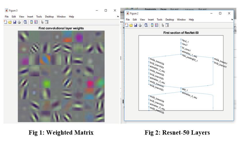
Fig 3: Network – Layer flow
Fig 3 describes about the Network Layer flow & class prediction by using classifier ofResnet50 (pre-trained
network model)
Fig 4: Confusion Matrix
Fig 4, describe about our data prediction level for melanoma skin cancer classification by using Resnet50.Medico-legal Update, July-September 2020, Vol.20, No. 3 355
Conclusion [4] Celebi ME, Kingravi HA, Uddin B, Iyatomi H,
Aslandogan YA, Stoecker WV, Moss RH. A
In this work, our aim is to finding the level of skin
methodological approach to the classification
cancer in human body based on pre-trained network of
of dermoscopy images. Computerized Medical
(Resnet-50-categories of 1000 objects) model and CNN
imaging and graphics. 2007 Sep 1;31(6):362-73.
features. And these CNN features dataset and query
image features are analyzed and its level is predicted [5] Deng Y, Manjunath BS, Shin H. Color image
by using deep learning, whether the query image which segmentation. InProceedings. 1999 IEEE Computer
belongs to which category either melanoma or not. In Society Conference on Computer Vision and
our pre-trained network model (Resnet-50) getting 85.18 Pattern Recognition (Cat. No PR00149) 1999 Jun
% accuracy. In future, creating the new network model 23 (Vol. 2, pp. 446-451). IEEE.
can be done for skin cancer prediction. [6] Jaleel JA, Salim S, Aswin RB. Computer aided
detection of skin cancer. In2013 International
Ethical Clearance: Taken from Indian Institute of Conference on Circuits, Power and Computing
Technology, Kanpur Technologies (ICCPCT) 2013 Mar 20 (pp. 1137-
Source of Funding: Self 1142). IEEE.
[7] Barata C, Ruela M, Francisco M, Mendonça T,
Conflict of Interest: Nil Marques JS. Two systems for the detection of
melanomas in dermoscopy images using texture
References and color features. IEEE Systems Journal. 2013 Jul
[1] Bumrungkun P, Chamnongthai K, Patchoo W. 29;8(3):965-79.
Detection skin cancer using SVM and snake model. [8] Sonia R. Melanoma image classification system
In2018 International Workshop on Advanced by NSCT features and Bayes classification.
Image Technology (IWAIT) 2018 Jan 7 (pp. 1-4). International Journal of Advances in Signal and
IEEE. Image Sciences. 2016 Dec 30;2(2):27-33.
[2] Mustafa S, Kimura A. A SVM-based diagnosis [9] Kumarapandian S. Melanoma Classification Using
of melanoma using only useful image features. Multiwavelet Transform and Support Vector
In2018 International Workshop on Advanced Machine. International Journal of MC Square
Image Technology (IWAIT) 2018 Jan 7 (pp. 1-4). Scientific Research. 2018 Sep 28;10(3):01-7.
IEEE. [10] Manikandan M. SKIN DETECTION UNDER
[3] Youssef A, Bloisi DD, Muscio M, Pennisi A, VARYING ILLUMINATION. International
Nardi D, Facchiano A. Deep convolutional pixel- Journal of MC Square Scientific Research. 2012
wise labeling for skin lesion image segmentation. Dec 15;4(1):84-95.
In2018 IEEE International Symposium on Medical
Measurements and Applications (MeMeA) 2018
Jun 11 (pp. 1-6). IEEE.You can also read