Dissection of Larval and Pupal Wings of Bicyclus anynana Butterflies - MDPI
←
→
Page content transcription
If your browser does not render page correctly, please read the page content below
Protocol
Dissection of Larval and Pupal Wings of Bicyclus
anynana Butterflies
Tirtha Das Banerjee 1, * and Antónia Monteiro 1,2
 1   Department of Biological Sciences, National University of Singapore, 14 Science Drive 4,
     Singapore 117543, Singapore; antonia.monteiro@nus.edu.sg
 2   Yale-NUS College, 10 College Avenue West, Singapore 138609, Singapore
 *   Correspondence: tirtha_banerjee@u.nus.edu

 Received: 9 December 2019; Accepted: 6 January 2020; Published: 10 January 2020

 Abstract: The colorful wings of butterflies are emerging as model systems for evolutionary and
 developmental studies. Some of these studies focus on localizing gene transcripts and proteins
 in wings at the larval and pupal stages using techniques such as immunostaining and in situ
 hybridization. Other studies quantify mRNA expression levels or identify regions of open chromatin
 that are bound by proteins at different stages of wing development. All these techniques require
 dissection of the wings from the animal but a detailed video protocol describing this procedure
 has not been available until now. Here, we present a written and accompanying video protocol
 where we describe the tools and the method we use to remove the larval and pupal wings of the
 African Squinting Bush Brown butterfly Bicyclus anynana. This protocol should be easy to adapt to
 other species.

 Keywords: wing dissection; Bicyclus anynana; butterflies

1. Introduction
     Multiple studies on butterflies are focused on understanding the evolutionary and developmental
genetics of their colorful wing patterns. Labs around the world have worked with different species to
address a variety of questions at the intersection of these fields. Examples include the discovery of
the involvement of the gene optix in wing pattern mimicry of Heliconius butterflies [1,2]; discovery of
doublesex as a mimicry supergene in Papilio polytes [3]; involvement of Wnt signaling in wing pattern
of butterflies such as Junonia coenia, Heliconius erato and Vanesssa cardui [4–6]; involvement of the
genes spalt and BarH-1 in the wing pigmentation of multiple Pieris and Colias species [7–9]; and the
involvement of calcium signaling in wing patterning in Junonia orithya [10] to mention a few.
     Bicyclus anynana has been a popular system to study wing patterning, especially the
eyespots, which are novel traits to the nymphalid lineage [11]. Many of the studies have been
focused on identifying the local expression of proteins such as Spalt [12], Engrailed/Invected [12],
Distal-less [13], Antennapedia [14,15], Notch [16], Cubitus-interruptus [17], Ecdysone Receptor [18],
Ultrabithorax [15,17], and the expression of gene transcripts such as hedgehog [14], apterous [19],
patched [16], wingless [20], doublesex [21] and decapentaplegic [22] in the developing wing. Gene
expression and RNAi studies have proposed that many wound healing network genes are expressed in
eyespots [23], that a positional-information mechanism is involved in the formation of the concentric
rings [16,20], and that a reaction-diffusion mechanism is involved in setting up the eyespot centers [22]
using gene expression and functional analysis via CRISPR-Cas9 [24,25]. All these studies require the
removal of wings from the bodies of larvae and/or pupae to examine patterns of gene expression.
Protocols describing the dissection of larval and pupal wings have been previously published [26,27].

Methods Protoc. 2020, 3, 5; doi:10.3390/mps3010005                                   www.mdpi.com/journal/mps
Methods Protoc. 2020, 3, 5 2 of 7
However, the protocols are brief and have no accompanying video, making it difficult for newcomers
in the field to follow.
In this paper, we describe the process of larval and pupal wing removal using a video and explain
the process along with all the tools and chemicals needed in the main text below. This protocol can
also supplement other similar experiments such as live cell imaging in vivo used to understand cell
differentiation and dynamics [28,29].
2. Experimental Design
2.1. Required Materials and Equipment
2.1.1. Materials
• Curved tweezers (Dumont; Dumont Switzerland, Montignez, Switzerland; Cat. No.: 11274-20);
Methods Protoc. 2020, 3, x FOR PEER REVIEW 2 of 7
• Fine straight tweezers (Dumont; Dumont Switzerland; Cat. No.: 11254-20);
• previously published(Thomas
Flat spatula [26,27]. However, the protocols
Scientific; Thomas are brief and have no
Scientific, accompanying video,
Swedesboro, NJ, USA; Cat. No.: 1208Y75);
making it difficult for newcomers in the field to follow.
• Regular straight
In this paper, tweezers
we describe (Dumont;
the process Dumont
of larval and Switzerland;
pupal wing removal using Cat. No.:
a video (see 0203-5-PO);
Supplementary Materials) and explain the process along with all the tools and chemicals needed in
• Superfine Vannas scissors 8 cm (World Precision Instruments; World Precision Instruments,
the main text below. This protocol can also supplement other similar experiments such as live cell
Sarasota,
imaging FL,toUSA;
in vivo used Cat.cell
understand No.: 501778);and dynamics [28,29].
differentiation
• Blade holder (Swann-Morton No. 4; Swann-Morton, Sheffield, UK; Cat. No.: 0934);
2. Experimental Design
• Blades (Swann-Morton No. 4; Swann-Morton, Sheffield, UK; Cat. No.: 0115);
2.1. Required Materials and Equipment TM
• Glass spot plate (PYREX ; Corning, Corning, NY, USA; Cat. No.: 722085);
• 2.1.1. Materials
Dissection silicone plate (Dragon Skin 30 Mould Making Silicone Rubber; Cat. No.: 0751635278417.
• Petri
Curvedplate;
tweezers (Dumont; Dumont Switzerland,
Sigma-Aldrich; Sigma-Aldrich,Montignez, Switzerland; Cat.
Singapore; Cat.No.:No.:
11274-20);
P5981-100EA);
• Fine straight tweezers (Dumont; Dumont Switzerland; Cat. No.: 11254-20);
• • Insect pins
Flat spatula (BioQuip;
(Thomas BioQuip,
Scientific; Rancho
Thomas Scientific, Dominguez,
Swedesboro, NJ, USA; CA, USA;
Cat. No.: Cat. No.: 1208B2).
1208Y75);
• Regular straight tweezers (Dumont; Dumont Switzerland; Cat. No.: 0203-5-PO);
•
2.1.2. Superfine
Equipment Vannas scissors 8 cm (World Precision Instruments; World Precision Instruments,
Sarasota, FL, USA; Cat. No.: 501778);
• •• Zeiss
Blade holder (Swann-Morton No. 4; Swann-Morton, Sheffield, UK; Cat. No.: 0934);
Dissection Microscope (Carl-Zeiss, Jena, Germany; Stemi 305)
Blades (Swann-Morton No. 4; Swann-Morton, Sheffield, UK; Cat. No.: 0115);
• Glass spot plate (PYREXTM; Corning, Corning, NY, USA; Cat. No.: 722085);
•
2.1.3. Reagentssilicone plate (Dragon Skin 30 Mould Making Silicone Rubber; Cat. No.:
Dissection
0751635278417. Petri plate; Sigma-Aldrich; Sigma-Aldrich, Singapore; Cat. No.: P5981-100EA);
• • NaCl
Insect pins (BioQuip; BioQuip, Sigma-Aldrich,
(Sigma-Aldrich; Rancho Dominguez, CA, USA; Cat. No.:
Singapore; 1208B2).
Cat. No.: S9888-500G);
• KEquipment
2.1.2. 2 HPO4 (Sigma-Aldrich; Sigma-Aldrich, Singapore; Cat. No.: P3786-500G);
• • KH Zeiss2 PO 4 (Sigma-Aldrich;
Dissection Sigma-Aldrich,
Microscope (Carl-Zeiss, Singapore;
Jena, Germany; Stemi 305) Cat. No.: 229806-250G);
• RNAseZap (Themo Fisher Scientific; Thermo Fisher Scientific, Waltham, MA, USA; Cat. No.:
2.1.3. Reagents
•
AM9780).
NaCl (Sigma-Aldrich; Sigma-Aldrich, Singapore; Cat. No.: S9888-500G);
• K2HPO4 (Sigma-Aldrich; Sigma-Aldrich, Singapore; Cat. No.: P3786-500G);
3. •Procedure
KH2PO4 (Sigma-Aldrich; Sigma-Aldrich, Singapore; Cat. No.: 229806-250G);
• RNAseZap (Themo Fisher Scientific; Thermo Fisher Scientific, Waltham, MA, USA; Cat. No.:
AM9780).
3.1. Preparation for Dissection
3. Procedure
1. Transfer 500 µL of 1 × PBS into each well of the spot glass plate.
2. 3.1. Preparation
Transferforaround
Dissection 100 mL of 1 × PBS into the dissection well plate.
3. 1. Wash
Transfer 500 µL of 1 × PBS into each well of the spot glass plate.
the dissection tools in 70% ethanol prior to dissection.
2. Transfer around 100 mL of 1 × PBS into the dissection well plate.
4. 3. Freeze
Wash the anaesthetize
dissection tools in the larvaeprior
70% ethanol and to pupae
dissection.on ice for 10–20 min.
4. Freeze anaesthetize the larvae and pupae on ice for 10–20 min.
CRITICAL
CRITICAL STEP:
STEP: If you If you are
are performing performing
experiments experiments
involving involving
RNA, it is recommended RNA, it is recommended
that all the equipment is wiped with RNAseZap.
that all the equipment is wiped with RNAseZap.
3.2. Dissection of Larval Wings
3.2.
1. Dissection of from
Pick one larva Larval Wings
the ice and carefully secure it in the dissection plate with the help of two
pins. One pin should be placed immediately posterior to the head capsule, and the second pin
1. Pick oneoflarva
at the end fromItthe
the abdomen. ice and carefully
is recommended secure
to stretch the it in placing
larva, before the dissection plate with the help of two pins.
the second pin,
One
to makepin
the should be placed
dissection and removal ofimmediately
wings easier. posterior to the head capsule, and the second pin at the
2. The wings are located around the second and third thoracic legs.2.1. Required Materials and Equipment
2.1.1. Materials
• Curved tweezers (Dumont; Dumont Switzerland, Montignez, Switzerland; Cat. No.: 11274-20);
• Fine straight tweezers (Dumont; Dumont Switzerland; Cat. No.: 11254-20);
• Flat spatula (Thomas Scientific; Thomas Scientific, Swedesboro, NJ, USA; Cat. No.: 1208Y75);
•
Methods Protoc.straight
Regular 2020, 3, 5
tweezers (Dumont; Dumont Switzerland; Cat. No.: 0203-5-PO); 3 of 7
• Superfine Vannas scissors 8 cm (World Precision Instruments; World Precision Instruments,
Sarasota, FL, USA; Cat. No.: 501778);
• Blade holder (Swann-Morton No. 4; Swann-Morton, Sheffield, UK; Cat. No.: 0934);
• end
Bladesof the abdomen.
(Swann-Morton It is recommended
No. 4; Swann-Morton, toCat.
Sheffield, UK; stretch the larva, before placing the second pin, to
No.: 0115);
• make the
Glass spot dissection
plate and removal
(PYREXTM; Corning, of USA;
Corning, NY, wings Cat.easier.
No.: 722085);
• Dissection silicone plate (Dragon Skin 30 Mould Making Silicone Rubber; Cat. No.:
2. The wings are located around the second and third thoracic legs.
0751635278417. Petri plate; Sigma-Aldrich; Sigma-Aldrich, Singapore; Cat. No.: P5981-100EA);
3. • Holdpins
Insect the(BioQuip;
epidermis ofRancho
BioQuip, the larva usingCA,
Dominguez, a straight tweezer
USA; Cat. No.: 1208B2).and using the Vannas scissors make an
2.1.2.incision,
Methods as 2020,
Protoc.
Equipment indicated
3, x FORin Figure
PEER 1A.
REVIEW 3 of 7
4. • After the incision
Zeiss Dissection Microscopetry(Carl-Zeiss,
to find Jena,
the hindwing
Germany; Stemi around
305) the third thoracic leg (Figure 1C). If you are
3. Holdwith
working the epidermis
a youngoffifth the instar
larva usinglarva,a straight
the wing tweezer
can beandidentified
using the Vannas
adjacentscissors make an
to a white lump of
2.1.3. Reagents
incision, as indicated in Figure 1A.
tissue around the thoracic leg (Figure 1D). This white tissue at the base of the wing will become
• 4. (Sigma-Aldrich;
NaCl After the incision try to find
Sigma-Aldrich, the hindwing
Singapore; around the third thoracic leg (Figure 1C). If you are
Cat. No.: S9888-500G);
• the
K2HPOtrachea that will invade the wing blade and is the preferred spot for handling the wing to
4 (Sigma-Aldrich; Sigma-Aldrich, Singapore; Cat. No.: P3786-500G);
working with a young fifth instar larva, the wing can be identified adjacent to a white lump of
• avoid touching the
theactual wing tissueCat.attached.
KH2PO4 (Sigma-Aldrich; Sigma-Aldrich, Singapore; No.: 229806-250G);
•
tissue around thoracic leg (Figure 1D). This white tissue at the base of the wing will become
RNAseZap (Themo Fisher Scientific; Thermo Fisher Scientific, Waltham, MA, USA; Cat. No.:
5. Make the
AM9780). cuts to the
trachea tissues/trachea
that will invade the attached
wing bladeon andboth sides
is the of thespot
preferred wingforand carefully
handling pulltoout the
the wing
wingavoid touching
(touching only thethe
actual wing
white tissueusing
tissue) attached.a fine tweezer.
3. Procedure
6. 5. Makethe
Transfer cuts to the tissues/trachea
hindwing to one of the attached
wells of onthe
both sidesplate.
glass of the wing and carefully pull out the
wing
3.1. Preparation (touching only the white tissue) using a fine tweezer.
for Dissection
7. The forewing is present just a few millimeters above the third thoracic leg (Figure 1A). Perform
6. Transfer the hindwing to one of the wells of the glass plate.
1. Transfer 500 µL of 1 × PBS into each well of the spot glass plate.
the
7. cuts
The as with the
forewing hindwing andfewpull out the wing using a fine tweezer.
2. Transfer around 100 mLis of present just
1 × PBS into theadissection
millimeters
well plate. above the third thoracic leg (Figure 1A). Perform
8. 3. Transfer the forewing to one of
the cuts as with the hindwing and pull out
Wash the dissection tools in 70% ethanol the
prior to wells ofthe
dissection. thewing
glassusing
plate.
a fine tweezer.
4. Freeze anaesthetize the larvae and pupae on ice for 10–20 min.
8. Transfer the forewing to one of the wells of the glass plate.
CRITICAL
CRITICAL STEP:
STEP: If you Be careful
are performing not toinvolving
experiments touch RNA,
the wing membrane as even a gentle contact
it is recommended
CRITICAL
that all the STEP:
equipment is wipedBe careful
with not to touch the wing membrane as even a gentle contact with
RNAseZap.
with
the the tweezer
tweezer can damage
can damage the wing.
the wing.
3.2. Dissection of Larval Wings
1. Pick one larva from the ice and carefully secure it in the dissection plate with the help of two
pins. One pin should be placed immediately posterior to the head capsule, and the second pin
at the end of the abdomen. It is recommended to stretch the larva, before placing the second pin,
to make the dissection and removal of wings easier.
2. The wings are located around the second and third thoracic legs.
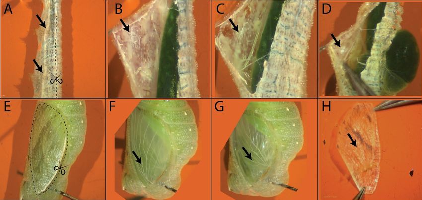
FigureFigure 1. Dissection
1. Dissection of larval
of larval andandpupal
pupalwings
wings of
of Bicyclus
Bicyclusanynana.
anynana. (A).(A).
Larval wings
Larval are located
wings are located
laterally (black arrows), dorsal to the second and third thoracic legs. The region for incision is
laterally (black arrows), dorsal to the second and third thoracic legs. The region for incision is marked
marked by a dotted line. (B). A larval forewing is located dorsally to the second thoracic leg. (C). A
by a dotted line. (B). A larval forewing is located dorsally to the second thoracic leg. (C). A larval
larval hindwing is located beside the third thoracic leg. (D). Early larval wings can be identified by
hindwing is located beside the third thoracic leg. (D). Early larval wings can be identified by finding
finding the white tissue (black arrow) around the thoracic legs. Furthermore, to release the pressure
the white
due tissue
to the (black
gut it isarrow) around the
recommended thoracic
to make legs. Furthermore,
an initial dorsal incision to releasewhich
(through the pressure
the gut due
can to the
gut it extend)
is recommended to make an initial dorsal incision (through which the gut can extend)
before the lateral incision is made. (E). For the dissection of pupal wings make incision as before the
lateralmarked
incisionbyisthe
made. (E). For the dissection of pupal wings make incision as marked
dotted line. (F). Pupal forewing. (G). Pupal hindwing. (H). Early (16–26 h afterby the dotted
pupation)
line. (F). Pupal pupal forewing.
forewing. (G). Pupal hindwing. (H). Early (16–26 h after pupation) pupal forewing.
3.3. Dissection of Pupal
3.3. Dissection Wings
of Pupal Wings
1. 1. Secure
Secure a pupaa pupa in the
in the dissectionplate
dissection platewith
with the
thehelp
helpofoftwo
two fine pins.
fine pins.
2. Make incisions using a fine blade at the region marked in Figure 1E.
2. Make incisions using a fine blade at the region marked in Figure 1E.
3. Remove the cuticle using a curved tweezer. The forewing should be visible at the surface of the
3. Remove the cuticle using a curved tweezer. The forewing should be visible at the surface of the
pupa (Figure 1F). If you are working with a wing that is less than 26 h old, the forewing might
pupabe (Figure 1F). Ifto
still attached you
theare working
cuticle (Figurewith
1H). aUsing
winga that is less
straight thanon
tweezer 26one
h old, thehold
hand, forewing might be
the cuticle
still attached
down andto the acuticle
using curved(Figure
tweezer 1H).
on theUsing a straight
other hand, gentlytweezer
dislodgeontheone
winghand, hold
from the the cuticle
cuticle,
scraping the wing from underneath in gentle nudges, and finally pull out the wing.
CRITICAL STEP: Make sure that the forewing is free from any attachment to the cuticle.•
2.1.1. NaCl (Sigma-Aldrich; Sigma-Aldrich, Singapore; Cat. No.: S9888-500G);
Materials
• K2HPO4 (Sigma-Aldrich; Sigma-Aldrich, Singapore; Cat. No.: P3786-500G);
• Curved tweezers (Dumont; Dumont Switzerland, Montignez, Switzerland; Cat. No.: 11274-20);
• KH2PO4 (Sigma-Aldrich; Sigma-Aldrich, Singapore; Cat. No.: 229806-250G);
• Fine straight tweezers (Dumont; Dumont Switzerland; Cat. No.: 11254-20);
• RNAseZap (Themo Fisher Scientific; Thermo Fisher Scientific, Waltham, MA, USA; Cat. No.:
• Flat spatula (Thomas Scientific; Thomas Scientific, Swedesboro, NJ, USA; Cat. No.: 1208Y75);
AM9780).
• Regular straight tweezers (Dumont; Dumont Switzerland; Cat. No.: 0203-5-PO);
•3. Procedure
Superfine Vannas scissors 8 cm (World Precision Instruments; World Precision Instruments,
Methods Protoc. FL,
Sarasota, 2020, 3, 5Cat. No.: 501778);
USA; 4 of 7
•3.1. Preparation
Blade holder (Swann-Morton
for Dissection No. 4; Swann-Morton, Sheffield, UK; Cat. No.: 0934);
• Blades (Swann-Morton No. 4; Swann-Morton, Sheffield, UK; Cat. No.: 0115);
•1. down
Transfer
Glass 500
spot
and
µL of
plate 1 × PBS
(PYREX
using aTM;into each well
Corning,
curved
of theNY,
Corning,
tweezer
spotUSA;
glassCat.
on the
plate.
No.: 722085);
other hand, gently dislodge the wing from the cuticle,
2.
• Transfer around 100 mL of 1 × PBS into the dissection
Dissection silicone plate (Dragon Skin 30 Mould Making well plate. Silicone Rubber; Cat. No.:
3. scraping
Wash the the
dissectionwing
tools from
in 70% underneath
ethanol prior to in gentle
dissection. nudges,
0751635278417. Petri plate; Sigma-Aldrich; Sigma-Aldrich, Singapore; Cat. No.: and finally pull
P5981-100EA); out the wing.
4.
• Freeze anaesthetize
Insect pins (BioQuip;the larvae and
BioQuip, pupae
Rancho on ice for 10–20
Dominguez, min. Cat. No.: 1208B2).
CA, USA;
CRITICAL
CRITICAL STEP:
STEP: If you Make sure
are performing that the
experiments forewing
involving RNA, itisis free from any
recommended attachment to the cuticle.
2.1.2. Equipment
that all the equipment is wiped with RNAseZap.
4. • After the forewing
Zeiss Dissection Microscopeis free, transfer
(Carl-Zeiss, the wing
Jena, Germany; to
Stemi 305) one of the wells of the glass plate using a flat
3.2. Dissection of Larval Wings
spatula.
2.1.3.Pick
Reagents
Hold the wing by the hinge region, and do not touch the rest of the wing blade with
1. one larva from the ice and carefully secure it in the dissection plate with the help of two
•
the
pins.
NaCl
tweezers.
Methods
One Protoc.
pin should
(Sigma-Aldrich;
Hold
2020,
be theimmediately
3,placed
x FOR wing
PEER
Sigma-Aldrich,
with
REVIEW the
posterior
Singapore;
tweezers
to the
Cat. No.:
against
head capsule,
S9888-500G); andthe spatula
the second pin until the spatula4 breaks
of 7 the
• liquid-air
at the end
K2HPO surface
of the abdomen.
4 (Sigma-Aldrich; interface. You
It is recommended
Sigma-Aldrich, can also
to stretch
Singapore; Cat.theuse the
larva,
No.: tweezers
before placing theto
P3786-500G); gently
second pin, help slide the wing into the
• to
KHmake
2PO4 the
4. dissection
After and
the forewing
(Sigma-Aldrich; removal of wings
is free,
Sigma-Aldrich, easier. Cat.
transfer
Singapore; the No.:
wing to one of the wells of the glass plate using a flat
229806-250G);
2.
glass
The
wells.
wings are located around the second and third thoracic legs.Waltham, MA, USA; Cat. No.:
• RNAseZap (Themo Fisher Scientific;
spatula. Hold the wing by the hingeThermo Fisher Scientific,
region, and do not touch the rest of the wing blade with the
5. To remove the hindwing (Figure 1G), make an incision around the wing using a fine blade and
AM9780).
tweezers. Hold the wing with the tweezers against the spatula until the spatula breaks the
carefully pull out a glassy (peripodial) membrane on top of the hindwing.
3. Procedure liquid-air surface interface. You can also use the tweezers to gently help slide the wing into the
6. After the glassmembrane
wells. is removed, make a cut at the wing-hinge region and pull out the wing using
3.1. Preparation
5. Toforremove
Dissectionthe hindwing (Figure 1G), make an incision around the wing using a fine blade and
a curved tweezer.
1. Transfer carefully
500 µL of 1 pull
× PBSout
intoaeach well (peripodial)
glassy of the spot glass plate.
membrane on top of the hindwing.
7. 2. After
Transfer the
aroundhindwing
100 mL of 1 ×isPBSfree,
into transfer
the dissection the wing to one of the wells of the glass plate using a
wellaplate.
6. After the membrane is removed, make cut at the wing-hinge region and pull out the wing
3. flat
Washspatula.
the dissection tools in 70% ethanol prior to dissection.
4.
using a curved tweezer.
Freeze anaesthetize the larvae and pupae on ice for 10–20 min.
7. After the hindwing is free, transfer the wing to one of the wells of the glass plate using a flat
CRITICAL
CRITICAL
spatula. STEP:
STEP: If you Be careful
are performing not toinvolving
experiments touch RNA,
the wing membrane
it is recommended as even a gentle contact
that all the equipment is wiped with RNAseZap.
with the tweezer can damage the wing.
CRITICAL STEP: Be careful not to touch the wing membrane as even a gentle contact with
3.2. Dissection of Larval Wings
4. 1.Expected the tweezer can damage the wing.
Results
Pick one larva from the ice and carefully secure it in the dissection plate with the help of two
pins. One pin should be placed immediately posterior to the head capsule, and the second pin
4. Expected Results
LarvalatWings and
the end of Pupal Wing
the abdomen. It is recommended to stretch the larva, before placing the second pin,
to make the dissection and removal of wings easier.
2. TheLarval
Larval Wings
wingswings and
are located Pupal
at around
an Wing
early
the developmental
second stage
and third thoracic legs. are marked by a lack of tracheal invasion in the
wing disc and a prominent
Larval wings at anwhite
early tissue at the proximal
developmental stage are part of the
marked by awing (Figure
lack of tracheal2A,B). Larval
invasion in thewings at
wing disc
a later stage are and a prominent
larger and markedwhite bytissue
theatinvasion
the proximal part of thetissue
of tracheal wing (Figure
along2A,B). Larval(Figure
the veins wings 2C,D).
at a later
Pupal wings stage are
around larger
18–24 h and
willmarked by the invasion
have prominent of tracheal
tracheal tissue
tissue along
in the the veins
wing blade(Figure 2C,D).
(Figure 2E,F). The
Pupal wings around 18–24 h will have prominent tracheal tissue in the wing blade (Figure 2E,F). The
wings at the pupal stages are much larger and fragile than at the larval stage. Care must be taken to
wings at the pupal stages are much larger and fragile than at the larval stage. Care must be taken to
preventprevent
damage to thetowing
damage tissue
the wing at this
tissue stage.
at this stage.
Figure
Figure 2. 2. and
Larval Larval and wings
Pupal Pupal of
wings of Bicyclus
Bicyclus anynana.
anynana. (A) Early
(A) Early larvallarval forewing
forewing showing
showing the
the prominent
prominent white tissue that will differentiate into the trachea. Wings should be handled in this
white tissue that will differentiate into the trachea. Wings should be handled in this region during
region during dissections; (B) Early larval hindwing; (C) Late larval forewing; (D) Late larval
dissections; (B) Early larval hindwing; (C) Late larval forewing; (D) Late larval hindwing; (E) Pupal
hindwing; (E) Pupal forewing; (F) Pupal hindwing.
forewing; (F) Pupal hindwing.Methods Protoc. 2020, 3, 5 5 of 7
5. Discussion
Butterflies are becoming a model system to understand the process of color pattern formation
in Biology. Over the past three decades numerous research papers have illuminated the processes
involved in eyespots development in the wings of butterflies such as Bicyclus anynana and Junonia
coenia [11,13,20,22,23,30,31]; color patterning and mimicry in Heliconius and Papilio butterflies [1–3];
and involvement of multiple signaling pathways in wing pigmentation in species belonging to the
genus Pieris, Junonia, and Colias [4,7,9,10]. Almost all of these studies involved the process of wing
dissections. The dissected wings can be used to localize proteins and gene transcripts involved in color
patterning [5,12] and for more advanced techniques such as RNA, FAIRE, and ATAC sequencing [2,23].
Wing dissections, hence, are indispensable for a full understanding of the evolution and development
of butterfly wing color patterns. Furthermore, experiments such as in vivo live cell imaging [28,29],
used to study cellular dynamics overlap with some the wing dissection steps such as removal of cuticle
and might benefit from the protocol mentioned here.
To conclude, we have provided a detailed description of the process of wing dissections in
a butterfly species which we believe will be helpful for newcomers in the field to adapt to their
own species.
6. Reagents Setup
Preparation of 10 × PBS Buffer
1. In a 1 L beaker, add 700 mL MilliQ water and reagents mentioned in Table 1:
2. Transfer the content to a 1 L measuring cylinder. Raise the volume to one liter using MilliQ water.
3. Mix the solution and transfer the content to a 1 L glass bottle.
4. Autoclave the solution at 121 ◦ C for 20 min and store the content at room temperature.
Table 1. Reagents for 10 × PBS (Phosphate Buffer Saline) buffer preparation.
Reagents Weight/Volume
NaCl 81.8 g
KH2 PO4 5.28 g
K2 HPO4 10.68 g
Note: To prepare 1 × PBS, add 10 mL of 10 × PBS buffer and 90 mL of MilliQ water.
Author Contributions: T.D.B. and A.M. wrote the manuscript; T.D.B. designed and performed the experiment
and developed the associated video article. All authors have read and agreed to the published version of
the manuscript.
Funding: This research was funded National Research Foundation, Singapore grant R-154-000-B57-281.
Acknowledgments: We thank all Monteiro Lab members for their constant support and the comments of three
anonymous reviewers that helped in improving the manuscript.
Conflicts of Interest: The authors declare no conflict of interest.
References
1. Reed, R.D.; Papa, R.; Martin, A.; Hines, H.M.; Kronforst, M.R.; Chen, R.; Halder, G.; Nijhout, H.F.;
Mcmillan, W.O. optix Drives the Repeated Convergent Evolution of Butterfly Wing Pattern Mimicry. Science
2011, 333, 1137–1141. [CrossRef] [PubMed]
2. Lewis, J.J.; Geltman, R.C.; Pollak, P.C.; Rondem, K.E.; Van Belleghem, S.M.; Hubisz, M.J.; Munn, P.R.;
Zhang, L.; Benson, C.; Mazo-Vargas, A.; et al. Parallel evolution of ancient, pleiotropic enhancers underlies
butterfly wing pattern mimicry. Proc. Natl. Acad. Sci. USA 2019, 116, 24174–24183. [CrossRef] [PubMed]
3. Kunte, K.; Zhang, W.; Tenger-Trolander, A.; Palmer, D.H.; Martin, A.; Reed, R.D.; Mullen, S.P.; Kronforst, M.R.
doublesex is a mimicry supergene. Nature 2014, 507, 229–232. [CrossRef] [PubMed]Methods Protoc. 2020, 3, 5 6 of 7
4. Martin, A.; Reed, R.D. Wnt signaling underlies evolution and development of the butterfly wing pattern
symmetry systems. Dev. Biol. 2014, 395, 367–378. [CrossRef] [PubMed]
5. Martin, A.; Reed, R.D. wingless and aristaless2 define a developmental ground plan for moth and butterfly
wing pattern evolution. Mol. Biol. Evol. 2010, 27, 2864–2878. [CrossRef]
6. Zhang, L.; Mazo-Vargas, A.; Reed, R.D. Single master regulatory gene coordinates the evolution and
development of butterfly color and iridescence. Proc. Natl. Acad. Sci. USA 2017, 114, 10707–10712. [CrossRef]
7. Stoehr, A.M.; Walker, J.F.; Monteiro, A. Spalt expression and the development of melanic color patterns in
pierid butterflies. EvoDevo 2013, 4, 6. [CrossRef]
8. Woronik, A.; Tunström, K.; Perry, M.W.; Neethiraj, R.; Stefanescu, C.; de la Paz Celorio-Mancera, M.;
Brattström, O.; Käkelä, R.; Hill, J.; Lehmann, P.; et al. A homeobox gene, BarH-1, underlies a female
alternative life-history strategy. bioRxiv 2018, 424879. [CrossRef]
9. Woronik, A.; Stefanescu, C.; Käkelä, R.; Wheat, C.W.; Lehmann, P. Physiological differences between female
limited, alternative life history strategies: The Alba phenotype in the butterfly Colias croceus. J. Insect Physiol.
2018, 107, 257–264. [CrossRef]
10. Ohno, Y.; Otaki, J.M. Spontaneous long-range calcium waves in developing butterfly wings. BMC Dev. Biol.
2015, 15, 17. [CrossRef]
11. Monteiro, A. Origin, Development, and Evolution of Butterfly Eyespots. Annu. Rev. Entomol. 2015, 60,
253–271. [CrossRef] [PubMed]
12. Monteiro, A.; Glaser, G.; Stockslager, S.; Glansdorp, N.; Ramos, D. Comparative insights into questions of
lepidopteran wing pattern homology. BMC Dev. Biol. 2006, 6, 52. [CrossRef] [PubMed]
13. Monteiro, A.; Chen, B.; Ramos, D.M.; Oliver, J.C.; Tong, X.; Guo, M.; Wang, W.K.; Fazzino, L.; Kamal, F.
Distal-Less Regulates Eyespot Patterns and Melanization in Bicyclus Butterflies. J. Exp. Zool. Part B Mol. Dev.
Evol. 2013, 320, 321–331. [CrossRef] [PubMed]
14. Saenko, S.V.; Marialva, M.S.P.P.; Beldade, P. Involvement of the conserved Hox gene Antennapedia in the
development and evolution of a novel trait. EvoDevo 2011, 2, 9. [CrossRef]
15. Matsuoka, Y.; Monteiro, A. Hox genes are essential for the development of novel serial homologous eyespots
on the wings of Bicyclus anynana butterflies. bioRxiv 2019, 814848. [CrossRef]
16. Beldade, P.; Peralta, C.M. Developmental and evolutionary mechanisms shaping butterfly eyespots. Curr.
Opin. Insect Sci. 2017, 19, 22–29. [CrossRef]
17. Monteiro, A.; Prudic, K.L. Multiple approaches to study color pattern evolution in butterflies. Trends Evol.
Biol. 2010, 2, 7–15. [CrossRef]
18. Bhardwaj, S.; Prudic, K.L.; Bear, A.; Dasgupta, M.; Wasik, B.R.; Tong, X.; Cheong, W.F.; Wenk, M.R. Sex
Differences in 20-Hydroxyecdysone Hormone Levels Control Sexual Dimorphism in Bicyclus anynana Wing
Patterns. Mol. Biol. Evol. 2018, 35, 465–472. [CrossRef]
19. Prakash, A.; Monteiro, A. apterous A specifies dorsal wing patterns and sexual traits in butterflies. Proc. R.
Soc. B Biol. Sci. 2018, 285, 20172685. [CrossRef]
20. Özsu, N.; Chan, Q.Y.; Chen, B.; Gupta, M.D.; Monteiro, A. Wingless is a positive regulator of eyespot color
patterns in Bicyclus anynana butterflies. Dev. Biol. 2017, 429, 177–185. [CrossRef]
21. Prakash, A.; Monteiro, A. Doublesex mediates the development of sex—Specific pheromone organs in Bicyclus
butterflies via multiple mechanisms. bioRxiv 2019, 686477. [CrossRef]
22. Connahs, H.; Tlili, S.; van Creij, J.; Loo, T.Y.J.; Banerjee, T.D.; Saunders, T.E.; Monteiro, A. Activation of
butterfly eyespots by Distal-less is consistent with a reaction-diffusion process. Development 2019, 146,
dev169367. [CrossRef] [PubMed]
23. Özsu, N.; Monteiro, A. Wound healing, calcium signaling, and other novel pathways are associated with the
formation of butterfly eyespots. BMC Genom. 2017, 18, 788. [CrossRef] [PubMed]
24. Banerjee, T.; Monteiro, A. CRISPR-Cas9 Mediated Genome Editing in Bicyclus anynana Butterflies. Methods
Protoc. 2018, 1, 16. [CrossRef]
25. Zhang, L.; Reed, R.D. A Practical Guide to CRISPR/Cas9 Genome Editing in Lepidoptera; Sekimura, T., Nijhout, H.,
Eds.; Springer: Berlin, Germany, 2017; pp. 155–172.
26. Brakefield, P.M.; Beldade, P.; Zwaan, B.J. Dissection of larval and pupal wings from the African butterfly
Bicyclus anynana. Cold Spring Harb. Protoc. 2009, 2009, pdb-prot5207. [CrossRef]
27. Yoshido, A.; Sahara, K.; Yasukochi, Y. Protocols for Cytogenetic Mapping of Arthropod Genomes; CRC Press: Boca
Raton, FL, USA, 2014; pp. 219–256. ISBN 9781466598164.Methods Protoc. 2020, 3, 5 7 of 7
28. Ohno, Y.; Otaki, J.M. Live cell imaging of butterfly pupal and larval wings in vivo. PLoS ONE 2015, 10,
e0128332. [CrossRef]
29. Iwata, M.; Ohno, Y.; Otaki, J.M. Real-time in vivo imaging of butterfly wing development: Revealing the
cellular dynamics of the pupal wing tissue. PLoS ONE 2014, 9, e89500. [CrossRef]
30. Carroll, S.B.; Gates, J.; Keys, D.N.; Paddock, S.W.; Grace, E.F.; Selegue, J.E.; Williams, J.A. Pattern Formation
and Eyespot Determination in Butterfly Wings. Science 1994, 265, 109–114. [CrossRef]
31. Keys, D.N.; Lewis, D.L.; Selegue, J.E.; Pearson, B.J.; Goodrich, L.V.; Johnson, R.L.; Gates, J.; Scott, M.P.;
Carroll, S.B. Recruitment of a hedgehog Regulatory Circuit in Butterfly Eyespot Evolution. Science 1999, 283,
532–534. [CrossRef]
© 2020 by the authors. Licensee MDPI, Basel, Switzerland. This article is an open access
article distributed under the terms and conditions of the Creative Commons Attribution
(CC BY) license (http://creativecommons.org/licenses/by/4.0/).You can also read