Tandproblemen bij chinchilla's - Dental pathology in chinchillas
←
→
Page content transcription
If your browser does not render page correctly, please read the page content below

Vlaams Diergeneeskundig Tijdschrift, 2010, 79                                                               Review 345

                                      Dental pathology in chinchillas

                                       Tandproblemen bij chinchilla’s
             1
              V. Derbaudrenghien, 2A. Van Caelenberg, 3K. Hermans, 2Ingrid Gielen, 3A. Martel
                             1
                             Vetcare Center, Alsembergsesteenweg 439, B-1653 Dworp
                         2
                          Department of Medical Imaging and Small Animal Orthopedics
                          3
                            Department of Pathology, Bacteriology and Poultry Diseases
             Faculty of Veterinary Medicine, Ghent University, Salisburylaan 133, B-9820 Merelbeke

                                        v_derbaudrenghien@netcourrier.com

    ABSTRACT
        Chinchillas are prone to develop a wide range of dental pathologies. The most common one is malocclu-
    sion, a condition in which the teeth are misaligned and/or incorrectly positioned in relation to one another.
    Odontomas, caries, tooth resorption, and periodontal and endodontic diseases have also been reported. This
    article presents an overview of the specific anatomy and the most common dental pathologies of chinchillas,
    including the various aspects of the clinical symptoms, diagnosis and treatment of these pathologies.

    SAMENVATTING
        Tandproblemen komen vaak voor bij chinchilla’s. Naast malocclusie worden ook odontoma’s, cariës, tandre-
    sorptie, periodontale en endodontale ziekten beschreven. In dit overzicht wordt eerst de normale tandanatomie be-
    schreven. Daarna komen de meest voorkomende pathologieën, de klinische symptomen, diagnose en de behandeling
    aan bod.

INTRODUCTION                                                   rently available chinchilla feeds in Belgian pet shops
                                                               meet the specific nutritional recommendations found in
    Originating from South America, chinchillas were           the scientific literature. The diets are generally too
domesticated in the early 20th century. Their natural          rich in protein and carbohydrates, with insufficient
diet, based on fibrous plants, is optimally digested           crude fiber content.
thanks to a well-developed cecum and coprophagy.                   Owners presenting their chinchilla with dental pro-
Nowadays they have entered our households as pets              blems on consultation typically complain about lowe-
and gradually became part of the veterinarian’s clien-         red feed intake, decreased stool production, reduced
tele.                                                          activity and weight loss. Consequential to dental pro-
    While field veterinarians will generally state that        blems, secondary pathologies can develop, which of-
oral pathology is widespread in pet chinchillas, preva-        ten cast a cloud over the prognosis.
lence research on the issue is very limited. The most ex-          Besides external evaluation of the facial and jaw
tensive study known so far was carried out by Cross-           area and intraoral inspection, medical imaging is a
ley in 1997 and 1999 in the United Kingdom, where              must for diagnosing dental pathologies in chinchillas.
chinchillas are commonly kept as pets (Crossley, 2001).        A usual chinchilla dental radiography protocol includes
The results of the study are stunning: of the 651 pre-         dorsoventral, right lateral and rostrocaudal views.
sumed healthy chinchillas subjected to examination,            Computed tomography (CT), magnetic resonance ima-
35% showed external evidence of dental abnormalities.          ging (MRI) and ultrasonography (US) can be used to
Conscious clinical examination identified a variety of         obtain complementary and/or more detailed informa-
conditions, including palpable deformity of the ventral        tion.
side of the mandible (39% of the patients), incisor                Once a diagnosis is established, and if the progno-
overgrowth (31%) and palpable abnormal cheek teeth             sis is favorable, treatment can be considered. It gene-
occlusion (28%). On radiological examination, incisor          rally involves drugs, diet change and surgery, if requi-
elongation, cheek teeth root elongation and coronal            red. The surgery may include trimming, occlusal
cheek teeth overgrowth were found in 100, 94 and               equilibration and the extraction of teeth. This is done
69% of the cases, respectively (Crossley, 1995).               under general anesthesia and should be postponed if the
    Domestication (including changes of diet, housing          animal’s condition is too poor. Possible complications
and reproduction compared to wild individuals), ge-            include tooth fracture, hemorrhage, tooth regrowth
netics and trauma are the main factors contributing to         and post-anesthetic ileus. Therefore special attention
dental problems in chinchillas, and unsuitable diets           must be paid to encourage eating, before and as quickly
are the number one cause. A market survey carried out          as possible after intervention. Postoperative care is es-
by the authors highlighted the fact that none of the cur-      sential; it consists of the administration of antibiotics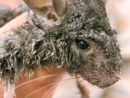
346                                                                      Vlaams Diergeneeskundig Tijdschrift, 2010, 79

Figure 1. Lateral, oblique rostrocaudal, and rostrocaudal view of a normal chinchilla skull.

                                                              necessary tooth material for continuous eruption to
                                                              compensate the ongoing tooth wear. The aradicular
                                                              hypsodont teeth in the chinchilla present particular
                                                              characteristics, perfectly matching their shape and
                                                              purpose. The periodontal ligament, for instance, does
                                                              not bridge the entire distance from bone to tooth as it
                                                              conventionally does. A middle or intermediate bundle
                                                              of collagen fibers attaches either to the alveolar bone
                                                              or to the cementum, but not to both. On the whole, the
                                                              aradicular hypsodont teeth in the chinchilla are less
                                                              solidly anchored.
                                                                  This adaptation may possibly provide added tooth
                                                              movement for growth (Wiggs and Lobprise, 1997; Van
                                                              Foreest, 1999). Chinchillas have large, chisel-shaped
Figure 2. Normal chinchilla. A. Ventrodorsal view of the      yellow to orange incisor teeth (Figure 1). This pig-
maxilla - B. Dorsoventral view of the mandibula. The          mentation, not yet present at birth, is due to a superfi-
cheek teeth arcades diverge from rostral to caudal.           cial layer of enamel (Capello and Gracis, 2005). Teeth
                                                              discoloration is common and not pathological (Ca-
and analgetics, and often force-feeding.                      pello and Gracis, 2005). Most rodents, including chin-
    The determining prognostic factor for dental di-          chillas, are simplicidentata with a standard single row
sease in chinchilla is the stage in which it is diagnosed.    of maxillary incisors (Wiggs and Lobprise, 1997). In
Too often, owners are unaware of the problem and              chinchillas, the radius of curvature of the mandibular
animals are presented in end-stage conditions, for            incisor is more than double that of the maxillary inci-
which palliative care or euthanasia are the only humane       sor teeth (Capello, 2008). At rest, most rodents place
options. In view of this fact, veterinarians have a cru-      their lower jaw retrognathically, in so doing, separating
cial role to play in raising the awareness of owners and      their incisor teeth. Chinchillas’ incisors grow at a yearly
educating them. This should include basic knowledge           rate of 5.5 to 6.5 cm (Hoefer, 1994). In comparison, this
of the species and its natural habitat, nutritional needs     rate is around 2 mm per week for guinea pigs and rats
and the importance of hay and gnawing opportunities.          (Osofsky and Verstraete, 2006). A large gap or diastema
A set of preventive measures drawn up for the owners’         exists between incisors and premolars. Since it is im-
use is presented in this document.                            possible to anatomically differentiate premolars from
                                                              molars, they are commonly referred to as “molari-
NORMAL DENTAL ANATOMY                                         forms” or “cheek teeth”. The latter have a folded struc-
                                                              ture with large grinding surfaces, as an adaptation to a
   The dental formula of chinchillas is 2(I 1/1, C 0/0,       voluminous food intake. As a consequence of the chin-
P 1/1, M 3/3) = 20 (Crossley, 1995). They have a              chilla’s strictly herbivorous diet, the occlusal surfaces
monophyodont, full elodont or aradicular hypsodont            are rough and uneven, with a succession of enamel
dentition. A single set of teeth is present during the        crests and dentinal grooves (Figure 2). The cheek teeth
whole life of the animal, without deciduous precursors        are evenly lined up with one another. Unlike in rabbits,
(Verstraete, 2003). A full aradicular hypsodont               the occlusal surfaces are (nearly) horizontal and do not
dentition is characterized by incisors and cheek teeth        present a “zigzag” pattern (Verhaert, 2004; Capello,
that continue to erupt during the whole life of the           2008). In normal dentition, the mandibular cheek teeth
animal. Aradicular teeth have no true root structure          apices should not reach the ventral mandibular border.
(Capello, 2008). Hence, chinchilla teeth have long            The maxilla of chinchillas is narrower than the man-
crowns with no anatomic roots. The embedded sub-              dible, and the occlusal plane is slightly angled from
gingival tooth segment is called the “reserve crown”,         buccal to lingual. This characteristic, however, is much
as opposed to the exposed or clinical crown (Wiggs            less pronounced than in guinea pigs (Lobprise, 2007;
and Lobprise, 1997). This ‘reserve crown’ provides the        Sulik, et al. 2007). Along with relatively little lateral
Vlaams Diergeneeskundig Tijdschrift, 2010, 79                                                                   347

movement, the temporomandibular joint tolerates a
large range of rostrocaudal motion (Verstraete, 2003).
During mastication, the mandibular condyles glide in
long gutter-like mandibular fossae, pulled by mastica-
tory muscles, mainly the masseter. There is no sub-
luxation of the articulation during physiological lower
jaw movements (Reiter, 2008).
                                                            Figure 3. Lateral view of a normal maxilla (A) and
DENTAL PATHOLOGIES                                          maxilla with cheek tooth malocclusion (B) in two chin-
                                                            chillas.
Malocclusion

   The condition of malocclusion can be described as
a misalignment of the teeth or an incorrect relationship
between the maxillary and the mandibular teeth. It is
the most widespread and common disorder in strictly
herbivorous rodents such as chinchillas and guinea
pigs. In chinchillas, malocclusion can be seen in ani-
mals as young as 6 months of age (Stroke et al., 1996).
As both the incisor and the cheek teeth of chinchillas
are continuously growing, one can easily imagine that
any disturbance in the normal attrition pattern is po-
tentially an open door to overgrowth problems and
malocclusion (Wiggs and Lobprise, 1997). Malocclu-
sion can be limited either to incisors only or to cheek     Figure 4. Advanced stage of cheek teeth malocclusion.
teeth only, or it can involve both incisors and cheek       Jaw of a chinchilla in lateral view. Notice the conse-
                                                            quential maxillary and mandibular bulging.
teeth.

Incisor malocclusion                                        Incisor-molariform or incisor-cheek teeth malocclusion
                                                            syndrome
    Mandibular incisors tend to grow in a dorsofacial
direction, while the intrinsically more curved maxillary        Typically occurring in species with full aradicular
incisors logically twist and curl into the oral cavity,     hypsodont dentition, this dental syndrome encom-
which means that the condition of the maxillary inci-       passes various components that may appear simulta-
sors can possibly deteriorate to the point where skull,     neously:
sinuses or ocular sockets are penetrated if the condition       • incisor malocclusion or elongation
is left untreated. As a result of teeth overgrowth, the         • “step-mouth”, “spikes”: molariform occlusal plane
animal will be unable to eat properly, it will mainly       irregularity with sharp points, typically on the mandi-
drop its food (“quidding”), and it will traumatise its      bular lingual and the maxillary buccal teeth facet
tongue and have excessive salivation (ptyalism or               • intraoral molariform elongation, with or without
“slobbers”). In contrast to rabbits, incisor malocclusion   elongation of clinical crowns; possibly lingual or buc-
seldom occurs as a single entity in rodents. Incisor mal-   cal deviation and possibly “bridging” over the tongue
occlusion may indeed be either consequential to, or oc-         • apical reserve crown elongation
cur alongside premolar-molar malocclusion, especially           • premolar and/or molar periapical changes, with
in older chinchillas, where the condition is generally      possible cortex perforation
linked with molariform abnormalities. In view of this           • periodontal disease, with increased premolar
fact, a thorough and full oral examination is a real        and/or molar mobility
must in patients with incisor malocclusion. Early age           • secondary soft tissue lesions of the mucosa; oral
incisor malocclusion, generally due to a genetic maxil-     pain
lary brachygnathia, is uncommon in rodents (Verstra-            • submandibular, maxillofacial or retrobulbar ab-
ete, 2003).                                                 scesses
                                                                • osteomyelitis (Verstraete, 2003).
Cheek teeth malocclusion                                        These aspects are not necessarily all present; each
                                                            patient develops its own set of problems.
   Cheek teeth malocclusion as a stand-alone problem
also occurs in chinchillas (Figure 3). Typically, the re-   Periodontal and endodontic disease
serve crown elongates, with extension of its apical
portion into the surrounding periapical tissues. Conse-        Although Actinomyces viscosus can induce primary
quently, distortions of the ventral mandibular border       periodontal disease in laboratory rats, this condition is
and the maxillary alveolar bullae can be observed (Ca-      uncommon in rodents, including chinchillas (Wiggs
pello and Caudoro, 2008) (Figure 4).                        and Lobprise, 1997; Legendre, 2003). Secondary pe-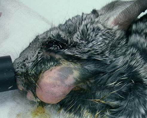
348                                                                     Vlaams Diergeneeskundig Tijdschrift, 2010, 79

riodontal disease, on the other hand, can regularly be       periapical bone remodeling needed to allow reserve
seen as a result of malocclusion or trauma. Aradicular       crown elongation could accidentally be redirected to-
hypsodont teeth are more susceptible to periodontal          wards the tooth itself (Crossley 1997). Mandibular ab-
loosening (and infection), as the particular periodontal     scesses are regularly seen in end-stage dental disease
ligament already provides extra tooth movement ca-           (Figure 5).
pacity. In tooth displacement consequent to crown                Odontomas are neoplastic processes in which well
elongation, and occasionally to trauma, the interproxi-      differentiated epithelial and mesenchymal cells of all
mal space is widened, allowing impaction of food and         different dental tissue types can be identified. These
debris (Crossley, 1995). As a result, abscesses easily
develop. Damaged incisors can lead to endodontic ab-
scessation, generally extending to the level of the pre-
molars (Legendre, 2003). Infectious processes or
trauma in rodents can bring about secondary gingivi-
tis, stomatitis with ulceration, and periodontitis (Van
Foreest, 1999).

Oral trauma

   A large range of oral fractures are described in
chinchillas, varying from simple mandibular symphy-
seal dislocation to complex jaw fractures with involve-
ment mainly of the incisor teeth. Oral fractures are ge-
nerally caused by gnawing on hard objects (Legendre,
2003).

Less common dental pathologies
                                                             Figure 5. Chinchilla in end-stage dental disease, showing
                                                             a mandibular abscess. External aspect after shaving of
    Caries, tooth resorption and odontomas have also         the left cheek.
been reported to occur in chinchillas (Crossley et al.,
1997).
    Caries appears as areas of brown staining on the oc-
clusal and/or interproximal tooth surfaces, resulting
from pigment uptake by the altered dentine. These le-
sions are much more common in captive animals than
in wild animals, probably as a combined result of diet,
salivary pH and tooth anatomy. Commercially availa-
ble chinchilla foods generally have much higher su-
crose and refined starch contents than the diets of wild
animals (Legendre, 2003). However, in chinchillas ca-
ries remain less common than other dental problems.
Some authors suggest that continuous molariform
growth and consequential constant attrition reduces          Figure 6. Latero-lateral x-ray of a chinchilla with gastro-
the occurrence of caries simply because the occlusal         intestinal stasis, impaction and tympani secondary to
surface is worn away too quickly for caries to occur         dental disease.
(Sone et al., 2004).
    Odontoclastic tooth resorption is set off by perio-
dontal inflammation. Initially, lacunae appear in the
tooth, into which granulation tissue moves in an en-
deavor to promote healing. Although repair sometimes
is done, the lost dentin is never replaced. Instead, a
composite of bone-like and cementum-like tissue fills
in the gap, with a resulting ankylosis of the tooth to the
jaw. The presence of deep periodontal pockets in many
specimens with this condition shows that the animals
have been affected by several episodes of inflammation
of the periodontal ligaments, which occurs during the
development of malocclusion. Hence, it could be con-
cluded that this periodontal inflammation triggers
odontoclastic resorption. However, the true pathoge-
nesis has not yet been determined and other hypothe-
sises remain plausible, including the possibility that the   Figure 7. Chinchilla with the “slobbers” condition.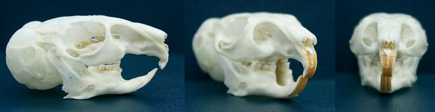
Vlaams Diergeneeskundig Tijdschrift, 2010, 79                                                                                  349

tumors of odontogenic origin have been diagnosed in                    taminated with soil and richer in silicate phytolith, all
young rodents, but are generally rare in chinchillas                   increasing its abrasive character. All of this means that
(Wagner et al., 1999).                                                 the commercial diets are much higher in energy and lo-
                                                                       wer in fiber content. Wild animals need to consume
Pathologies secondary to dental disease                                large volumes of the relatively low-energy mountain
                                                                       vegetation to meet their nutritional requirements. As
    As chewing becomes too painful or impossible,                      animals in captivity chew smaller quantities of less
anorexia progressively appears, with subsequent                        abrasive food, dental elongation can appear. All re-
weight loss seen in 79% of the animals. Since the ani-                 commendations regarding the specific requirements
mals reduce their fiber intake or, worse, fail to eat, the             for chinchillas point in the direction of high overall die-
digestive system cannot function properly. Fecal drop-                 tary fiber content. On a dry matter basis, the pellets
pings typically become smaller; tympany and consti-                    should contain 18 to 20% crude protein, 15 to 35%
pation can occur (Figure 6). Oral discomfort or pain,                  crude fiber and 4% fat (Hand and Thatcher, 2000).
abnormal occlusion and/or restricted jaw movement in-                  Crude fiber structure should be taken into considera-
duce excessive salivation (“slobbers”), which brings                   tion, as well-structured feeds increase the level of in-
about perioral and forelimb saliva staining, and/or pe-                gestion activity and feeding, with positive impact on at-
rioral skin disease (Crossley et al., 1997) (Figure 7).                trition rates (Wolf et al., 2003). A large variety of
Maxillary reserve crown elongation can specifically                    pellets, treats, mineral supplements and hay are avai-
cause pressure, deformation and even obstruction of the                lable for the consumer to choose from. Within the fra-
bony lacrimal canal (Crossley and Roxburgh, 1999).                     mework of this article, a market survey of foods spe-
Tears are not evacuated, which results in epiphora and                 cifically available for chinchillas in Belgium was
altered periorbital hair patterns. Moreover, secondary                 carried out (Table 1). Only feeds labeled on the pac-
eye pathology and periocular skin disease can develop                  kage as “complete foods” were considered. Products
(Anonymous, 1988; Crossley, 1995). Reserve crowns                      available on the internet were not surveyed.
occasionally grow into the orbital area, inducing local                    When comparing the nutritional composition of
inflammation, possible retrobulbar abscedation and                     these feeds to the above mentioned advised require-
exophthalmia.                                                          ments, it appears that none of the pet foods for chin-
                                                                       chillas available in Belgium meet the above standards.
FACTORS PREDISPOSING TO DENTAL DISEASE                                 All feeds are either lacking in the crude protein and
                                                                       crude fiber that are essential to avoid or limit dental
    Various factors, including husbandry and genetic                   problems, or they barely reach the lower limit of the re-
and traumatic influences, have been identified as play-                quirements. “Chinchilla Complete®” and “Pellets®”
ing a role in the development of dental problems in                    are the only ones providing reasonable amounts of
chinchillas.                                                           crude fiber. Among the feeds available, only half of
    Insufficient or incomplete cheek tooth wear as a re-               them (“Chinchilla Complete®”, “Pellets®”, “Selective®
sult of an unsuitable diet is the primary cause of den-                and “Chinchilla Duo®”) contain similarly looking pel-
tal pathology in chinchillas (Capello, 2008). In capti-                lets. The pellets in the others have a heterogeneous ap-
vity, these rodents are habitually fed with grain or                   pearance, with various different ingredients the animal
pellets, and their access to hay is generally limited. The             can consciously choose between. Practically, this trans-
vegetal ingredients processed in these feeds are culti-                lates into chinchillas eating a lot of high-starch and su-
vated under far more optimal conditions than the ve-                   crose components, and leaving the harder and less
getation found in their natural mountain environment.                  tasty bits, such as alfalfa pellets, behind. This results in
The mountain vegetation is more fibrous, tougher, con-                 a dramatic drop in the overall crude fiber intake, espe-

Table 1. Overview of the nutritional composition of chinchilla foods available on the Belgian market (May 2009).

Brand                                Versele laga                   Vitakraft     Supreme Pet Foods       Beduco      Recommended
                                                                                                                       requirements

Commercial      Chinchilla    Chinchilla     Snack     Chinchilla    Pellets     Charlie     Selective      Deli
name            complete       Nature        Nature      Duo                    Chinchilla                 Nature
                                                                                  food
Nutritional composition (% of dry matter)
Crude protein       16           14.5          11         15.5         16          16           16           16         18 to 20%
Crude fat           3.5           3            3           3          2.8          3            3            3             4%
Crude ash            8            7            3           9           10          7            7            6               -
Crude fiber         20            14           10          16         18.5         16           19           14         15 to 35%

Target animal   Chinchilla    Chinchilla    Chinchilla Chinchilla   Chinchilla Chinchilla    Chinchilla   Chincilla
                                             Rabbit      Degu
                                            Guinea pig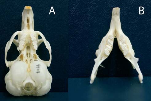
350                                                                       Vlaams Diergeneeskundig Tijdschrift, 2010, 79

cially if the owners refill the feeding pot, throwing             • defecation: changes in quantity, shape and aspect
away the leftovers. Finally, none of the packages pro-         of droppings
vide guidelines regarding the daily recommended                   • general health: disease history, weight loss, sali-
quantities to provide, nor is anything mentioned con-          vation, ocular discharge, fur changes.
cerning the importance of providing hay.
    Chinchillas, as all rodents, need to have access to        General clinical symptoms
various materials they can chew on without risk of
trauma. In addition to their indispensible wearing effect          Reduced activity, lower food intake and decreased
on the teeth, these gnawing opportunities will contri-         stool production are by far the most common signs of
bute to increased general activity levels and to avoiding      dental disease in chinchillas. In addition, wet fur around
boredom and its related stress. In chinchillas, stress is      the mouth, chin and forepaws can be observed as a re-
typically exteriorized in “fur-chewing”, a behavioral          sult of pain-induced increased pawing and ptyalism
condition in which the animal bites off areas of its own       (Capello and Gracis, 2005). Initially, the animal may
or some other animal’s fur (Ponzio et al., 2007).              have a selective appetite, generally preferring softer bits
    The domesticated chinchilla was established out            and leaving out high fiber content foods (Hoefer, 1994).
of a very small original group of 11 wild-caught indi-         When chinchillas receive mixed pellets, for the owner,
viduals exported from Chile to California in 1923              this early stage may practically mean finding more al-
(Tremblay, 2000). In this situation, the founder effect        falfa pellets as leftovers. Some animals present bruxism
must probably be considered to be a significant factor.        or teeth grinding due to discomfort (Crossley, 1995).
This translates into a loss of genetic variation, which        Frequent later sequels of dental disease in chinchillas
has an effect on gene frequency and the prevalence of          are weight loss and emaciation, however hard they are
genetic diseases. In this small population, one can as-        to detect in this densely-furred animal, where only re-
sume there is an increased risk for genetic drift and in-      gular weighing ensures effective weight follow-up (Ca-
breeding. Tooth development being in some degree ge-           pello, 2008). Yet another factor makes the discovery of
netically controlled, genetic drift could explain the          the problem even harder. Animals with sore teeth, jaws
higher incidence of dental problems in captive-bred            or oral mucosa are often unable to prehend, chew or
animals (Kraft, 1994; Silverman and Tell, 2005).               swallow food properly. As the chinchilla’s painful ea-
    Every so often, dental problems can be triggered by        ting attempts generally result in food scattering, the
trauma. This can bring about dental tissue damage,             bowls do become empty, despite the very low food in-
including tooth bud relocation. Subsequently, the cor-         take. While food intake decreases, fecal droppings ini-
responding lower and upper teeth are no longer cor-            tially become smaller. Later, fecal output may totally
rectly aligned, and abnormal tooth elongation can ap-          cease (Crossley, 1995). Chinchillas with teeth problems
pear. Most commonly, the incisors are affected (Reiter,        no longer use their mouth for grooming, with the re-
2008).                                                         sulting dull-looking and tangled fur.

DIAGNOSING TOOTH PROBLEMS IN CHIN-                             Clinical examination
CHILLAS
                                                                  As the physical presentation of a condition invol-
History                                                        ving the teeth can vary, a holistic approach, including
                                                               an assessment of the general condition, external eva-
    Although early identification is crucial, detecting        luation of the facial and jaw area and, finally, intraoral
dental conditions in chinchillas is tricky, even more so       inspection, is essential for a correct screening of the
for non-trained or non-experienced owners. On the              animal.
one hand, most signs of dental disease in rodents are             Weight loss is the most frequent sign identified du-
non-specific and difficult to see; on the other hand, pa-      ring general physical examination. A thorough visual
tients with tooth abnormalities can be asymptomatic            inspection of the facial area and the jaw should include:
during the initial phases of the disease. Commonly,               • overall shape and symmetry of the head and
chinchillas indeed show clinical signs only in an ad-          mouth
vanced stage of dental disease (Crossley, 1995; Bren-             • position of the mandible and mouth opening, and
ner et al., 2005). In view of this fact, regular follow-       observation of the masticatory movements
up and education of owners by the veterinarian are                • external appearance of the incisors
essential in early diagnosis. In order to identify oral dis-      • nasal plateau and perinasal area
orders, the history should cover various aspects such             • position and shape of the eyes, the periocular
as:                                                            area.
    • housing: type of housing, risk of trauma, recent            Following inspection, a careful maxillofacial pal-
changes                                                        pation should be done, including evaluation of the
    • feeding habits: food type(s); quantities and fre-        mandibular lymph nodes, mandibular/sublingual
quency, recent changes in feeding habits                       glands, zygomatic arch, orbit, temporomandibular
    • eating patterns: quantities eaten (overall and of        joints and mandibles. For the mandibles, special at-
each type of food), leftovers, changes in preference of        tention should be given to evaluating the surface for
foods/ingredients, changes in chewing patterns                 bone deformities. Incisor and cheek teeth occlusion as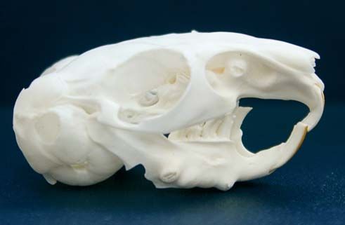
Vlaams Diergeneeskundig Tijdschrift, 2010, 79                                                                     351

well as jaw movement capacity must also be evaluated
(Capello and Gracis, 2005). Chinchillas are known to
be pretty tough rodents with quite high pain thres-
holds. The absence of pain reaction when examining
the animal can in no circumstance exclude a dental
condition. Epiphora and ventral mandible bone defor-
mities, on the other hand, are two typical clinical signs
that, in the vast majority of cases, will direct the clini-
cian towards dental problems. These symptoms are
indeed typical telltale signs of maxillary and mandi-
bular cheek teeth deformation, respectively (Capello,
2008). Epiphora is due either to obstruction of the la-
crimal canal or to pain-induced increased tear produc-
tion (typically with mucosal or bone pain). It can ap-
pear as a stand-alone symptom or be accompanied by
exophthalmos and/or conjunctivitis. These two condi-
tions develop when the orbit floor and the zygomatic
process of the maxilla become involved. In this case,
severe ptyalism is often noticed (Crossley et al., 1997).
Dacryocystitis – or nasolacrimal sac infection – can ap-
pear secondary to lacrimal canal obstruction, provoking
pain, swelling of the inner lower eyelid and epiphora.
Commonly, these infections are associated with Sta-
phylococcus aureus, Streptococcus pneumoniae or
Pseudomonas species. Blepharospasms with partial
closing of the eye are sometimes present (Capello and
Gracis, 2005). In any case, ocular (or nasal) discharge
should include dental disease in its differential diag-       Figure 8. Oral exam of an anesthetised chinchilla; pouch
nosis (Crossley, 1995). Regularly found on clinical           dilator and incisor speculum in place.
examination, facial abscesses appear as unilateral swel-
lings. It is generally taken for granted that a cheek tooth   al. 2005). Intraoral examination should include chec-
is responsible, whereas in chinchillas, incisor-related       king:
endodontic abscedation can actually reach up to the               • the integrity and mobility of each single tooth
premolar level (Legendre, 2003).                                  • the appearance of the occlusal surfaces: enamel
    Oral inspection of the conscious chinchilla is com-       crests, dentinal grooves, spikes, occlusal angle
plicated by the animal’s size (small mouth opening,               • the orientation and length of the clinical crowns,
narrow oral cavity, long tongue) and the fact that it is      the size of the interproximal spaces, the feed impaction
rather difficult to restrain. Although it may possibly re-        • the presence of periodontal pockets, the appea-
sult in a lot of struggling, escape endeavors and fur         rance of the periodontal tissues.
slips, oral inspection should however be attempted du-            In Crossley’s study (Crossley, 2001), incisor over-
ring every physical examination as a first step in eva-       growth was found in 55% of the cases. In one-third of
luating the oral health of the animal. To do so, the chin-    the affected animals, abnormal incisor wear patterns
chilla is ideally restrained in physiological standing        could be identified, when in cheek teeth this was only
position and examination is performed by means of an          the case for 16% of the patients. Less than 10% of the
otoscope or a lighted bivalve pediatric speculum, care-       chinchillas examined showed spikes and feed impac-
fully inserted in the mouth (Osofsky and Verstraete,          tion (Crossley, 1995). The clinical examination might
2006). If the patient is too agitated, it should be deeply    be misleading, and a relatively minor molariform mal-
sedated or anesthetized (for information on sedation          occlusion should be considered an important clinical
and anesthesia, please see below). Nevertheless, the          finding.
procedure remains difficult, and this probably accounts
for the small amount of information available descri-         Anesthesia
bing dental lesions in chinchillas. When the animals are
anesthetized/sedated, incisor speculums and cheek                 Dental interventions on chinchillas require general
pouch dilators of the smallest size can be used in chin-      anesthesia to control stress and pain, to minimize ope-
chillas for optimal visualization (Brenner, et al. 2005;      ration time and to allow the surgeon to work in the sa-
Capello and Gracis, 2005) (Figure 8). Once the instru-        fest conditions possible. A pre-anesthetic evaluation is
ments are in place, feed residues are first removed. A        highly recommended. Especially in very weak pa-
proper examination of the oral cavity is done by sys-         tients, full-body radiography can help in evaluating the
tematically inspecting each quadrant and tooth. The use       overall condition and, more specifically, to see if there
of periodontal probes is questionable, because of the         is secondary gastro-intestinal stasis. In the event of an
trauma they can induce (Verstraete, 2003; Brenner, et         inconclusive pre-anesthetic evaluation, and if an animal
352 Vlaams Diergeneeskundig Tijdschrift, 2010, 79
is too debilitated to undergo surgery, this surgery tal medical imaging (Brenner et al., 2005).
should be postponed when possible. Post-anesthetic In comparison with direct visual inspection, images
ileus is a common complication in rodents, therefore obtained through oral endoscopy are of much better
long-lasting fasting is contraindicated and special at- quality and the capacity for detecting oral lesions is
tention should be given to encourage the animal to eat, greatly enhanced. Chinchillas are ideal candidates for
both before and as quickly as possible after the inter- oral endoscopy as the horizontal occlusal surface faci-
vention. In any case, fasting should not exceed one litates the examination. This examination includes tho-
hour in young chinchillas to avoid hypoglycemia. Due rough inspection of the appearance of the gingival and
to their small size and difficult or impossible intuba- oral mucosa, the length of the cheek teeth clinical
tion, it can be difficult to induce anesthesia in rodents. crowns, the appearance of the occlusal surface, the
Longer and more intrusive procedures require inhala- size of the interproximal space, and the appearance of
tion anesthesia after induction. This method implies the each tooth. Abnormalities such as crown elongation, al-
use of a tight-fitting anesthetic mask, which is repea- teration of the enamel crests and dentinal grooves, lin-
tedly put on and off, thus both enabling anesthesia of gual or buccal deviation of the cheek teeth (with or
the animal and enabling the surgeon to work (Osofsky without lesions of the mucosa), and widening of the in-
and Verstraete, 2006). A small nasal mask can possibly terproximal spaces with consequent feed impaction
be used. Injection anesthesia does not allow fine, quick can be seen (Capello and Gracis, 2005). At present,
control over the anesthetic depth and it also requires a radiography remains the principal diagnostic tool. Va-
mask for the oxygen supply. The following anesthesia rious lesions such as dental resorptive lesions, bone re-
protocols can be used (Carpenter, 2005): sorption and lysis, cortical perforation, apical elonga-
isoflurane 2-5 % induction; 0.25-4.0% maintenance tion, tooth fractures and missing teeth can be properly
(anesthesia of choice/or short interventions) diagnosed with it. However, it is important to empha-
ketamine 40 mg/kg + Acepromazine 0.5 mg/kg IM size the fact that a normal radiography does not exclude
ketamine 20-40 mg/kg + Diazepam 1-2 mg/kg IM the presence of pathologies. Small or specific hard tis-
The consequential shortcomings such as prolon- sue lesions can indeed be missed, though still found on
ged surgery time, risk of inhalation of the anesthetic gas autopsy (Gracis, 2008). Magnification radiography
by the operator, and potential unnecessary pain and techniques, using units with small focal spots (0.1mm)
stress for the animal can be bypassed through nasal in- and a 100-mA capacity can compensate for size. A
tubation Legendre, 2003). The monitoring of rodents usual chinchilla skull and dental radiography study in-
during anesthesia should focus especially on respira- cludes right lateral, dorsoventral and rostro-caudal
tion, heart rate and body temperature. Hypoventilation views. An ideal laterolateral (LL) positioning results in
and apnea are common; satisfactory ventilation can be a perfect overlapping of the left and right tympanic bul-
maintained by adjusting the head position and anes- lae, optic foramen, orbits, temporomandibular joints
thetic depth. Insufficient anesthesia increases pain and and ventral mandibular margins (Verstraete, 2003; Gra-
stress, which often translates into tachycardia. Heat cis, 2008). In the LL projection, seen by the majority
packs ought to be used, as small animals are prone to of authors as the most useful one, both incisor and
hypothermia (Osofsky and Verstraete, 2006). cheek teeth are evaluated. The maxillary incisors’ re-
serve crown should not reach further than two-thirds of
Medical imaging the length of the diastema, while that of the mandibu-
lar incisors ought not to grow beyond the second cheek
Studies have shown that during mouth inspection in teeth (Wiggs and Lobprise, 1997). The incisors occlu-
conscious and in anesthetized rabbits, respectively, sal surface is normally chisel-shaped; the wearing an-
only 35 and 50% of oral lesions are detected. Chin- gle is generally more pronounced in maxillary teeth.
chillas and guinea pigs are no exception to this rule. Flat occlusal surfaces are indicative of dental disease
Disregarding the fact that visual observation is com- and/or intervention. Possible maxillary incisor hard
plicated by the small oral cavity and the narrow mouth palate perforation consequent to overgrowth can be
opening, one should keep in mind that the tooth portion seen on this projection (Capello and Gracis, 2005;
that is actually visible above the gingival margins (i.e. Gracis, 2008). Typically, the chinchilla cheek teeth oc-
the clinical crown) represents only a small part of the clusal plane should be flat, nearly horizontal and pa-
tooth, the tip of the iceberg. Most of the tooth structure rallel to the ventral mandibular border on a LL projec-
is located below the gingival margin and is not visible tion. The surface should form a regular smooth
during oral examination (Crossley and Miguelez, 2001; palisade. In order to evaluate whether the animal is suf-
Lobprise, 2007). In view of this fact, medical imaging fering from maxillary cheek tooth elongation, a virtual
is an absolute must in diagnosing and understanding line is drawn from the tympanic bullae hilus to the dor-
dental pathology in rodents in general, and in chin- sal aspect of the maxillary incisor tooth (Figure 9). Api-
chillas in particular (Gracis, 2008). The imaging tech- ces above this line are indicative for disease. Mandi-
niques used currently in rodent dentistry include oral bular cheek teeth apices reach close to the ventral
endoscopy, radiology, computed tomography and mag- mandibular cortex, which should be thin, smooth and
netic resonance imaging. Unless the animal is really without deformities. The presence of bone deformities
calm and used to physical manipulation, sedation or or so-called “bulging” is extremely suggestive for man-
anesthesia is preferable for precise and effective den- dibular cheek teeth elongation. Finally, the loss of pa-Vlaams Diergeneeskundig Tijdschrift, 2010, 79 353
Figure 9. Radiograph of a chinchilla skull with graphic Figure 10. Lateral view of a chinchilla with pronounced
overlay, showing the above limit for maxillary teeth api- cheek teeth and incisive teeth malocclusion. At the level
ces. of the cheek teeth abnormal occlusal plane, abnormal
delineation and growth direction of the cheek teeth roots,
and bulging and thinned mandibular cortices were seen.
rallelism between the maxillae and mandibles in this Severe maxillary incisor curling with crowns and roots
view is a third indicator for coronal elongation (Figure impinging on the hard palate.
10). Dorsoventral (DV) projections generally are fa-
vored over ventro-dorsal (VD) because they are done
in ventral recumbency, thus decreasing respiratory radiologic interpretation. For this reason, two obliques
complication risks. Good quality DV radiographies (left and right) as well as intraoral projections are re-
should have a perfect symmetry (between the right commended for an in-depth evaluation of the mandi-
and left sides of the skull). Through this projection, the bles, maxillae and teeth (Verstraete, 2003; Gracis,
maxillary and mandibular bone edges as well as the 2008). Lateral oblique (LO) views are useful for eva-
skull-mandible connection and the orbital cavities can luating the mandibular and maxillary cheek teeth api-
be properly evaluated. Abnormalities such as cheek ces and reserve crowns. The mandibular cheek teeth of
teeth elongation or orientation changes can be quite ea- one side (closest to the film) and the contralateral
sily pointed out (Figures 11 and 12). On the other maxillary cheek are visualized. Individual incisor api-
hand, DV views are not suitable for incisor evaluation. ces are generally clearer in this projection than on the
Rostrocaudal (RC) projection is taken with animals in LL. Intraoral projections, where non-screen films are
dorsal recumbency (Verhaert, 2004; Capello and Gra- placed directly in the mouth of the patient, are also
cis, 2005). The mouth is closed and the head straighte- mentioned in the literature. This technique generally
ned up at an angle of 90°C. Bohmer suggests open- provides radiographies with very fine resolution and
mouth RC for specific assessment of maxillary cheek high diagnostic value (Verhaert, 2004; Capello and
teeth overgrowth and temporomandibular joints Gracis, 2005; Gracis, 2008). However, to properly
(Bohmer, 2001a, 2001b). RC views provide valuable place small films in the equally small oral cavities of
complementary information, facilitating a three-di- chinchillas is a real challenge, and it sometimes cannot
mensional understanding of the possible pathology. be done.
The existence of spurs and spikes, bridging, coronal Even though radiography remains the primary diag-
and apical elongation, or cortical perforation can be nostic tool in chinchilla dentistry, it often fails in the
highlighted, and the angles of the cheek teeth and the early detection of pathologies such as cheek tooth
occlusion surface area can be measured. elongation. The reasons for this shortcoming lie essen-
The small size of these rodents combined with the tially in the difficulty of avoiding superposition when
dental quadrant superposition increase the difficulty of taking skull x-rays, as well as in the insufficient detail354 Vlaams Diergeneeskundig Tijdschrift, 2010, 79
Fig. 11
Figures 11 and 12. Lateral and dorso-ventral view of a
chinchilla with extensive overgrowth of the mandibular
and maxillary cheek teeth, spike formation and secon-
dary incisor teeth pathology and exophthalmia.
and soft tissue contrast it produces, which does not ena-
ble the detection of the subtle tissue changes of early-
stage dental disease (Crossley et al., 1998; Capello and
Caudoro, 2008). Nonetheless, despite the fact that
chinchillas will often only show clinical signs in a
quite advanced stage, early diagnosis remains absolu-
tely crucial. Furthermore, early screening and conse-
quent breeding pool selection paves the way to a heal-
thier future chinchilla population (Crossley et al., Fig. 12
1998). Various studies have highlighted the improved
diagnostic capabilities of CT in comparison with tra- even there the higher cost is often a major hindrance
ditional radiography for rabbits and rodents, including (Brenner et al., 2005; Gracis, 2008).
chinchillas (Gracis, 2008). Especially in small species
such as these, CT offers: TREATMENT
• enhanced sensitivity, especially for temporoman-
dibular joints and tympanic bullae Incisor teeth treatment
• greater capacity to detect fine or nearly imper-
ceptible skeletal changes Before addressing incisor tooth problems in chin-
• a much higher level of detail of the teeth chillas, it is crucial to bear in mind that incisor over-
• a higher level of detection of both soft and hard tis- growth seldom appears as a stand-alone problem in ro-
sue modifications (Brenner et al., 2005; Crossley et al., dents. In any case, a thorough mouth examination
1998). needs to be carried out to identify possible underlying
CT of a normal chinchilla skull allows clear visua- causes and allow a correct diagnosis and prognosis.
lization of the mandible, maxilla, zygomatic arch, tem- In the case of overgrowth, or when they are partially
poromandibular joint, parietal and nasal bones, tym- broken as a result of trauma, incisors need trimming or
panic bullae, sinuses and teeth. Abnormalities such as tooth-height reduction (Legendre, 2003; Capello and
coronal and root elongation, deformities and/or perfo- Gracis, 2005). Prior to trimming, and with the aim of
ration of the cheek bone, as well as caries can be seen avoiding injury to the soft tissues of lip and tongue, a
even in the early stages (Capello and Caudoro, 2008) split tongue depressor is positioned caudally to the in-
(Figure 13). Moreover, the three-dimensional recon- cisors, in the diastema space (Legendre, 2003). Tooth-
struction and rendering features of modern CT are height reduction of incisors is ideally carried out with
highly valuable for the additional information they a cylindrical diamond bur fitted onto a high-speed
provide that is relevant for diagnosis and treatment, as dental handpiece. Dremel® tools or cutting disks moun-
well as for educating the owners (Capello, 2008). Fi- ted on a surgical handpiece are to be avoided, as they
nally, MRI and US are used every now and then for the are oversized and easily induce soft tissue trauma.
specific evaluation of soft tissues such as muscles, sa- Cutters or nail clippers are contraindicated as they of-
livary glands and the orbital area (Gracis, 2008). In ten induce diagonal fractures and tooth splitting, pos-
comparison with MRI and CT, US offers the double ad- sibly with pulp exposure (Legendre, 2003). This very
vantage that it is relatively cheaper and it is more ap- painful consequence can lead to periapical pathosis
plicable to field conditions. Despite the obvious ad- (Verstraete, 2003). Reduction ought to be performed
vantages presented by modern medical imaging, these delicately to avoid too extensive trimming and, above
techniques are unfortunately still scarce and generally all, thermal damage to the pulp. When nasal intubation
only available in research or university structures, and is used, cooling fluid can be considered on conditionVlaams Diergeneeskundig Tijdschrift, 2010, 79 355
Figure 13. CT of the skull of a chinchilla with extended cheek teeth overgrowth. CT images (from left to right, taken
from the nares to the orbital area) show elongation of maxillary and mandibular cheek teeth roots, the presence of
cheek teeth spikes in both the labial and the buccal directions, and thinning of the mandibular cortex.
that the oropharynx is packed. While trimming, it is im- (Osofsky and Verstraete, 2006). Patience, delicacy and
portant to restore the incisors’ normal chisel-shaped oc- careful handling are essential when operating on pa-
clusal angulation in order to restore them to the most tients of this size. Various complications such as tooth
normal condition possible. This procedure is carried out fracture, hemorrhage and tooth regrowth do occur.
on the basis of need, generally every 3 to 6 weeks. Pul- Tooth fractures, and in particular root fractures, com-
pal exposure is a possible complication of the inter- monly happen, leaving behind fragments which can ge-
vention, which requires partial pulpectomy and pulp nerally not be recovered. These fragments can cause
capping. The pulp cavity opening is filled with inter- tooth regrowth and abscedation. In the event of root
mediate restorative material (IRM®, Dentsply Interna- fracture without regrowth, an x-ray is advisable a few
tional, York, USA). Composites or other harder fillings weeks after surgery to verify the absence of dysplastic
are not recommended as they disturb the physiological apical growth, which requires surgery. Small bleedings
tooth abrasion (Lobprise, 2007). In some cases, incisor frequently crop up during surgery, but are generally
malocclusion or overgrowth cannot be corrected by harmless. Unless an iatrogenic alveolar bone fracture
trimming alone, or the required intervention frequency takes place during extraction, profuse hemorrhage is
to achieve an acceptable condition is not feasible. In uncommon (Legendre, 2003). Chinchillas adapt quite
these circumstances, although far more seldom than in well to incisor extraction, although less so than rabbits,
rabbits, incisor extraction can be considered in chin- as they tend to chew more with their incisors than la-
chillas (Lobprise, 2007; Capello, 2008). Due to the gomorphs (Capello, 2008).
very long incisors, the intervention is often tricky but
can generally be done via non-surgical extraction (Ver- Cheek tooth treatment
straete, 2003; Osofsky and Verstraete, 2006). As for any
other tooth, extraction is based on luxation. A small Occlusal leveling of cheek teeth clinical crowns in-
luxator or a bent hypodermic needle of appropriate size cludes height reduction as well as the removal of spikes
is brought carefully into the periodontal space. Once in and spurs. The same remarks regarding the choice of in-
place, a constant pressure is applied in the apical di- struments for incisor reduction apply here as well (Ver-
rection for 20 to 30 seconds. This procedure is alter- straete, 2003). A full dental check-up, including dental
natively repeated at the mesial and distal tooth facet, radiography is strongly advised prior to trimming in or-
i.e. the sides of the teeth. Wiggling of the tooth should der to have an overall picture of the mouth (Capello and
be avoided as this is precisely the type of effort that the Gracis, 2005). While leveling the teeth, the practitioner
periodontal ligament is designed to resist. Gradually, should give attention to restoring the physiological ho-
the luxator moves deeper and deeper, the periodontal rizontal occlusal plane, as well as the incisor occlusion.
ligament ruptures and the tooth becomes loose (Wiggs If needed, a normal skull can be used as reference du-
and Lobprise, 1997; Verstraete, 2006). Before fully ring the intervention (Verstraete, 2003). The same pre-
extracting the tooth, it is first pushed back and twisted cautions as for incisors need to be taken regarding pulp
slightly. This crucial step produces damage o the ger- exposure. In chinchillas, cheek teeth coronal reduction
minal tissue, which will prevent the tooth from growing can be hindered by reason of gingival proliferation co-
back (Legendre, 2003). existing with the elongation of the clinical crown. Gin-
When incisor extraction is done as part of incisor givectomy is needed when coronal reduction is required
malocclusion treatment, the removal of all 4 incisors is beyond the pathological gingival margin (Capello and
advised. On the other hand, extraction of the opposing Gracis, 2005). Some cases of incapacity to close the
incisor is not needed when just 1 incisor is removed. In mouth after occlusal adjustment have been reported,
the latter case, the remaining incisors are sufficiently but only in guinea pigs (Legendre, 2003).
worn off through the lateral occlusion movements Cheek tooth extraction should be considered in356 Vlaams Diergeneeskundig Tijdschrift, 2010, 79
Table 2. Antibiotics, analgetics and anti-inflammatory quent feed impaction and fistulation to the skin surface
drugs that can be used for chinchillas. Recommended are described (Legendre, 2003). In addition, over-
active substances and posology (Quensberry, 2003). growth of the opposite teeth is frequent and will require
life-long regular trimming. Local instability can appear
Active substance Posology in neighboring teeth with possible reoccurring perio-
Carprofen 4 mg/kg per os (po) SID
dontal disease and step-mouth development.
Buprenorphine 0.05 mg/kg subcutaneous (sc) As the mandibular and maxillary cheek teeth are not
BID-TID exactly positioned facing each other, extraction of the
Enrofloxacine 5-15 mg/kg po, intramuscular (im) opposing teeth provides no solution. Quite to the con-
or (sc) BID trary, this intervention frequently even exacerbates the
Metronidazole 10-20 mg/kg po BID malocclusion problem (Legendre, 2003). In the event
Marbofloxacine 5 mg/kg im sc po BID of abscedation, specific treatment including drainage
Meloxicam 0.2 mg /kg im sc po BID and in-depth debridement is required. Abscesses and
osteomyelitis caused by periapical lesions or involving
both periodontal and endodontal pathologies will ge-
cases involving ectopic, fractured or unstable teeth nerally not heal unless the diseased tooth is extracted
(Legendre, 2003). It can be done using either an intra- or any other cause identified and dealt with. Some
oral or an extraoral technique (Verstraete, 2003). authors suggest packing the abscess cavity with cal-
The intraoral non-surgical method can be applied to cium hydroxide paste instead. However, this method is
any cheek tooth and is in principle the less traumatic not advisable as it does not address the cause and ex-
one. It requires an experienced and skilled hand and is poses the animal to tissue necrosis due to the caustic
not applicable in cases involving tooth ankylosis or nature of calcium hydroxide. A long-term antibiogram-
pronounced elongation (Legendre, 2003). The techni- based antibiotherapy is in any case required. Antibio-
que is similar to that used for any tooth. Special atten- tic impregnated polymethyl-methacrylate beads can
tion is required to avoid tooth fracture. Once the peri- also be used (Verstraete, 2003).
odontal ligament is severed, the straight walls of the
healthy aradicular hypsodont cheek tooth facilitates Postoperative care
its final removal (Legendre, 2003).
The extraoral surgical approach is an option only for Postoperative care is of major importance when
mandibular cheek teeth (Capello, 2008). An incision is chinchillas are hospitalized for dental problems. In
made in the cheek over the ventro-lateral portion of the any event, antibiotics and analgetics should be provi-
mandibula. A gingival flap of the affected tooth is lif- ded, especially to extraction patients on account of the
ted aside to expose alveolar bone. This bone is cut traumatic nature of the procedure, and to debilitated
away by means of a bone chisel or bur. Care should be animals which are more prone to infections (Table 2).
taken at this level to avoid local nerves and blood ves- It is essential to proceed with caution when choo-
sels. The accessible part of the periodontal ligament is sing the antibiotic type, dose and period of admini-
severed and the bare roots loosened with luxators. stration for rodents, as life-threatening complications
Once this is done, final extraction can either be per- such as diarrhea may arise (Van Foreest, 1999), (Ver-
formed through the buccotomy opening, or else the straete, 2003). Moreover, anti-inflammatory drugs are
tooth can repulsed into the oral cavity as in equine den- required in the event of periodontal inflammation or
tistry. The affected area is curetted. Finally, the defect any other ongoing inflammatory process. Secondary
is filled with osteoconductive material, and the soft tis- stomatologic (gingivitis, stomatitis, ptyalism, etc.) or
sues are sutured (Legendre, 2003). In order to prevent ophthalmologic pathologies (lacrimal duct obstruc-
infection, and unless significant hemorrhage is present, tion, dacryocystitis, conjunctivitis, retrobulbar absces-
it is recommended to flush the sockets and leave them ses, etc.) need specific treatment when present. They
open for drainage (Wiggs and Lobprise, 1997). The ex- generally clear up once the initial problem is taken care
traction of aradicular hypsodont cheek teeth remains a of. In the case of moist dermatitis secondary to ptya-
challenging and quite risky intervention. Besides the lism, it is advised to clip the perioral area. In this case,
narrow access and the limited space in which to ma- antibiotics should be provided, as cases of associated
neuver, chinchilla cheek teeth are also fairly close to Pseudomonas infection have been reported (Wiggs
one another and their embedded portion is much larger and Lobprise, 1997). While patients generally tend to
than the supragingival portion. Tooth fracture during resume eating within a few hours after the intervention,
extraction is a common complication. Furthermore, as some chinchillas with painful mouths remain anorec-
the bone plates are very thin, the space between alveoli tic. If the situation persists, force-feeding may be re-
and the orbit / nasal cavity (maxillary teeth) or the cor- quired. Critical Care® (Oxbow Animal Health, Mur-
tical margin of the mandibula (mandibular cheek teeth) dock, USA), or simply pureed vegetables or fruits like
is extremely small. Needless to say, in these circum- apples and carrots can be given through a syringe. In
stances the risk for iatrogenic damage is real, and even many instances, although not all, force-feeding stimu-
more so in the case of bone lysis secondary to dental lates the gastrointestinal system, and the animals start
pathology (Verstraete, 2003; Osofsky and Verstraete, eating spontaneously (Wiggs and Lobprise, 1997). For
2006). Incomplete healing of the socket with conse- patients admitted in very poor condition, this conva-Vlaams Diergeneeskundig Tijdschrift, 2010, 79 357
lescence diet can be provided for a longer period of for instance, consulting because they are not sure if
time, possibly in addition to spontaneous eating. Once “that white thing there is normal or not…” . Likewise,
the condition of the animal improves, the diet can gra- prolonged anorexia and poor or absent dropping pro-
dually be replaced by a more abrasive, low-energy duction are not of an alarming nature for a lot of ow-
and voluminous one (Legendre, 2003). Bringing the ners.
chinchilla back as soon as possible into its own envi- The authors believe the veterinarians can play a ma-
ronment, with the familiar noises and odors, and where jor education role in this matter.
it can benefit from TLC (Tender, Loving and Care), is The owners could, for example, consult the veteri-
another positive factor in stimulating appetite and ac- narian before or directly after the purchase of a chin-
celerating recovery (Van Foreest, 1999). When the pa- chilla, to obtain the necessary information on how to
tient is handed over to the owner, the owner should be keep their new pet in perfect health. In this best case sce-
reminded of possible post-operative complications and nario, the veterinarian can take the opportunity to edu-
specific signs to watch for in coming days (Wiggs and cate the owner regarding the specific tendency of chin-
Lobprise, 1997). Ideally, a recheck visit should be chillas to develop dental pathology. The information
planned 10 to 14 days following the intervention to en- provided ought to cover basic knowledge of the species
sure that the healing is going as planned. From that and its natural environment, nutritional requirements and
point on, regular controls are often required (Van Fo- especially the importance of fodder and fiber, as well as
reest, 1999). If a one-off surgery and dietary change can aspects of housing, including the provision of gnawing
be curative in patients where inappropriate feed was the opportunities. Owners should be made aware of the
underlying cause of the dental problem, re-elongation need for a regular dental check-up plan, in the same way
resumes in a number of cases because the animal is as dogs and cats have their annual health visit. Further-
unable or not eager to have a normal chewing pattern. more, one could come up with a set of “golden rules” or
In this case, dental intervention may be needed every preventive measures that can be applied to minimize the
4 to 8 weeks (Legendre, 2003). onset of dental problems. These could, for instance, in-
clude:
Prognosis • avoid breeding with animals with a family history
of dental pathology
The prognosis for dental disease in rodents is ge- • consider hay as the chinchillas’ primary food,
nerally more cautious than for rabbits (Capello, 2008). which should always be available
The stage in which the dental disease is diagnosed re- • hay intake can be stimulated by offering a variety
mains the crucial and determining point for prognosis of the types currently available
in chinchillas. Sadly, because owners’ awareness of the • limit pellets to 2-3 tablespoons a day per animal
problem is often limited or inexistent, at first presen- • prefer feed in which the pellets are all the same,
tation chinchillas are far too frequently diagnosed with with high crude fiber content
severe to end-stage malocclusion, which requires re- • be sure to provide gnawing materials in sufficient
current dental intervention or, for which in the worst quantity and variety. Wooden sticks (preferably un-
cases, only palliative care can be offered. The fact that treated willow, beech, hazelnut, or fruit tree), coconut
chinchillas seem to have a much higher pain tolerance shells or cardboard, for instance, can be offered on a re-
threshold than guinea pigs, for instance, unfortunately gular basis (Tremblay, 2000)
works against them, as they will often only show cli- • avoid high sucrose content treats
nical symptoms in the late stages of the disease (Ca- • consult a veterinarian without delay if the quantity
pello, 2008). The prognosis is most guarded for chin- of droppings declines, if the chinchilla starts slavering a
chillas presented with poor overall health conditions, lot, if it often drops its feed, or if it eats less or becomes
marked anisognathism or gingival proliferation (Le- fussy regarding its hay (Osofsky and Verstraete, 2006).
gendre, 2002). By contrast, patients brought in for un-
complicated mastication difficulties have higher In cases of poor dental condition, the owners should
chances of being definitively cured. However, in view be assisted on how to manage the problem and reduce
of the fact that dental pathologies can present various its progression (Crossley, 1995; Legendre, 2003). Diet
forms and complication levels, it is advisable to for- changes are to be suggested, ranging from more abra-
mulate a prognosis on a case-by-case basis (Capello, sive food to soaked pellets or adapted semi-liquid for-
2008). mulations in the worst cases. At this stage it is impor-
tant to inform the clients that if a diet change can in
Disease prevention and client education some cases resolve the problem, in other, more severe
situations, it will not, and more invasive measures
Even though some owners are obviously making ef- such as surgery may have to be considered. For these
forts to learn how to properly care for their animal and more serious cases, the veterinarian needs to emphasize
to monitor it on a regular basis, the veterinary client’s the fact that dental diseases in chinchillas are often of
awareness regarding animal health in general, and a chronic nature and do require regular intervention or
chinchillas in particular, is astonishingly poor in the a long-term commitment on the part of the owner
vast majority of cases. It is not exceptional to have ow- (Osofsky and Verstraete, 2006). In cases where the
ners presenting pets with obvious incisor overgrowth, animals are extremely weak, are suffering despite me-You can also read