Early prediction of acute necrotizing pancreatitis by artificial intelligence: a prospective cohort analysis of 2387 cases
←
→
Page content transcription
If your browser does not render page correctly, please read the page content below
www.nature.com/scientificreports

                OPEN             Early prediction of acute
                                 necrotizing pancreatitis by artificial
                                 intelligence: a prospective
                                 cohort‑analysis of 2387 cases
                                 Szabolcs Kiss 1,2,3, József Pintér4, Roland Molontay 4,5, Marcell Nagy 4, Nelli Farkas2,6,
                                 Zoltán Sipos2, Péter Fehérvári2,7, László Pecze2, Mária Földi 1,2,3, Áron Vincze8,
                                 Tamás Takács9, László Czakó9, Ferenc Izbéki10, Adrienn Halász1,10, Eszter Boros10,
                                 József Hamvas11, Márta Varga12, Artautas Mickevicius13, Nándor Faluhelyi14,
                                 Orsolya Farkas14, Szilárd Váncsa2,15, Rita Nagy2,3,15, Stefania Bunduc 15,16,
                                 Péter Jenő Hegyi15,17, Katalin Márta15,17, Katalin Borka15,18, Attila Doros15,19,
                                 Nóra Hosszúfalusi15,20, László Zubek15,21, Bálint Erőss 15,17, Zsolt Molnár 15,21,22,
                                 Andrea Párniczky2,3, Péter Hegyi 2,15,17,33, Andrea Szentesi1,2,33* & Hungarian Pancreatic
                                 Study Group17*

                                 Pancreatic necrosis is a consistent prognostic factor in acute pancreatitis (AP). However, the clinical
                                 scores currently in use are either too complicated or require data that are unavailable on admission
                                 or lack sufficient predictive value. We therefore aimed to develop a tool to aid in necrosis prediction.
                                 The XGBoost machine learning algorithm processed data from 2387 patients with AP. The confidence
                                 of the model was estimated by a bootstrapping method and interpreted via the 10th and the 90th
                                 percentiles of the prediction scores. Shapley Additive exPlanations (SHAP) values were calculated
                                 to quantify the contribution of each variable provided. Finally, the model was implemented as an
                                 online application using the Streamlit Python-based framework. The XGBoost classifier provided an

                                 1
                                  Doctoral School of Clinical Medicine, Faculty of Medicine, University of Szeged, Szeged 6720,
                                 Hungary. 2Institute for Translational Medicine, Szentágothai Research Centre, Medical School, University of Pécs,
                                 Szigeti út 12., II. Emelet, Pécs 7624, Hungary. 3Heim Pál National Pediatric Institute, Üllői út 86, Budapest 1089,
                                 Hungary. 4Human and Social Data Science Lab, Budapest University of Technology and Economics, Műegyetem
                                 rkp. 3, Budapest 1111, Hungary. 5Stochastics Research Group, Hungarian Academy of Sciences, Budapest
                                 University of Technology and Economics, Egry József u. 1, Budapest 1111, Hungary. 6Institute of Bioanalysis,
                                 Medical School, University of Pécs, Honvéd u. 1, Pécs 7624, Hungary. 7Department of Biomathematics and
                                 Informatics, University of Veterinary Medicine, István u. 2, Budapest 1078, Hungary. 8Division of Gastroenterology,
                                 First Department of Medicine, Medical School, University of Pécs, Ifjúság út 13, Pécs 7624, Hungary. 9Department of
                                 Medicine, University of Szeged, Kálvária sgt. 57, Szeged 6725, Hungary. 10Department of Internal Medicine, Szent
                                 György Teaching Hospital of County Fejér, Seregélyesi út 3, Székesfehérvár 8000, Hungary. 11Bajcsy-Zsilinszky
                                 Hospital, Maglódi út 89‑91, Budapest 1106, Hungary. 12Department of Gastroenterology, BMKK Dr Rethy
                                 Pal Hospital, Gyulai út 18, Békéscsaba 5600, Hungary. 13Vilnius University Hospital Santaros Clinics, Clinics of
                                 Abdominal Surgery, Nephrourology and Gastroenterology, Faculty of Medicine, Vilnius University, Santariškių
                                 g. 2, 08410 Vilnius, Lithuania. 14Department of Medical Imaging, Medical School, University of Pécs, Ifjúság út
                                 13, Pécs 7624, Hungary. 15Centre for Translational Medicine, Semmelweis University, Üllői út 26, Budapest 1085,
                                 Hungary. 16Doctoral School, Carol Davila University of Medicine and Pharmacy, Bulevardul Eroii Sanitari
                                 8, 050474 Bucharest, Romania. 17Division of Pancreatic Diseases, Heart and Vascular Center, Semmelweis
                                 University, Baross u. 23, Budapest 1082, Hungary. 182nd Department of Pathology, Semmelweis University, Üllői
                                 út 93, Budapest 1091, Hungary. 19Department of Transplantation and Surgery, Semmelweis University, Baross
                                 u. 23, Budapest 1082, Hungary. 20Department of Internal Medicine and Hematology, Semmelweis University,
                                 Szentkirályi u. 46, Budapest 1088, Hungary. 21Department of Anaesthesiology and Intensive Therapy, Semmelweis
                                 University, Üllői út 78, Budapest 1082, Hungary. 22Department of Anaesthesiology and Intensive Therapy, Poznan
                                 University of Medical Sciences, ul. św. Marii Magdaleny 14, 61861 Poznan, Wielkopolska, Poland. 33These authors
                                 contributed equally: Péter Hegyi and Andrea Szentesi. *A list of authors and their affiliations appears at the end of
                                 the paper. *email: szentesiai@gmail.com

Scientific Reports |   (2022) 12:7827                | https://doi.org/10.1038/s41598-022-11517-w                                                   1

                                                                                                                                               Vol.:(0123456789)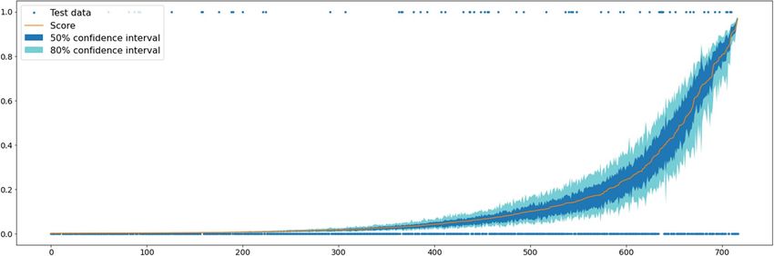
www.nature.com/scientificreports/
AUC value of 0.757. Glucose, C-reactive protein, alkaline phosphatase, gender and total white blood
cell count have the most impact on prediction based on the SHAP values. The relationship between
the size of the training dataset and model performance shows that prediction performance can be
improved. This study combines necrosis prediction and artificial intelligence. The predictive potential
of this model is comparable to the current clinical scoring systems and has several advantages over
them.
Acute pancreatitis (AP) affects about 34 per 100,000 people per year, and it is the most frequent gastrointesti-
nal disease requiring acute h ospitalization1,2. The overall mortality is around 3%3,4; however, in about 10–20%
of AP cases, acute necrotizing pancreatitis (ANP) develops, thus further increasing the risk of morbidity and
mortality5,6. The overall mortality of ANP is approximately 15–20%, of which there is a further twofold increase
in a third of ANP cases where the necrotic tissue becomes i nfected7,8.
Early appraisal of severity and prognosis is crucial in AP, particularly on clinical admission, to identify patients
at risk of developing life-threatening complications. In these cases, close monitoring and early intervention may
prevent organ dysfunction and a fatal outcome9,10.
It has long been known that necrosis is a consistent prognostic factor in A P9. The diagnosis of this local
complication strongly relies on contrast-enhanced computer tomography (CECT) because it has a much higher
sensitivity to detect ANP than u ltrasonography7. Despite being the gold standard method for diagnosing ANP,
CECT has many disadvantages: (1) ANP usually becomes apparent only 72 h after the onset of symptoms; (2)
early and inappropriate CECT may prolong hospitalization; and (3) it is not accessible in every case11. There is
therefore a need for other methods to supplement ANP assessment.
As the underlying pathophysiology of AP becomes more and more familiar by the accumulation of scientific
data, several potential therapeutic targets have been i dentified12,13. Since some of these specific therapies may be
available soon, prompt initiation of treatment after early identification of ANP could be even more important.
Since ANP is associated with life-threatening complications and increased mortality and it is the principal
determinant of the incidence of secondary infection in A P14, researchers have endeavored to find an accurate
clinical scoring system or biomarker that can predict ANP, the severe disease course or mortality itself. As regards
ANP, these systems are either too complicated or require data that is unavailable in the initial stage of hospitali-
zation or lack sufficient sensitivity and specificity. They are therefore rarely used in everyday clinical practice.
As artificial intelligence (AI) can overcome the limitations provided by the complexity of the data and time-
dependent variables, the number of AI tools is increasing in medicine15. AI applications in pancreatic diseases
are also evolving q uickly16. Four AI models aimed to predict the severity of AP on clinical admission, all of which
seems to outperform the conventional prediction scores17–19. Despite their promising preliminary results, these
AI tools are limited by the overlap between the patient group used for model preparation and internal validation
and the relatively low patient number.
This study has two main goals: first, to overcome these limitations and build an AI model that provides an
accurate prediction for ANP development; and, second, to create an online tool from the model that could aid
physicians in the early prognosis of AP.
Methods
This study was reported following the Transparent Reporting of a multivariable prediction model for Individual
Prognosis or Diagnosis (TRIPOD) S tatement20. Ethics approval was obtained from the Hungarian Medical
Research Council’s Scientific and Research Ethics Committee (22254-1/2012/EKU, 17787-8/2020/EÜIG). Written
informed consent was obtained from all participants before enrolment. The study was conducted in accordance
with the Helsinki Declaration.
Data source and eligibility criteria. The analyzed dataset was collected by the Hungarian Pancreatic
Study Group between 2012 and 2019. There were 2461 adult patients enrolled in the patient registry from 30
centers across 13 countries (Appendix A). All patients fulfilled two out of three AP diagnostic criteria based
on the revised Atlanta classification21. Data were collected by physicians and trained clinical administrators on
admission and each day during the whole hospital stay and were stored both on paper and electronically. Rel-
evant clinical data underwent a four-level quality check system before analysis.
In all cases deemed eligible a CECT was performed during hospitalization to assess pancreatic necrosis
formation. Exclusion criteria were as follows: (1) no pancreas imaging had been performed; and (2) the mere
suspicion of necrosis formation by imaging, which was not confirmed later by CECT.
Groups, outcomes, and predictors analyzed. Eligible participants were divided into two groups: (1)
pancreatic necrosis formation was confirmed by a radiologist by CECT during hospitalization; and (2) absence
of necrosis development. The dataset was analyzed and compared accordingly.
ANP was defined as lack of parenchymal enhancement or findings of peripancreatic necrosis such as an acute
necrotic collection on C ECT22. Other local (acute peripancreatic fluid collection and pseudocyst) and systemic
(new-onset diabetes, heart failure, renal failure, and respiratory failure) complications and disease severity were
defined based on the revised Atlanta c lassification21. Data on in-hospital mortality, length of hospital stay, and
etiology of AP were also collected.
The assessed predictors of ANP were gender, age, body mass index (BMI), and laboratory parameters meas-
ured in the first 24 h of clinical admission. The following were evaluated: alanine transaminase, albumin, amylase,
Scientific Reports | (2022) 12:7827 | https://doi.org/10.1038/s41598-022-11517-w 2
Vol:.(1234567890)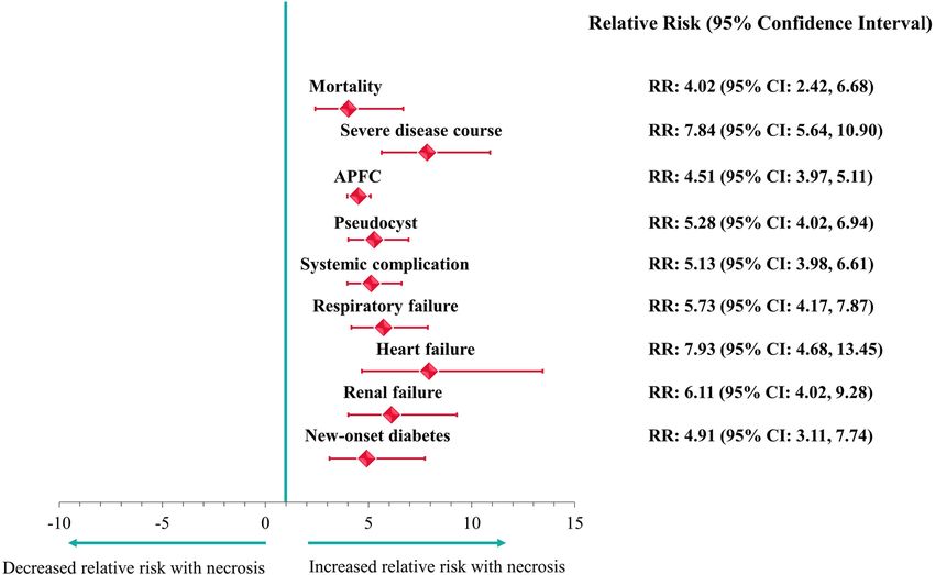
www.nature.com/scientificreports/
Figure 1. Flowchart representing the process of developing the model.
alkaline phosphatase (ALP), aspartate transaminase, blood urea nitrogen, calcium, C-reactive protein (CRP),
creatinine, direct bilirubin, gamma-glutamyl transferase (GGT), glucose, estimated glomerular filtration rate
(eGFR), glycated hemoglobin (HbA1c), hematocrit, hemoglobin, lactate dehydrogenase (LDH), lipase, potas-
sium, procalcitonin, red blood cell count, sodium, thrombocyte, total bilirubin, total cholesterol, total protein,
total white blood cell count (WBC), and triglyceride.
Predictive modelling. The process of predictive modelling is depicted in Fig. 1. Thirty-one variables have
been used for modelling. Data quality is provided in Appendix A. Missing data were handled with a k-nearest-
neighbor-based data imputer algorithm (KNNImputer)23. The SMOTE algorithm24 was used to deal with the
imbalance in class distribution (number of patients with and without ANP).
Random Forest, Logistic Regression, Catboost, XGBoost, and LightGBM were tested for modelling to identify
the best performing machine learning a lgorithm25–28. The catboost, xgboost, lightgbm, and scikit-learn Python
packages were applied. The optimal model was chosen based on the receiver operating characteristic (ROC)
curve and area under the ROC curve (AUC) value after performing four-fold cross-validation. The confidence
of the best performing model was estimated with a bootstrapping method, namely by re-sampling the training
dataset and training a hundred independent copies of the model on these datasets. The confidence of the model
prediction was interpreted with the aid of the 10th and the 90th percentiles of the prediction scores.
Shapley Additive exPlanations (SHAP) values were calculated29 to locally explain the model prediction and
to quantify the contribution of each variable provided. Finally, the model was deployed as an online application
using the Streamlit Python-based framework.
Other statistical analyses. The presence of sampling bias was tested by assessing the representativeness
between the cohort analyzed and the whole cohort (Appendix A). The prediction parameters were also com-
pared between patients with and without ANP with the Kolmogorov–Smirnov test and the Chi-squared test.
ANP was tested as a risk factor for mortality, severe AP, and local and systemic complications by calculating risk
ratios (RR) with the corresponding 95% confidence interval (CI).
Ethics approval. Ethics approval was obtained from the Hungarian Medical Research Council’s Scien-
tific and Research Ethics Committee (22254-1/2012/EKU, 17787-8/2020/EÜIG). The study was conducted in
accordance with the Helsinki Declaration.
Consent to participate. Written informed consent was obtained from all participants before enrolment.
Consent for publication. The corresponding author accepts responsibility for releasing this material on
behalf of all co-authors.
Scientific Reports | (2022) 12:7827 | https://doi.org/10.1038/s41598-022-11517-w 3
Vol.:(0123456789)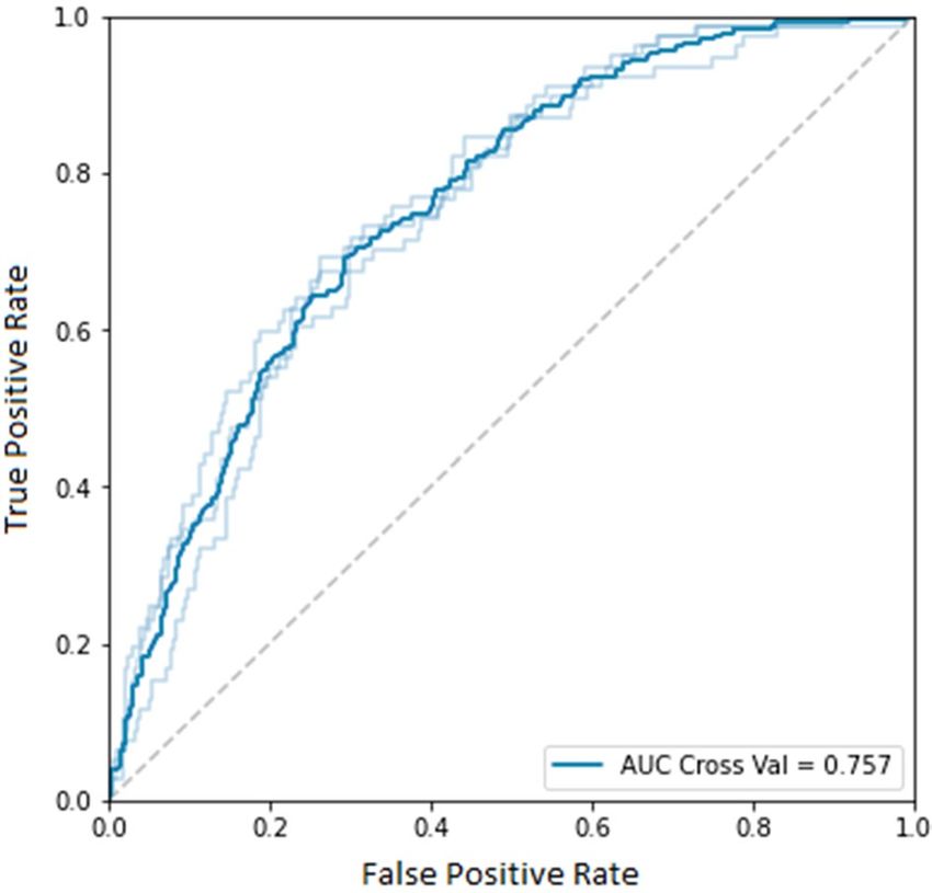
www.nature.com/scientificreports/
Variable Value (n = 2387)
Age in years, median (IQR) 57 (44–69)
Male, n (%) 1357 (56.85%)
BMI, median (IQR) 27.14 (23.88–31.25)
Etiology, n (%)
Biliary 955 (40.01%)
Alcoholic 484 (20.28%)
Hypertriglyceridaemia 81 (3.39%)
Biliary and alcoholic 39 (1.63%)
Biliary and hypertriglyceridaemia 13 (0.54%)
Alcoholic and hypertriglyceridaemia 58 (2.43%)
Post-ERCP 67 (2.81%)
Idiopathic 432 (18.10%)
Other 258 (10.81%)
Revised Atlanta classification
Mild, n (%) 1714 (71.81%)
Moderate, n (%) 551 (23.08%)
Severe, n (%) 122 (5.11%)
Mortality, n (%) 66 (2.76%)
Length of stay in days, median (IQR) 8 (6–12)
Patients with local complication, n (%) 623 (26.19%)
APFC, n (%) 510 (21.37%)
Pseudocyst, n (%) 179 (7.50%)
Acute necrotic collection, n (%) 233 (9.76%)
Patients with systemic complication, n (%) 202 (8.46%)
Respiratory failure, n (%) 136 (5.70%)
Heart failure, n (%) 52 (2.18%)
Renal failure, n (%) 83 (3.48%)
New-onset diabetes, n (%) 75 (3.14%)
Table 1. Characteristics of the analyzed study population. APFC acute peripancreatic fluid collection, BMI
body mass index, ERCP endoscopic retrograde cholangiopancreatography, IQR interquartile range.
Results
Characteristics of the cohort analyzed. 2387 of the 2461 patients with AP proved to be eligible for
the analysis. Characteristics of this population are summarized in Table 1. In 9.76% of the cases, ANP was con-
firmed. There was a statistically significant difference between patients with and without ANP as regards age,
gender, and BMI (Appendix B Supplementary Figs. 16–18). A detailed analysis of the results as regards other
biomarkers can be found in Appendix B.
ANP was associated with a significantly higher risk for mortality, severe disease course, and all the investigated
local and systemic complications (Fig. 2). ANP was also associated with longer hospitalization (9.13 ± 6.21 days
vs. 20.78 ± 19.70 days, p < 0.001).
Model selection and model performance. After an evaluation of the machine learning algorithms, an
XGBoost classifier was identified as the best performing model with an AUC value of 0.757 (standard deviation:
0.012) on cross-validation (Fig. 3). The relationship between the size of the data set and the model performance
is depicted in Fig. 4. The steady increase of AUC values implies that our model has not yet reached its maximal
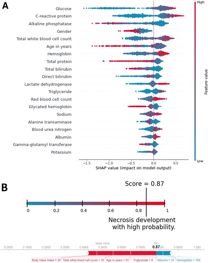
prediction performance. Internal validation implies that our model has higher reliability near the endpoints of
the prediction spectrum since the confidence intervals are narrower (Fig. 5).
The assessment of the impact on the model output showed that glucose, CRP, ALP, gender, and WBC have the
five highest SHAP values. The most influential predictors are shown in Fig. 6 Panel A. Our assessment showed
that the predictive potential depends on the number of biomarkers provided. The models built on the top k most
influential predictors according to their SHAP values show an increasing performance as regards the predictive
potential; however, the extent of this improvement decreases with the number of variables provided (Fig. 7).
Application. The current version of the model can be accessed at http://necro-app.org/. At least five of the
available predictors must be provided to use the application. This limit was applied based on the relation between
the size of the dataset and the desired accuracy30. The application is aided by a built-in BMI calculator and vali-
dations to filter out invalid values. The model offers a numerical probability value between 0 and 1. The higher
the number, the higher the risk for ANP becomes. These numerical values are also supplied with a textual inter-
pretation. For educational purposes, the effect of the biomarkers on prediction is also indicated (Fig. 6 Panel B).
Scientific Reports | (2022) 12:7827 | https://doi.org/10.1038/s41598-022-11517-w 4
Vol:.(1234567890)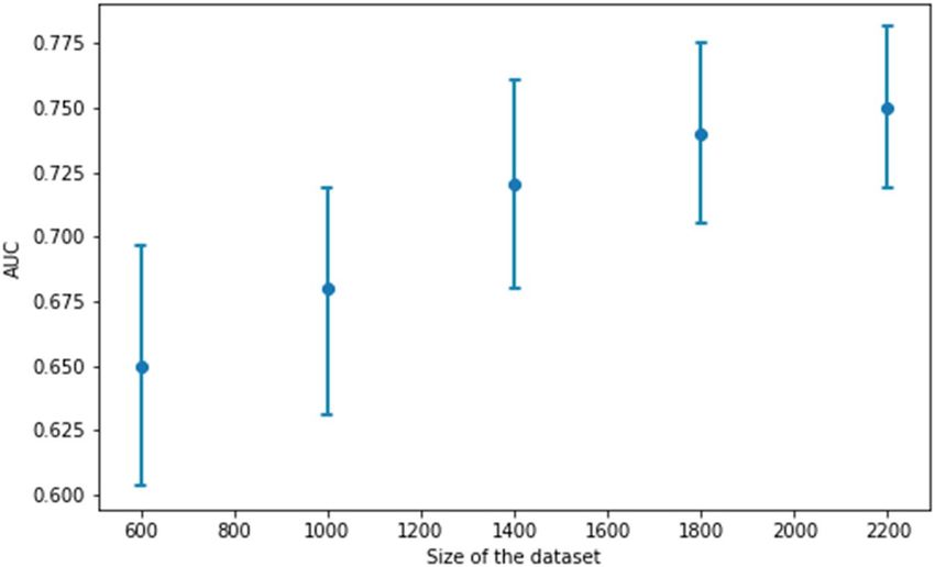
www.nature.com/scientificreports/
Figure 2. Association between necrosis development and other complications in acute pancreatitis.
Figure 3. Receiver operating characteristic (ROC) curve for the XGBoost model.
By checking an extra field, the application assigns a confidence interval in addition to the numerical value. This
adds further clarification to the predicted necrosis probability; however, it takes extra time.
Discussion
The current study describes the first AI model designed to predict ANP. In addition to creating this model, we
also implemented it as an easily accessible online tool. In addition to these, ANP was extensively described in a
large, prospective, multicenter, cohort study.
Our cohort in the context of previous data. With the occurrence of ANP in around one-tenth of
patients, our results are comparable with previously reported data31,32. The importance of ANP in determin-
ing the disease course and outcome is well-known33,34. Schepers et al. found that 38% of the patients with ANP
developed respiratory, cardiovascular or renal system failure35. In our cohort, necrosis was also associated with a
four to eight-fold increased risk of local and systematic complications, severe disease course, and mortality. We
also confirmed their observation regarding prolonged hospitalization indicating the impact of ANP on short-
Scientific Reports | (2022) 12:7827 | https://doi.org/10.1038/s41598-022-11517-w 5
Vol.:(0123456789)
www.nature.com/scientificreports/
Figure 4. The relationship between the size of the data set and the model performance. The blue dot represents
the area under the ROC curve value and the vertical lines show the corresponding confidence intervals.
Figure 5. The predicted necrosis probabilities with the corresponding 50% (between the 25th and 50th
percentiles) and 80% confidence (between the 10th and 90th percentiles).
term (i.e.: in-hospital) outcomes. However, the importance of pancreatic necrosis development also lies in the
long-term complications.
Recent studies investigated this topic and shed light on long-term outcomes. A meta-analysis of long-term
follow-up studies found that the pooled prevalence of exocrine pancreatic insufficiency (EPI) after ANP is
between 41 and 58% depending on the extent of n ecrosis36. In a cohort study by Maatman et al., this ratio was
only 19%22. The discrepancy in the frequency can be attributed to that. While the meta-analysis accounted for EPI
during both the hospital stay and follow-up, the cohort assessed EPI after the resolution of AP. Furthermore, the
retrospective nature of data has an inherent limitation, which can also explain this difference. In addition to the
increased frequency of EPI, they found endocrine insufficiency in 35% of the patients with a median follow-up
of 46 months. Despite the fact that our study covered the time of hospitalization, our results imply that necrosis
formation increases the risk of new-onset diabetes.
Currently existing clinical scores as predictors of necrosis. Since ANP is a potent prognostic factor
for the short-term severity of AP and could forecast long-term consequences, it would be ideal for identifying
these patients as soon as possible. The prediction of ANP was attempted by numerous scoring systems and
biomarkers37; however, each of them has its own limitations. The Balthazar Computer Tomography Severity
Index (CTSI) possesses a higher positive predictive value for necrosis than most commonly used prediction
methods38, e.g. the Ranson score and the APACHE II score, but it is limited by the availability of CECT. It must
be noted that ANP usually becomes apparent after two to three days after disease onset, and that prevents on-
admission prediction in certain cases. The application of other scoring systems without mandatory CECT is
restricted by their complexity. The Ranson score has eleven factors, which have to be assessed on admission and
Scientific Reports | (2022) 12:7827 | https://doi.org/10.1038/s41598-022-11517-w 6
Vol:.(1234567890)www.nature.com/scientificreports/
Figure 6. (A) The features with the highest impact on model output based on the SHAP values. The higher the
predictor is on the list, the bigger the impact on model output. Each patient is represented by a dot. The x-axis
represents the extent of the impact on prediction. The color of the dot shows the feature value (e.g. the red color
implies higher values). (B) An example of prediction and its textual interpretation. The lower picture highlights
the effect of individual predictors and the final necrosis probability provided by the model.
after 48 hours39. The APACHE II score is superior to the scores noted above in terms of flexibility and speed;
however, its sensitivity and specificity are far lower40.
Two prospective studies compared CTSI, Ranson score, and APACHE II score in predicting necrosis
development41,42. Despite limitation in terms of patient number and the slightly different AUC values for necro-
sis, they concluded that the positive predictive value decreases in the following order: CTSI, Ranson score,
and APACHE II. It must be emphasized that these scoring systems are strongly limited by the conversion of
continuous variables to binary ones and this topic should be investigated by more mathematical models with
better accuracy42.
Artificial intelligence in the prognosis of acute pancreatitis. Artificial intelligence has appeared
on the scene as a very intriguing modality of data-based decision support, and these models are extensively
iseases43. In the last decade, multiple
researched in numerous areas of medicine, including pancreaticobiliary d
P16. Most of these models were designed to predict the occurrence of a
AI algorithms have been developed in A
Scientific Reports | (2022) 12:7827 | https://doi.org/10.1038/s41598-022-11517-w 7
Vol.:(0123456789)www.nature.com/scientificreports/
Figure 7. The models build on the k predictors with the highest SHAP value.
specific complication or disease severity. The most commonly used score in critical care is the APACHE II score;
however, three AP severity AI models have been reported to outperform this score 17–19. The AI model developed
by Keogan et al. was compared to the CTSI and Ranson scores, both of which were found inferior in terms of
P44. It should be noted that this study assessed the disease severity with LOH and not
predicting the severity of A
with the revised Atlanta classification. Despite the positive results, these prediction systems, except for the arti-
ficial neural network by Mofidi et al.19, are limited by the overlap between the data used for model training and
the validation. Furthermore, these models need another step after validation. Despite the tremendous efforts and
scientific results, much of this knowledge has not been applied in everyday clinical p ractice45. In order to bring
these complex models to the bedside, they need to be implemented as easy to use and broadly accessible tools46.
The current study was not designed to predict severity but to assess the probability of necrosis formation on
clinical admission. Although we had a different outcome, we aimed to overcome the limitations of most previous
models and to find a way to use our AI model. As suggested by Shung et al., AI-assisted tools have to overcome
many challenges46. First of all, we must have high-quality data. This issue was addressed in our study with a
four-level data quality check system. The second main challenge is ongoing data maintenance. Our model was
constructed such that the new data could be incorporated after validation. Since the predictive potential of the
model shows an increasing trend, this could contribute to better accuracy. Algorithmic understanding is also a
key factor. The help of physicians, who will eventually use the AI model, is crucial to confirm the performance of
such a tool. Furthermore, practitioners could help in differentiating between valid prediction with actual signals
and distorted predictions masked by confounding v ariables46. Our web-based application shows the weighted
impact of the individual biomarkers in each decision. This tool thus meets these expectations. Consequently,
the next step will be screening for these confounding factors while continuously incorporating new data and
monitoring the feasibility of the bedside application of this model.
Strengths and limitations. Our study has multiple strengths and some limitations. Although the predic-
tive potential of this model is similar to that of currently available predictive scoring systems, it has multiple
advantages over them. It provides risk assessment with any five of the predictors in our study, which are com-
monly assessed in daily practice. Therefore, this better reflects everyday clinical practice. To the best of our
knowledge, this is the first AI model to strive to predict the development of ANP on clinical admission. We
designed our model on a much larger population, as compared to the already existing prognostic AI models
in AP, and there was no overlap between the original and validation population. Furthermore, we placed great
emphasis on the interpretation of the model for physicians and its implementation by creating an online applica-
tion. Nevertheless, in addition to predictive model development, ANP was extensively analyzed.
In addition to these strengths, the present study has several limitations. Firstly, as we move further from the
endpoint of the prediction spectrum, the confidence of the model becomes wider, and prediction becomes less
reliable. Secondly, the cross-validated AUC value of our XGBoost model is currently in the fair range47. Thirdly,
data imputation can also introduce bias. Most of these limitations can be overcome. Based on our analyses, we
could reach better predictive potential by increasing the training sample size and more data could provide more
accurate imputation as well. Therefore, by using the application, making further predictions with more data, the
model itself could improve.
It should be highlighted that AI models should not be considered as a substitute for human intelligence16.
These tools, including our model, were designed to facilitate physicians’ decision-making and every prediction
should be interpreted in accordance with the clinical picture.
Scientific Reports | (2022) 12:7827 | https://doi.org/10.1038/s41598-022-11517-w 8
Vol:.(1234567890)www.nature.com/scientificreports/
Implication for practice and research. Development of ANP is associated with several short- and long-
term complications, e.g. endocrine insufficiency, but CECT is not performed solely and exclusively to confirm
necrosis in AP. Therefore, by knowing the high risk for necrosis development, we can identify a group of patients
who need closer follow-up. Nevertheless, this model can aid physicians when CECT is either contraindicated or
not available. Also, as soon as new therapies emerge, early identification of ANP will become even more impor-
tant. Further research is needed on other potential predictive factors, which could be incorporated in the current
model to further improve predictions.
Conclusion
This study is the first to combine prediction of necrosis development and artificial intelligence in AP. The pre-
dictive potential of this model is comparable to the already existing clinical scoring systems and the model is
expected to further improve with use. The easy-to-use web application supported by the interpretation of the
prediction facilitates early, on-admission prediction of necrosis and allows continuous data maintenance and
algorithmic understanding.
Data availability
The datasets generated and/or analysed during the current study are available from the corresponding author
on reasonable request.
Received: 14 December 2021; Accepted: 7 April 2022
References
1. Boxhoorn, L. et al. Acute pancreatitis. Lancet (London, England) 396, 726–734. https://doi.org/10.1016/s0140-6736(20)31310-6
(2020).
2. Xiao, A. Y. et al. Global incidence and mortality of pancreatic diseases: A systematic review, meta-analysis, and meta-regression of
population-based cohort studies. Lancet Gastroenterol. Hepatol. 1, 45–55. https://doi.org/10.1016/s2468-1253(16)30004-8 (2016).
3. Berger, Z. et al. Acute pancreatitis in Chile. A multicenter study on epidemiology, etiology and clinical outcome. Retrospective
analysis of clinical files. Pancreatology 20, 637–643. https://doi.org/10.1016/j.pan.2020.04.016 (2020).
4. Párniczky, A. et al. Prospective, multicentre, nationwide clinical data from 600 cases of acute pancreatitis. PLoS ONE 11, e0165309.
https://doi.org/10.1371/journal.pone.0165309 (2016).
5. Aranda-Narvaez, J. M., Gonzalez-Sanchez, A. J., Montiel-Casado, M. C., Titos-Garcia, A. & Santoyo-Santoyo, J. Acute necrotizing
pancreatitis: Surgical indications and technical procedures. World J. Clin. Cases 2, 840–845. https://doi.org/10.12998/wjcc.v2.i12.
840 (2014).
6. Jha, A. K., Goenka, M. K., Kumar, R. & Suchismita, A. Endotherapy for pancreatic necrosis: An update. JGH Open 3, 80–88. https://
doi.org/10.1002/jgh3.12109 (2019).
7. Bugiantella, W. et al. Necrotizing pancreatitis: A review of the interventions. Int. J. Surg. 28(Suppl 1), S163-171. https://doi.org/
10.1016/j.ijsu.2015.12.038 (2016).
8. Colvin, S. D., Smith, E. N., Morgan, D. E. & Porter, K. K. Acute pancreatitis: An update on the revised Atlanta classification.
Abdominal Radiol. (New York) 45, 1222–1231. https://doi.org/10.1007/s00261-019-02214-w (2020).
9. Balthazar, E. J., Robinson, D. L., Megibow, A. J. & Ranson, J. H. Acute pancreatitis: Value of CT in establishing prognosis. Radiology
174, 331–336. https://doi.org/10.1148/radiology.174.2.2296641 (1990).
10. Ding, N., Guo, C., Li, C., Zhou, Y. & Chai, X. An artificial neural networks model for early predicting in-hospital mortality in acute
pancreatitis in MIMIC-III. Biomed. Res. Int. 2021, 6638919. https://doi.org/10.1155/2021/6638919 (2021).
11. IAP/APA evidence-based guidelines for the management of acute pancreatitis. Pancreatology 13, e1–e15. https://d oi.o
rg/1 0.1 016/j.
pan.2013.07.063 (2013).
12. Li, J., Zhou, R., Zhang, J. & Li, Z. F. Calcium signaling of pancreatic acinar cells in the pathogenesis of pancreatitis. World J. Gas-
troenterol. 20, 16146–16152. https://doi.org/10.3748/wjg.v20.i43.16146 (2014).
13. Petersen, O. H., Gerasimenko, J. V., Gerasimenko, O. V., Gryshchenko, O. & Peng, S. The roles of calcium and ATP in the physiol-
ogy and pathology of the exocrine pancreas. Phys. Rev. 101, 1691–1744. https://doi.org/10.1152/physrev.00003.2021 (2021).
14. Bradley, E. L. 3rd., Murphy, F. & Ferguson, C. Prediction of pancreatic necrosis by dynamic pancreatography. Ann. Surg. 210,
495–504. https://doi.org/10.1097/00000658-198910000-00010 (1989).
15. Hu, H.-T. et al. Artificial intelligence assists identifying malignant versus benign liver lesions using contrast-enhanced ultrasound.
J. Gastroenterol. Hepatol. https://doi.org/10.1111/jgh.15522 (2021).
16. Gorris, M., Hoogenboom, S. A., Wallace, M. B. & van Hooft, J. E. Artificial intelligence for the management of pancreatic diseases.
Digest. Endosc. 33, 231–241. https://doi.org/10.1111/den.13875 (2021).
17. Andersson, B., Andersson, R., Ohlsson, M. & Nilsson, J. Prediction of severe acute pancreatitis at admission to hospital using
artificial neural networks. Pancreatology 11, 328–335. https://doi.org/10.1159/000327903 (2011).
18. Pearce, C. B., Gunn, S. R., Ahmed, A. & Johnson, C. D. Machine learning can improve prediction of severity in acute pancreatitis
using admission values of APACHE II score and C-reactive protein. Pancreatology 6, 123–131. https://doi.org/10.1159/000090032
(2006).
19. Mofidi, R., Duff, M. D., Madhavan, K. K., Garden, O. J. & Parks, R. W. Identification of severe acute pancreatitis using an artificial
neural network. Surgery 141, 59–66. https://doi.org/10.1016/j.surg.2006.07.022 (2007).
20. Collins, G. S., Reitsma, J. B., Altman, D. G. & Moons, K. G. Transparent Reporting of a multivariable prediction model for Indi-
vidual Prognosis or Diagnosis (TRIPOD): The TRIPOD statement. Ann. Intern. Med. 162, 55–63. https://d oi.o
rg/1 0.7 326/m 14-0 697
(2015).
21. Banks, P. A. et al. Classification of acute pancreatitis–2012: Revision of the Atlanta classification and definitions by international
consensus. Gut 62, 102–111. https://doi.org/10.1136/gutjnl-2012-302779 (2013).
22. Maatman, T. K. et al. The continuum of complications in survivors of necrotizing pancreatitis. Surgery 168, 1032–1040. https://
doi.org/10.1016/j.surg.2020.07.004 (2020).
23. Troyanskaya, O. et al. Missing value estimation methods for DNA microarrays. Bioinformatics 17, 520–525. https://doi.org/10.
1093/bioinformatics/17.6.520%JBioinformatics (2001).
24. Chawla, N. V., Bowyer, K. W., Hall, L. O. & Kegelmeyer, W. P. SMOTE: Synthetic minority over-sampling technique. J. Artif. Intell.
Res. 16, 321–357 (2002).
25. Chen, T. & Guestrin, C. in Proceedings of the 22nd ACM SIGKDD International Conference on Knowledge Discovery and Data
Mining 785–794 (Association for Computing Machinery, 2016).
Scientific Reports | (2022) 12:7827 | https://doi.org/10.1038/s41598-022-11517-w 9
Vol.:(0123456789)www.nature.com/scientificreports/
26. Dorogush, A. V., Ershov, V. & Gulin, A. J. A. CatBoost: Gradient boosting with categorical features support. abs/1810.11363 (2018).
27. Pedregosa, F. et al. Scikit-learn: Machine learning in Python. J. Mach. Learn. Res. 12, 2825 (2012).
28. Ke, G. et al. Proceedings of the 31st International Conference on Neural Information Processing Systems. 3149–3157. (Curran Associ-
ates Inc., 2017).
29. Lundberg, S. M. & Lee, S.-I. NIPS.
30. Althnian, A. et al. Impact of dataset size on classification performance: An empirical evaluation in the medical domain. Appl. Sci.
11, 796 (2021).
31. Baron, T. H., DiMaio, C. J., Wang, A. Y. & Morgan, K. A. American gastroenterological association clinical practice update: Man-
agement of pancreatic necrosis. Gastroenterology 158, 67-75.e61. https://doi.org/10.1053/j.gastro.2019.07.064 (2020).
32. Boumitri, C., Brown, E. & Kahaleh, M. Necrotizing pancreatitis: Current management and therapies. Clin. Endosc. 50, 357–365.
https://doi.org/10.5946/ce.2016.152 (2017).
33. Aparna, D., Kumar, S. & Kamalkumar, S. Mortality and morbidity in necrotizing pancreatitis managed on principles of step-up
approach: 7 years experience from a single surgical unit. World J. Gastrointest. Surg. 9, 200–208. https://doi.org/10.4240/wjgs.v9.
i10.200 (2017).
34. Harris, H. W., Barcia, A., Schell, M. T., Thoeni, R. F. & Schecter, W. P. Necrotizing pancreatitis: A surgical approach independent
of documented infection. HPB (Oxford) 6, 161–168. https://doi.org/10.1080/13651820410033634 (2004).
35. Schepers, N. J. et al. Impact of characteristics of organ failure and infected necrosis on mortality in necrotising pancreatitis. Gut
68, 1044–1051. https://doi.org/10.1136/gutjnl-2017-314657 (2019).
36. Huang, W. et al. Exocrine pancreatic insufficiency following acute pancreatitis: Systematic review and meta-analysis. Dig. Dis. Sci.
64, 1985–2005. https://doi.org/10.1007/s10620-019-05568-9 (2019).
37. Leonard-Murali, S. et al. Necrotizing pancreatitis: A review for the acute care surgeon. Am. J. Surg. 221, 927–934. https://doi.org/
10.1016/j.amjsurg.2020.08.027 (2021).
38. Leung, T. K. et al. Balthazar computed tomography severity index is superior to Ranson criteria and APACHE II scoring system
in predicting acute pancreatitis outcome. World J. Gastroenterol. 11, 6049–6052. https://doi.org/10.3748/wjg.v11.i38.6049 (2005).
39. Abu-Eshy, S. A., Abolfotouh, M. A., Nawar, E. & AbuSabib, A. R. Ranson’s criteria for acute pancreatitis in high altitude: Do they
need to be modified?. Saudi J. Gastroenterol. 14, 20–23. https://doi.org/10.4103/1319-3767.37797 (2008).
40. Lankisch, P. G. et al. The APACHE II score is unreliable to diagnose necrotizing pancreatitis on admission to hospital. Pancreas
24, 217–222. https://doi.org/10.1097/00006676-200204000-00002 (2002).
41. Khanna, A. K. et al. Comparison of Ranson, Glasgow, MOSS, SIRS, BISAP, APACHE-II, CTSI Scores, IL-6, CRP, and procalcitonin
in predicting severity, organ failure, pancreatic necrosis, and mortality in acute pancreatitis. HPB Surg. 2013, 367581. https://doi.
org/10.1155/2013/367581 (2013).
42. Papachristou, G. I. et al. Comparison of BISAP, Ranson’s, APACHE-II, and CTSI scores in predicting organ failure, complications,
and mortality in acute pancreatitis. Am. J. Gastroenterol. 105, 435–441. https://doi.org/10.1038/ajg.2009.622 (2010) ((quiz 442)).
43. Chen, P.-T. et al. Applications of artificial intelligence in pancreatic and biliary diseases. J. Gastroenterol. Hepatol. 36, 286–294.
https://doi.org/10.1111/jgh.15380 (2021).
44. Keogan, M. T. et al. Outcome analysis of patients with acute pancreatitis by using an artificial neural network. Acad. Radiol. 9,
410–419. https://doi.org/10.1016/s1076-6332(03)80186-1 (2002).
45. Hegyi, P., Erőss, B., Izbéki, F., Párniczky, A. & Szentesi, A. Accelerating the translational medicine cycle: The Academia Europaea
pilot. Nat. Med. 27, 1317–1319. https://doi.org/10.1038/s41591-021-01458-8 (2021).
46. Shung, D. L. & Sung, J. J. Y. Challenges of developing artificial intelligence-assisted tools for clinical medicine. J. Gastroenterol.
Hepatol. 36, 295–298. https://doi.org/10.1111/jgh.15378 (2021).
47. El Khouli, R. H. et al. Relationship of temporal resolution to diagnostic performance for dynamic contrast enhanced MRI of the
breast. J. Magn. Reason. Imaging 30, 999–1004. https://doi.org/10.1002/jmri.21947 (2009).
Acknowledgements
The research was supported by project grants K131996 to PH, FK131864 to AM, K128222 to LC and University of
Pécs Medical School Research Fund (300909) to AS. The work of JP, RM and MN was supported by the Ministry
of Innovation and the National Research, Development and Innovation Office within the framework of the Arti-
ficial Intelligence National Laboratory Programme. The funders had no effect on the concept, data collection and
analysis, or writing of the manuscript. We wish to thank all contributing members of the Hungarian Pancreatic
Study Group, a full list of them can be found in the Supplementary Information, Appendix A.
Author contributions
S.K.: conceptualisation, project administration, investigation, data curation, visualization, writing-original draft;
J.P.: formal analysis, methodology, visualization, writing-original draft; R.M.: formal analysis, methodology, visu-
alization, writing-original draft; M.N.: formal analysis, methodology, visualization, writing-original draft; N.F.:
conceptualisation, formal analysis, methodology, visualization, writing-original draft; Z.S.: formal analysis, meth-
odology, visualization, writing-original draft; P.F.: formal analysis, methodology, visualization, writing-review
& editing; L.P.: formal analysis, methodology, visualization, writing-review & editing; M.F.: conceptualisation,
project administration, methodology, investigation, data curation, visualization, writing-original draft; Á.V.:
data curation, writing-review & editing; T.T.: data curation, writing-review & editing; L.C.: investigation, data
curation, writing-review & editing; F.I.: data curation, writing-review & editing; A.H.: data curation, investiga-
tion, writing-review & editing; E.B.: data curation, investigation, writing-review & editing; J.H.: data curation,
writing-review & editing; M.V.: data curation, writing-review & editing; A.M.: conceptualisation, data curation,
writing-review & editing; N.F.: conceptualisation, data curation, writing-review & editing; O.F.: conceptualisation,
data curation, writing-review & editing; S.V.: conceptualisation, investigation, data curation, writing-review &
editing; R.N.: investigation, data curation, writing-review & editing; S.B.: data curation, investigation, writing-
review & editing; P.J.H.: data curation, investigation, data curation, writing-review & editing; K.M.: investiga-
tion, data curation, writing-review & editing; K.B.: investigation, writing-review & editing; A.D.: investigation,
writing-review & editing; N.H.: investigation, writing-review & editing; L.Z.: investigation, writing-review &
editing; B.E.: investigation, data curation, writing-review & editing; Z.M.: investigation, writing-review & editing;
A.P.: investigation, data curation, writing-review & editing; P.H.: conceptualisation; supervision; investigation,
data curation, funding acquisition, writing-original draft; A.S.: conceptualisation; supervision; investigation, data
curation, funding acquisition, writing-original draft. All authors certify that they have participated sufficiently
Scientific Reports | (2022) 12:7827 | https://doi.org/10.1038/s41598-022-11517-w 10
Vol:.(1234567890)www.nature.com/scientificreports/
in the work to take public responsibility for the content, including participation in the concept, design, analysis,
writing, or revision of the manuscript.
Funding
Open access funding provided by University of Pécs. The research was supported by project grants K131996 to
PH, FK131864 to AM, K128222 to LC and University of Pécs Medical School Research Fund to AS. The work
of JP, RM and MN was supported by the Ministry of Innovation and the National Research, Development and
Innovation Office within the framework of the Artificial Intelligence National Laboratory Programme. The
funders had no effect on the concept, data collection and analysis, or writing of the manuscript.
Competing interests
The authors declare no competing interests.
Additional information
Supplementary Information The online version contains supplementary material available at https://doi.org/
10.1038/s41598-022-11517-w.
Correspondence and requests for materials should be addressed to A.S.
Reprints and permissions information is available at www.nature.com/reprints.
Publisher’s note Springer Nature remains neutral with regard to jurisdictional claims in published maps and
institutional affiliations.
Open Access This article is licensed under a Creative Commons Attribution 4.0 International
License, which permits use, sharing, adaptation, distribution and reproduction in any medium or
format, as long as you give appropriate credit to the original author(s) and the source, provide a link to the
Creative Commons licence, and indicate if changes were made. The images or other third party material in this
article are included in the article’s Creative Commons licence, unless indicated otherwise in a credit line to the
material. If material is not included in the article’s Creative Commons licence and your intended use is not
permitted by statutory regulation or exceeds the permitted use, you will need to obtain permission directly from
the copyright holder. To view a copy of this licence, visit http://creativecommons.org/licenses/by/4.0/.
© The Author(s) 2022
Hungarian Pancreatic Study Group
Szabolcs Kiss1,2,3, Nelli Farkas2,6, Zoltán Sipos2, Péter Fehérvári2,7, László Pecze2, Mária
Földi1,2,3, Áron Vincze8, Tamás Takács9, László Czakó9, Ferenc Izbéki10, Adrienn Halász1,10,
Eszter Boros10, József Hamvas11, Márta Varga12, Artautas Mickevicius13, Nándor Faluhelyi14,
Orsolya Farkas14, Szilárd Váncsa2,15, Rita Nagy2,3,15, Stefania Bunduc15,16, Péter Jenő
Hegyi15,17, Katalin Márta15,17, Katalin Borka15,18, Attila Doros15,19, Nóra Hosszúfalusi15,20,
László Zubek15,21, Bálint Erőss15,17, Zsolt Molnár15,21,22, Andrea Párniczky2,3, Péter
Hegyi2,15,17, Andrea Szentesi1,2, Judit Bajor8, Szilárd Gódi8, Patrícia Sarlós8, József Czimmer8,
Imre Szabó8, Gabriella Pár8, Anita Illés8, Roland Hágendorn8, Balázs Csaba Németh9,
Balázs Kui9, Dóra Illés9, László Gajdán10, Veronika Dunás‑Varga10, Roland Fejes10, Mária
Papp23, Zsuzsanna Vitális23, János Novák24, Imola Török25, Melania Macarie25, Elena
Ramírez‑Maldonado26, Ville Sallinen27, Shamil Galeev28, Barnabás Bod29, Ali Tüzün Ince30,
Dániel Pécsi2, Péter Varjú2, Márk Félix Juhász2,3, Klementina Ocskay2,15, Alexandra Mikó2,31 &
Zsolt Szakács2,32
2
Institute for Translational Medicine, Szentágothai Research Centre, Medical School, University of Pécs, Szigeti út
12., II. Emelet, Pécs 7624, Hungary. 3Heim Pál National Pediatric Institute, Üllői út 86, Budapest 1089, Hungary.
15
Centre for Translational Medicine, Semmelweis University, Üllői út 26, Budapest 1085, Hungary. 17Division of
Pancreatic Diseases, Heart and Vascular Center, Semmelweis University, Baross u. 23, Budapest 1082, Hungary.
21
Department of Anaesthesiology and Intensive Therapy, Semmelweis University, Üllői út 78, Budapest 1082,
Hungary. 22Department of Anaesthesiology and Intensive Therapy, Poznan University of Medical Sciences, ul. św.
Marii Magdaleny 14, 61861 Poznan, Wielkopolska, Poland. 23Division of Gastroenterology, Department of Internal
Medicine, University of Debrecen, Debrecen, Hungary. 24Pándy Kálmán Hospital of Békés County, Gyula, Hungary.
25
County Emergency Clinical Hospital of Târgu Mures‑Gastroenterology Clinic, University of Medicine, Pharmacy,
Sciences and Technology “George Emil Palade”, Targu Mures, Romania. 26General Surgery, Consorci Sanitari
del Garraf, Sant Pere de Ribes, Barcelona, Spain. 27Department of Transplantation and Liver Surgery, Helsinki
University Hospital, University of Helsinki, Helsinki, Finland. 28Saint Luke Clinical Hospital, St. Petersburg, Russia.
29
Dr. Bugyi István Hospital, Szentes, Hungary. 30Hospital of Bezmialem Vakif University, School of Medicine,
Istanbul, Turkey. 31Department of Medical Genetics, Medical School, University of Pécs, Pécs, Hungary. 32First
Department of Medicine, Medical School, University of Pécs, Pécs, Hungary.
Scientific Reports | (2022) 12:7827 | https://doi.org/10.1038/s41598-022-11517-w 11
Vol.:(0123456789)You can also read