2018 ESC Guidelines for the diagnosis and management of syncope
←
→
Page content transcription
If your browser does not render page correctly, please read the page content below
European Heart Journal (2018) 00, 1–69                                                                           ESC GUIDELINES
                                                doi:10.1093/eurheartj/ehy037

            2018 ESC Guidelines for the diagnosis and
            management of syncope
            The Task Force for the diagnosis and management of syncope of
            the European Society of Cardiology (ESC)
            Developed with the special contribution of the European Heart
            Rhythm Association (EHRA)
            Endorsed by: European Academy of Neurology (EAN), European
            Federation of Autonomic Societies (EFAS), European Federation of
            Internal Medicine (EFIM), European Union Geriatric Medicine Society
            (EUGMS), European Society of Emergency Medicine (EuSEM)

            Authors/Task Force Members: Michele Brignole* (Chairperson) (Italy),
            Angel Moya* (Co-chairperson) (Spain), Frederik J. de Lange (The Netherlands),
            Jean-Claude Deharo (France), Perry M. Elliott (UK), Alessandra Fanciulli (Austria),
            Artur Fedorowski (Sweden), Raffaello Furlan (Italy), Rose Anne Kenny (Ireland),
            Alfonso Martın (Spain), Vincent Probst (France), Matthew J. Reed (UK),
            Ciara P. Rice (Ireland), Richard Sutton (Monaco), Andrea Ungar (Italy), and
            J. Gert van Dijk (The Netherlands)

            * Corresponding authors: Michele Brignole, Department of Cardiology, Ospedali Del Tigullio, Via Don Bobbio 25, IT-16033 Lavagna, (GE) Italy. Tel: þ39 0185 329 567,
            Fax: þ39 0185 306 506, Email: mbrignole@asl4.liguria.it; Angel Moya, Arrhythmia Unit, Hospital Vall d’Hebron, P Vall d’Hebron 119-129, ES-08035 Barcelona, Spain.
            Tel: þ34 93 2746166, Fax: þ34 93 2746002, Email: amoyamitjans@gmail.com.
            ESC Committee for Practice Guidelines (CPG) and National Cardiac Societies document reviewers: listed in the Appendix.
            1
              Representing the European Academy of Neurology (EAN)
            2
              Representing the European Federation of Internal Medicine (EFIM)
            3
              Representing the European Society of Emergency Medicine (EuSEM)
            ESC entities having participated in the development of this document:
            Associations: European Heart Rhythm Association (EHRA)
            Councils: Council on Cardiovascular Nursing and Allied Professions, Council for Cardiology Practice, Council on Cardiovascular Primary Care
            Working Groups: Myocardial and Pericardial Diseases
            The content of these European Society of Cardiology (ESC) Guidelines has been published for personal and educational use only. No commercial use is authorized. No part of the
            ESC Guidelines may be translated or reproduced in any form without written permission from the ESC. Permission can be obtained upon submission of a written request to Oxford
            University Press, the publisher of the European Heart Journal and the party authorized to handle such permissions on behalf of the ESC (journals.permissions@oxfordjournals.org).
            Disclaimer. The ESC Guidelines represent the views of the ESC and were produced after careful consideration of the scientific and medical knowledge and the evidence available
            at the time of their publication. The ESC is not responsible in the event of any contradiction, discrepancy and/or ambiguity between the ESC Guidelines and any other official recom-
            mendations or guidelines issued by the relevant public health authorities, in particular in relation to good use of healthcare or therapeutic strategies. Health professionals are encour-
            aged to take the ESC Guidelines fully into account when exercising their clinical judgment, as well as in the determination and the implementation of preventive, diagnostic or
            therapeutic medical strategies; however, the ESC Guidelines do not override, in any way whatsoever, the individual responsibility of health professionals to make appropriate and
            accurate decisions in consideration of each patient’s health condition and in consultation with that patient and, where appropriate and/or necessary, the patient’s caregiver. Nor do
            the ESC Guidelines exempt health professionals from taking into full and careful consideration the relevant official updated recommendations or guidelines issued by the competent
            public health authorities, in order to manage each patient’s case in light of the scientifically accepted data pursuant to their respective ethical and professional obligations. It is also the
            health professional’s responsibility to verify the applicable rules and regulations relating to drugs and medical devices at the time of prescription.

            C The European Society of Cardiology 2018. All rights reserved. For permissions please email: journals.permissions@oxfordjournals.org
            V

Downloaded from https://academic.oup.com/eurheartj/advance-article-abstract/doi/10.1093/eurheartj/ehy037/4939241
by guest
on 23 March 2018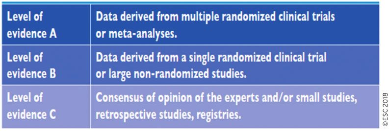
2 ESC Guidelines
Document Reviewers: Adam Torbicki (CPG Review Coordinator) (Poland), Javier Moreno (CPG Review
Coordinator) (Spain), Victor Aboyans (France), Stefan Agewall (Norway), Riccardo Asteggiano (Italy),
Jean-Jacques Blanc (France), Natan Bornstein1 (Israel), Serge Boveda (France), Héctor Bueno (Spain),
Haran Burri (Switzerland), Antonio Coca (Spain), Jean-Philippe Collet (France), Giorgio Costantino2
(Italy), Ernesto Dıaz-Infante (Spain), Victoria Delgado (The Netherlands), Faas Dolmans
(The Netherlands), Oliver Gaemperli (Switzerland), Jacek Gajek (Poland), Gerhard Hindricks (Germany),
Josef Kautzner (Czech Replublic), Juhani Knuuti (Finland), Piotr Kulakowski (Poland),
Ekaterini Lambrinou (Cyprus), Christophe Leclercq (France), Philippe Mabo (France), Carlos A. Morillo
(Canada), Massimo Francesco Piepoli (Italy), Marco Roffi (Switzerland), Win K. Shen (USA),
Iain A. Simpson (UK), Martin Stockburger (Germany), Peter Vanbrabant3 (Belgium),
Stephan Windecker (Switzerland), and Jose Luis Zamorano (Spain)
The disclosure forms of all experts involved in the development of these Guidelines are available on the
ESC website http://www.escardio.org/guidelines.
...................................................................................................................................................................................................
Keywords Guidelines • Syncope • Transient loss of consciousness • Vasovagal syncope • Reflex
syncope • Orthostatic hypotension • Cardiac syncope • Sudden cardiac death • Electrophysiological
study • Prolonged ECG monitoring • Tilt testing • Carotid sinus massage • Cardiac pacing • Implantable
cardioverter defibrillator • Syncope unit • Emergency department
..
Table of Contents .. 4.2.4.1 In-hospital monitoring . . . . . . . . . . . . . . . . . . . . . . . . . . . . 23
.. 4.2.4.2 Holter monitoring . . . . . . . . . . . . . . . . . . . . . . . . . . . . . . . . 23
..
1. Preamble . . . . . . . . . . . . . . . . . . . . . . . . . . . . . . . . . . . . . . . . . . . . . . . . . . . . . . . . . . 4 .. 4.2.4.3 Prospective external event recorders. . . . . . . . . . . . . . 23
2. Introduction . . . . . . . . . . . . . . . . . . . . . . . . . . . . . . . . . . . . . . . . . . . . . . . . . . . . . . . 5 .. 4.2.4.4 Smartphone applications . . . . . . . . . . . . . . . . . . . . . . . . . . 23
..
2.1 What is new in the 2018 version? . . . . . . . . . . . . . . . . . . . . . . . . . . . . . . . 6 .. 4.2.4.5 External loop recorders. . . . . . . . . . . . . . . . . . . . . . . . . . . 23
3. Definitions, classification, and pathophysiology . . . . . . . . . . . . . . . . . . . . . . 7
.. 4.2.4.6 Remote (at home) telemetry . . . . . . . . . . . . . . . . . . . . . . 23
..
3.1 Definitions . . . . . . . . . . . . . . . . . . . . . . . . . . . . . . . . . . . . . . . . . . . . . . . . . . . . . 7 .. 4.2.4.7 Implantable loop recorders . . . . . . . . . . . . . . . . . . . . . . . 23
3.2 Classification and pathophysiology of syncope and transient
.. 4.2.4.8 Diagnostic criteria . . . . . . . . . . . . . . . . . . . . . . . . . . . . . . . . 23
..
loss of consciousness . . . . . . . . . . . . . . . . . . . . . . . . . . . . . . . . . . . . . . . . . . . . . . 8 .. 4.2.5 Video recording in suspected syncope . . . . . . . . . . . . . . . . . . . . 25
3.2.1 Syncope. . . . . . . . . . . . . . . . . . . . . . . . . . . . . . . . . . . . . . . . . . . . . . . . . . 8
.. 4.2.5.1 In-hospital video recording . . . . . . . . . . . . . . . . . . . . . . . . 25
..
3.2.2 Non-syncopal forms of (real or apparent) transient .. 4.2.5.2 Home video recording . . . . . . . . . . . . . . . . . . . . . . . . . . . . 25
loss of consciousness. . . . . . . . . . . . . . . . . . . . . . . . . . . . . . . . . . . . . . . . . . . 8
.. 4.2.6 Electrophysiological study . . . . . . . . . . . . . . . . . . . . . . . . . . . . . . . . 25
..
4. Diagnostic evaluation and management according to .. 4.2.6.1 Asymptomatic sinus bradycardia – suspected
..
risk stratification . . . . . . . . . . . . . . . . . . . . . . . . . . . . . . . . . . . . . . . . . . . . . . . . . . . . 10 .. sinus arrest causing syncope . . . . . . . . . . . . . . . . . . . . . . . . . . . . . . 25
4.1 Initial evaluation . . . . . . . . . . . . . . . . . . . . . . . . . . . . . . . . . . . . . . . . . . . . . . . 10 .. 4.2.6.2 Syncope in bifascicular bundle branch block
..
4.1.1 Diagnosis of syncope. . . . . . . . . . . . . . . . . . . . . . . . . . . . . . . . . . . . . 11 .. (impending high-degree atrioventricular block) . . . . . . . . . . . . 25
4.1.2 Management of syncope in the emergency department .. 4.2.6.3 Suspected tachycardia . . . . . . . . . . . . . . . . . . . . . . . . . . . . . 25
..
based on risk stratification . . . . . . . . . . . . . . . . . . . . . . . . . . . . . . . . . . . . . 13 .. 4.2.7 Endogenous adenosine and other biomarkers . . . . . . . . . . . . . 27
4.2 Diagnostic tests . . . . . . . . . . . . . . . . . . . . . . . . . . . . . . . . . . . . . . . . . . . . . . . 17 .. 4.2.7.1 Adenosine (triphosphate) test and plasma
4.2.1 Carotid sinus massage. . . . . . . . . . . . . . . . . . . . . . . . . . . . . . . . . . . . 17 ... concentration . . . . . . . . . . . . . . . . . . . . . . . . . . . . . . . . . . . . . . . . . . . . 27
..
4.2.2 Orthostatic challenge . . . . . . . . . . . . . . . . . . . . . . . . . . . . . . . . . . . . 18 .. 4.2.7.2 Cardiovascular biomarkers. . . . . . . . . . . . . . . . . . . . . . . . . 27
4.2.2.1 Active standing . . . . . . . . . . . . . . . . . . . . . . . . . . . . . . . . . . . 18 .. 4.2.7.3 Immunological biomarkers . . . . . . . . . . . . . . . . . . . . . . . . . 27
..
4.2.2.2 Tilt testing. . . . . . . . . . . . . . . . . . . . . . . . . . . . . . . . . . . . . . . . 20 .. 4.2.8 Echocardiography . . . . . . . . . . . . . . . . . . . . . . . . . . . . . . . . . . . . . . . 27
4.2.3 Basic autonomic function tests. . . . . . . . . . . . . . . . . . . . . . . . . . . . 21 .. 4.2.8.1 Exercise stress echocardiography . . . . . . . . . . . . . . . . . . 27
..
4.2.3.1 Valsalva manoeuvre. . . . . . . . . . . . . . . . . . . . . . . . . . . . . . . 21 .. 4.2.9 Exercise stress testing. . . . . . . . . . . . . . . . . . . . . . . . . . . . . . . . . . . . 28
4.2.3.2 Deep breathing . . . . . . . . . . . . . . . . . . . . . . . . . . . . . . . . . . . 21 .. 4.2.10 Coronary angiography . . . . . . . . . . . . . . . . . . . . . . . . . . . . . . . . . . . 28
..
4.2.3.3 Other autonomic function tests . . . . . . . . . . . . . . . . . . . 21 .. 5. Treatment. . . . . . . . . . . . . . . . . . . . . . . . . . . . . . . . . . . . . . . . . . . . . . . . . . . . . . . . 28
4.2.3.4 Twenty-four-hour ambulatory and home
.. 5.1 General principles of treatment of syncope. . . . . . . . . . . . . . . . . . . . . 28
..
blood pressure monitoring. . . . . . . . . . . . . . . . . . . . . . . . . . . . . . . 21 .. 5.2 Treatment of reflex syncope. . . . . . . . . . . . . . . . . . . . . . . . . . . . . . . . . . . 29
4.2.4 Electrocardiographic monitoring (non-invasive
.. 5.2.1 Education and lifestyle modifications . . . . . . . . . . . . . . . . . . . . . . 29
..
and invasive) . . . . . . . . . . . . . . . . . . . . . . . . . . . . . . . . . . . . . . . . . . . . . . . . . 23 .. 5.2.2 Discontinuation/reduction of hypotensive therapy . . . . . . . . 31
Downloaded from https://academic.oup.com/eurheartj/advance-article-abstract/doi/10.1093/eurheartj/ehy037/4939241
by guest
on 23 March 2018
ESC Guidelines 3
5.2.3 Physical counter-pressure manoeuvres . . . . . . . . . . . . . . . . . . . 31
.. 6.1.1 Comorbidity and polypharmacy . . . . . . . . . . . . . . . . . . . . . . . . . . 44
..
5.2.4 Tilt training . . . . . . . . . . . . . . . . . . . . . . . . . . . . . . . . . . . . . . . . . . . . . . 31 .. 6.1.2 Falls . . . . . . . . . . . . . . . . . . . . . . . . . . . . . . . . . . . . . . . . . . . . . . . . . . . . . 45
..
5.2.5 Pharmacological therapy . . . . . . . . . . . . . . . . . . . . . . . . . . . . . . . . . 31 .. 6.1.3 Cognitive assessment and physical performance tests. . . . . . 45
5.2.5.1 Fludrocortisone . . . . . . . . . . . . . . . . . . . . . . . . . . . . . . . . . . . 31 .. 6.2 Syncope in paediatric patients. . . . . . . . . . . . . . . . . . . . . . . . . . . . . . . . . . 46
..
5.2.5.2 Alpha-agonists. . . . . . . . . . . . . . . . . . . . . . . . . . . . . . . . . . . . . 32 .. 6.2.1 Diagnostic evaluation . . . . . . . . . . . . . . . . . . . . . . . . . . . . . . . . . . . . 46
5.2.5.3 Beta-blockers . . . . . . . . . . . . . . . . . . . . . . . . . . . . . . . . . . . . . 32 .. 6.2.2 Therapy . . . . . . . . . . . . . . . . . . . . . . . . . . . . . . . . . . . . . . . . . . . . . . . . . 46
..
5.2.5.4 Other drugs. . . . . . . . . . . . . . . . . . . . . . . . . . . . . . . . . . . . . . . 32 .. 7. Psychogenic transient loss of consciousness and its evaluation. . . . . . . 46
5.2.5.5 Emerging new therapies in specific subgroups . . . . . . . 32 .. 7.1 Diagnosis . . . . . . . . . . . . . . . . . . . . . . . . . . . . . . . . . . . . . . . . . . . . . . . . . . . . . 46
..
5.2.6 Cardiac pacing . . . . . . . . . . . . . . . . . . . . . . . . . . . . . . . . . . . . . . . . . . . 32 .. 7.1.1 Historical criteria for attacks. . . . . . . . . . . . . . . . . . . . . . . . . . . . . . 46
5.2.6.1 Evidence from trials in suspected or .. 7.1.2 Documentation of key features during an attack . . . . . . . . . . . 46
..
certain reflex syncope and electrocardiogram- .. 7.1.2.1 Management of psychogenic pseudosyncope . . . . . . . 47
documented asystole . . . . . . . . . . . . . . . . . . . . . . . . . . . . . . . . . . . . . 32 .. 8. Neurological causes and mimics of syncope . . . . . . . . . . . . . . . . . . . . . . . . 47
..
5.2.6.2 Evidence from trials in patients with carotid .. 8.1 Clinical conditions. . . . . . . . . . . . . . . . . . . . . . . . . . . . . . . . . . . . . . . . . . . . . 47
sinus syndrome. . . . . . . . . . . . . . . . . . . . . . . . . . . . . . . . . . . . . . . . . . . 33 .. 8.1.1 Autonomic failure . . . . . . . . . . . . . . . . . . . . . . . . . . . . . . . . . . . . . . . 47
..
5.2.6.3 Evidence from trials in patients with tilt-induced .. 8.1.2 Epilepsy and ictal asystole . . . . . . . . . . . . . . . . . . . . . . . . . . . . . . . . 47
vasovagal syncope . . . . . . . . . . . . . . . . . . . . . . . . . . . . . . . . . . . . . . . . 33
.. 8.1.3 Cerebrovascular disorders . . . . . . . . . . . . . . . . . . . . . . . . . . . . . . . 48
..
5.2.6.4 Evidence from trials in patients with .. 8.1.4 Migraine. . . . . . . . . . . . . . . . . . . . . . . . . . . . . . . . . . . . . . . . . . . . . . . . . 49
adenosine-sensitive syncope . . . . . . . . . . . . . . . . . . . . . . . . . . . . . . 34
.. 8.1.5 Cataplexy . . . . . . . . . . . . . . . . . . . . . . . . . . . . . . . . . . . . . . . . . . . . . . . 49
..
5.2.6.5 Choice of pacing mode . . . . . . . . . . . . . . . . . . . . . . . . . . . . 34 .. 8.1.6 Drop attacks . . . . . . . . . . . . . . . . . . . . . . . . . . . . . . . . . . . . . . . . . . . . 49
5.2.6.6 Selection of patients for pacing and
.. 8.2 Neurological tests. . . . . . . . . . . . . . . . . . . . . . . . . . . . . . . . . . . . . . . . . . . . . 49
..
proposed algorithm . . . . . . . . . . . . . . . . . . . . . . . . . . . . . . . . . . . . . . 34 .. 8.2.1 Electroencephalography . . . . . . . . . . . . . . . . . . . . . . . . . . . . . . . . . 50
5.3 Treatment of orthostatic hypotension and orthostatic intolerance
.. 8.2.2 Brain computed tomography and magnetic
..
syndromes . . . . . . . . . . . . . . . . . . . . . . . . . . . . . . . . . . . . . . . . . . . . . . . . . . . . . . . 37 .. resonance imaging . . . . . . . . . . . . . . . . . . . . . . . . . . . . . . . . . . . . . . . . . . . . 50
..
5.3.1 Education and lifestyle measures. . . . . . . . . . . . . . . . . . . . . . . . . . 37 .. 8.2.3 Neurovascular studies . . . . . . . . . . . . . . . . . . . . . . . . . . . . . . . . . . . 50
5.3.2 Adequate hydration and salt intake . . . . . . . . . . . . . . . . . . . . . . . 37 .. 8.2.4 Blood tests . . . . . . . . . . . . . . . . . . . . . . . . . . . . . . . . . . . . . . . . . . . . . . 50
..
5.3.3 Discontinuation/reduction of vasoactive drugs . . . . . . . . . . . . 37 .. 9. Organizational aspects . . . . . . . . . . . . . . . . . . . . . . . . . . . . . . . . . . . . . . . . . . . . 50
5.3.4 Counter-pressure manoeuvres. . . . . . . . . . . . . . . . . . . . . . . . . . . 37 .. 9.1 Syncope (transient loss of consciousness) management unit . . . . . 50
..
5.3.5 Abdominal binders and/or support stockings . . . . . . . . . . . . . . 37 .. 9.1.1 Definition of a syncope unit . . . . . . . . . . . . . . . . . . . . . . . . . . . . . . 50
5.3.6 Head-up tilt sleeping . . . . . . . . . . . . . . . . . . . . . . . . . . . . . . . . . . . . . 37 .. 9.1.2 Definition of syncope specialist . . . . . . . . . . . . . . . . . . . . . . . . . . . 50
..
5.3.7 Midodrine . . . . . . . . . . . . . . . . . . . . . . . . . . . . . . . . . . . . . . . . . . . . . . . 37 .. 9.1.3 Goal of a syncope unit . . . . . . . . . . . . . . . . . . . . . . . . . . . . . . . . . . . 50
5.3.8 Fludrocortisone . . . . . . . . . . . . . . . . . . . . . . . . . . . . . . . . . . . . . . . . . 37 .. 9.1.4 Model of a syncope unit . . . . . . . . . . . . . . . . . . . . . . . . . . . . . . . . . . 52
..
5.3.9 Additional therapies . . . . . . . . . . . . . . . . . . . . . . . . . . . . . . . . . . . . . 38 .. 9.1.5 Access and referrals to a syncope unit . . . . . . . . . . . . . . . . . . . . 52
5.3.10 Emerging new pharmacological therapy in specific .. 9.1.6 Outcomes and quality indicators . . . . . . . . . . . . . . . . . . . . . . . . . 52
..
subgroups. . . . . . . . . . . . . . . . . . . . . . . . . . . . . . . . . . . . . . . . . . . . . . . . 38 .. 9.2 The clinical nurse specialist in the syncope unit. . . . . . . . . . . . . . . . . . 52
5.4 Cardiac arrhythmias as the primary cause . . . . . . . . . . . . . . . . . . . . . . 38 .. 9.2.1 Definition . . . . . . . . . . . . . . . . . . . . . . . . . . . . . . . . . . . . . . . . . . . . . . . 52
..
5.4.1 Syncope due to intrinsic sinoatrial or atrioventricular .. 9.2.2 Role and skills of the clinical nurse specialist . . . . . . . . . . . . . . . 53
conduction system disease . . . . . . . . . . . . . . . . . . . . . . . . . . . . . . . . . . . . 38 .. 10. Key messages. . . . . . . . . . . . . . . . . . . . . . . . . . . . . . . . . . . . . . . . . . . . . . . . . . . . 54
..
5.4.1.1 Sinus node disease. . . . . . . . . . . . . . . . . . . . . . . . . . . . . . . . . 38 .. 11. Gaps in evidence and areas for future research . . . . . . . . . . . . . . . . . . . . 54
5.4.1.2 Atrioventricular conduction system disease. . . . . . . . . 39
.. 12. ‘What to do’ and ‘what not to do’ messages from the
..
5.4.1.3 Bundle branch block and unexplained syncope . . . . . . 39 .. Guidelines . . . . . . . . . . . . . . . . . . . . . . . . . . . . . . . . . . . . . . . . . . . . . . . . . . . . . . . . . . 56
5.4.2 Syncope due to intrinsic cardiac tachyarrhythmias . . . . . . . . . 40
.. 13. Supplementary Data and Web Practical Instructions. . . . . . . . . . . . . . . 58
..
5.4.2.1 Paroxysmal supraventricular tachycardia. . . . . . . . . . . . 41 .. 14. Appendix . . . . . . . . . . . . . . . . . . . . . . . . . . . . . . . . . . . . . . . . . . . . . . . . . . . . . . . 58
5.4.2.2 Paroxysmal ventricular tachycardia . . . . . . . . . . . . . . . . . 41
.. 15. References . . . . . . . . . . . . . . . . . . . . . . . . . . . . . . . . . . . . . . . . . . . . . . . . . . . . . . 59
..
5.5 Treatment of syncope secondary to structural cardiac, ..
cardiopulmonary, and great vessel disease . . . . . . . . . . . . . . . . . . . . . . . . . 42
..
..
5.6 Treatment of unexplained syncope in patients at high risk of ..
.. Abbreviations and acronyms
sudden cardiac death . . . . . . . . . . . . . . . . . . . . . . . . . . . . . . . . . . . . . . . . . . . . . 42 ..
5.6.1 Definition . . . . . . . . . . . . . . . . . . . . . . . . . . . . . . . . . . . . . . . . . . . . . . . 42 .. ABPM Ambulatory blood pressure monitoring
..
5.6.2 Left ventricular systolic dysfunction . . . . . . . . . . . . . . . . . . . . . . . 42 .. AF Atrial fibrillation
5.6.3 Hypertrophic cardiomyopathy . . . . . . . . . . . . . . . . . . . . . . . . . . . 43 .. ARVC Arrhythmogenic right ventricular cardiomyopathy
..
5.6.4 Arrhythmogenic right ventricular cardiomyopathy. . . . . . . . . 43 .. ATP Adenosine triphosphate
5.6.5 Patients with inheritable arrhythmogenic disorders . . . . . . . . 43 .. AV Atrioventricular
..
5.6.5.1 Long QT syndrome. . . . . . . . . . . . . . . . . . . . . . . . . . . . . . . . 43 .. AVID Antiarrhythmics versus Implantable
5.6.5.2 Brugada syndrome. . . . . . . . . . . . . . . . . . . . . . . . . . . . . . . . . 44 .. Defibrillators trial
..
5.6.5.3 Other forms . . . . . . . . . . . . . . . . . . . . . . . . . . . . . . . . . . . . . . 44 .. BBB Bundle branch block
6. Special issues . . . . . . . . . . . . . . . . . . . . . . . . . . . . . . . . . . . . . . . . . . . . . . . . . . . . . 44 .. BNP B-type natriuretic peptide
..
6.1 Syncope in patients with comorbidity and frailty . . . . . . . . . . . . . . . . 44 . BP Blood pressure
Downloaded from https://academic.oup.com/eurheartj/advance-article-abstract/doi/10.1093/eurheartj/ehy037/4939241
by guest
on 23 March 20184 ESC Guidelines
b.p.m. Beats per minute
... 1. Preamble
..
CI Confidence interval ..
CI-CSS Cardioinhibitory carotid sinus syndrome
.. Guidelines summarize and evaluate available evidence with the aim of
.. assisting health professionals in selecting the best management strat-
CPG Committee for Practice Guidelines ..
CRT-D Cardiac resynchronization therapy defibrillator
.. egies for an individual patient with a given condition. Guidelines and
.. their recommendations should facilitate decision making of health
CSM Carotid sinus massage ..
CSS Carotid sinus syndrome
.. professionals in their daily practice. However, the final decisions con-
..
DCM Dilated cardiomyopathy .. cerning an individual patient must be made by the responsible health
.. professional(s) in consultation with the patient and caregiver as
DDD-PM Dual chamber pacemaker ..
ECG Electrocardiogram/electrocardiographic .. appropriate.
.. A great number of guidelines have been issued in recent years by
ED Emergency department ..
EEG Electroencephalogram .. the European Society of Cardiology (ESC), as well as by other soci-
.. eties and organisations. Because of the impact on clinical practice,
EFAS European Federation of Autonomic Societies ..
EFIM European Federation of Internal Medicine .. quality criteria for the development of guidelines have been estab-
.. lished in order to make all decisions transparent to the user. The rec-
EHRA European Heart Rhythm Association ..
ENS European Neurological Society .. ommendations for formulating and issuing ESC Guidelines can be
.. found on the ESC website (http://www.escardio.org/Guidelines-&-
EPS Electrophysiological study ..
ESC European Society of Cardiology .. Education/Clinical-Practice-Guidelines/Guidelines-development/Writ
.. ing-ESC-Guidelines). ESC Guidelines represent the official position of
EUGMS European Union Geriatric Medicine Society ..
EuSEM European Society of Emergency Medicine .. the ESC on a given topic and are regularly updated.
.. Members of this Task Force were selected by the ESC, including
HBPM Home blood pressure monitoring ..
HCM Hypertrophic cardiomyopathy
.. representation from its relevant ESC sub-specialty groups, in order
.. to represent professionals involved with the medical care of patients
HR Heart rate ..
ICD Implantable cardioverter defibrillator
.. with this pathology. Selected experts in the field undertook a com-
.. prehensive review of the published evidence for management of a
ILR Implantable loop recorder ..
ISSUE International Study on Syncope of
.. given condition according to ESC Committee for Practice Guidelines
.. (CPG) policy. A critical evaluation of diagnostic and therapeutic pro-
Unknown Etiology ..
L-DOPA L-3,4-dihydroxyphenylalanine
.. cedures was performed, including assessment of the risk–benefit
..
LOC Loss of consciousness .. ratio. The level of evidence and the strength of the recommendation
.. of particular management options were weighed and graded accord-
LQTS Long QT syndrome ..
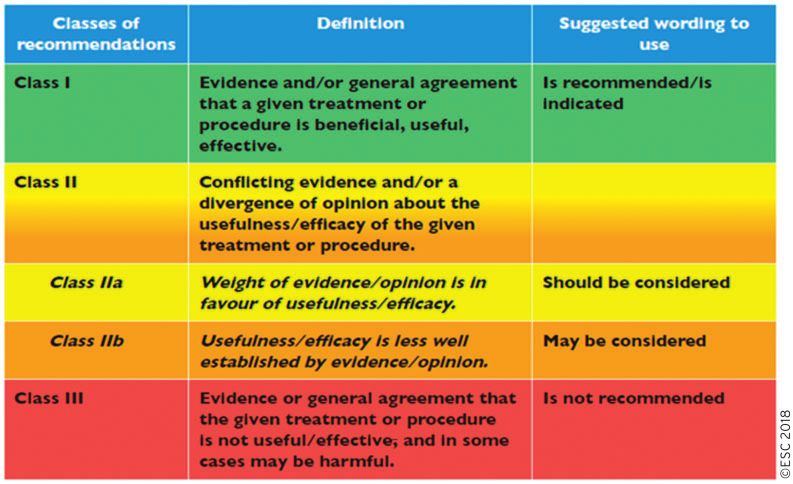
LVEF Left ventricular ejection fraction .. ing to predefined scales, as outlined in Tables 1 and 2.
.. The experts of the writing and reviewing panels provided declar-
MRI Magnetic resonance imaging ..
NYHA New York Heart Association .. ation of interest forms for all relationships that might be perceived as
.. real or potential sources of conflicts of interest. These forms were
OH Orthostatic hypotension ..
PC-Trial Physical Counterpressure Manoeuvres Trial .. compiled into one file and can be found on the ESC website (http://
.. www.escardio.org/guidelines). Any changes in declarations of interest
PCM Physical counter-pressure manoeuvres ..
PNES Psychogenic non-epileptic seizures .. that arise during the writing period were notified to the ESC and
.. updated. The Task Force received its entire financial support from
POST Prevention of Syncope Trial ..
POTS Postural orthostatic tachycardia syndrome .. the ESC without any involvement from the healthcare industry.
.. The ESC CPG supervises and coordinates the preparation of new
PPS Psychogenic pseudosyncope ..
RCT Randomized controlled trial .. Guidelines. The Committee is also responsible for the endorsement
.. process of these Guidelines. The ESC Guidelines undergo extensive
SCD Sudden cardiac death ..
SNRT Sinus node recovery time .. review by the CPG and external experts. After appropriate revisions
.. the Guidelines are approved by all the experts involved in the Task
SU Syncope unit ..
SUP Syncope Unit Project ... Force. The finalized document is approved by the CPG for publica-
SVT Supraventricular tachycardia
.. tion in the European Heart Journal. The Guidelines were developed
.. after careful consideration of the scientific and medical knowledge
TIA Transient ischaemic attack ..
t.i.d. Ter in die (three times daily)
.. and the evidence available at the time of their dating.
.. The task of developing ESC Guidelines also includes the creation of
TLOC Transient loss of consciousness ..
TNG Trinitroglycerin
.. educational tools and implementation programmes for the recommen-
..
VA Ventricular arrhythmia .. dations including condensed pocket guideline versions, summary slides,
.. booklets with essential messages, summary cards for non-specialists
VF Ventricular fibrillation ..
VT Ventricular tachycardia .. and an electronic version for digital applications (smartphones, etc.).
.. These versions are abridged and thus, if needed, one should always
VVS Vasovagal syncope ..
. refer to the full-text version, which is freely available via the ESC
Downloaded from https://academic.oup.com/eurheartj/advance-article-abstract/doi/10.1093/eurheartj/ehy037/4939241
by guest
on 23 March 2018ESC Guidelines 5
Table 1 Classes of recommendations
Table 2 Levels of evidence
website and hosted on the EHJ website. The National Societies of the
.. is also the health professional’s responsibility to verify the rules and
..
ESC are encouraged to endorse, translate and implement all ESC .. regulations applicable to drugs and devices at the time of prescription.
Guidelines. Implementation programmes are needed because it has
..
..
been shown that the outcome of disease may be favourably influenced ..
..
by the thorough application of clinical recommendations.
... 2. Introduction
Surveys and registries are needed to verify that real-life daily prac- ..
tice is in keeping with what is recommended in the guidelines, thus .. The first ESC Guidelines for the management of syncope were pub-
..
completing the loop between clinical research, writing of guidelines, .. lished in 2001, with subsequent versions in 2004 and 2009. In March
disseminating them and implementing them into clinical practice. .. 2015, the ESC CPG considered that there were enough new data to
..
Health professionals are encouraged to take the ESC Guidelines fully .. justify the production of new Guidelines.
into account when exercising their clinical judgment, as well as in the .. The most important aspect characterizing this document is the
..
determination and the implementation of preventive, diagnostic, or .. composition of the Task Force, which is truly multidisciplinary.
therapeutic medical strategies. However, the ESC Guidelines do not .. Cardiologists form a minority of the panel; experts in emergency
..
override in any way whatsoever the individual responsibility of health .. medicine, internal medicine and physiology, neurology and auto-
professionals to make appropriate and accurate decisions in consider- .. nomic diseases, geriatric medicine, and nursing cover all aspects of
..
ation of each patient’s health condition and in consultation with that pa- .. management of the various forms of syncope and transient loss of
tient or the patient’s caregiver where appropriate and/or necessary. It .. consciousness (TLOC).
Downloaded from https://academic.oup.com/eurheartj/advance-article-abstract/doi/10.1093/eurheartj/ehy037/4939241
by guest
on 23 March 20186 ESC Guidelines
Compared with the previous versions of these Guidelines, the .. The document aims to be patient-orientated and focused on ther-
..
2018 document contains Supplementary Data as an integral part. .. apy, and to reduce the risk of recurrence and the life-threatening
While the print text mainly aims to give formal evidence-based rec-
.. consequences of syncope recurrence. For this purpose, even in the
..
ommendations according to the standardized rules of the ESC, this .. absence of strong evidence from trials, we give as much advice as
new web-only feature allows expansion of the content to practical
.. possible on the most appropriate therapy based on the practical ex-
..
issues, and aims to fill the gap between the best available scientific evi- .. pertise of the members of the Task Force (‘Our patients seek solutions,
dence and the need for dissemination of these concepts into clinical
.. not only explanations’). When possible, we provide therapeutic and
..
practice (‘We have the knowledge, we need to teach it’). Thanks to the .. decision-making algorithms.
..
Supplementary Data further explanation on specific points is given, .. Finally, we recognize that one major challenge in syncope manage-
and thanks to the Web Practical Instructions advice is given on how .. ment is the reduction of inappropriate admissions and inappropriate
..
to evaluate patients with loss of consciousness (LOC), and how to .. use of tests while maintaining the safety of the patient. We give strong
perform and interpret tests properly; whenever possible, we provide .. focus to pathways and organizational issues (‘We have the knowledge;
..
tracings, videos, flow charts, and checklists. .. we need to apply it’). In particular, we propose a care pathway for the
.
Figure 1 What is new in the 2018 syncope Guidelines? AA = antiarrhythmic; AF = atrial fibrillation; ARVC = arrhythmogenic right ventricular car-
diomyopathy; CSM = carotid sinus massage; ECG = electrocardiogram; ED = emergency department; LVEF = left ventricular ejection fraction; EPS =
electrophysiological study; HCM = hypertrophic cardiomyopathy; ICD = implantable cardioverter defibrillator; ILR = implantable loop recorder;
OH = orthostatic hypotension; PCM = physical counter-pressure manoeuvres; POTS = postural orthostatic tachycardia syndrome; PPS = psycho-
genic pseudosyncope; SNRT = sinus node recovery time; SU = syncope unit; SVT = supraventricular tachycardia; VT = ventricular tachycardia.
Downloaded from https://academic.oup.com/eurheartj/advance-article-abstract/doi/10.1093/eurheartj/ehy037/4939241
by guest
on 23 March 2018ESC Guidelines 7
Central illustration New/revised concepts in the management of syncope. ECG = electrocardiogram; ED = emergency department; ICD =
implantable cardioverter defibrillator; SCD = sudden cardiac death.
..
management of patients with TLOC from their arrival in the emer- .. • TLOC is defined as a state of real or apparent LOC with loss of
gency department (ED), and give practical instructions on how to set .. awareness, characterized by amnesia for the period of uncon-
.. sciousness, abnormal motor control, loss of responsiveness, and
up outpatient syncope clinics (syncope units) aimed at reducing hos- ..
pitalization, under- and misdiagnoses, and costs. .. a short duration.
..
.. The two main groups of TLOC are ‘TLOC due to head trauma’
..
2.1 What is new in the 2018 version? .. and ‘non-traumatic TLOC’ (Figure 2). Traumatic TLOC will not be
The changes in recommendations made in the 2018 version com-
.. considered further in this document, so TLOC will be used to mean
..
pared with the 2009 version, the new recommendations, and the .. non-traumatic TLOC.
most important new/revised concepts are summarized in Figure 1.
.. The clinical features characterizing TLOC are usually derived from his-
..
.. tory taking from patients and eyewitnesses. Specific characteristics that
.. aid diagnosis are outlined in section 3 of the Web Practical Instructions.
..
.. TLOC groups are defined using pathophysiology: the qualifying cri-
3. Definitions, classification, and .. terion for syncope is cerebral hypoperfusion; for epileptic seizures, it
..
pathophysiology .. is abnormal excessive brain activity; and for psychogenic TLOC it is
.. the psychological process of conversion. The syncope definition rests
..
3.1 Definitions .. on pathophysiology because no set of clinical features encompasses
• Syncope is defined as TLOC due to cerebral hypoperfusion, char- .. all forms of syncope while also excluding all epileptic seizures and
acterized by a rapid onset, short duration, and spontaneous com-
..
.. psychogenic TLOC events.
plete recovery. ..
.. • The adjective presyncope is used to indicate symptoms and signs
Syncope shares many clinical features with other disorders; it
..
.. that occur before unconsciousness in syncope. Note that the
therefore presents in many differential diagnoses. This group of dis- .. noun presyncope is often used to describe a state that resembles
orders is labelled TLOC.
.. the prodrome of syncope, but which is not followed by LOC.
Downloaded from https://academic.oup.com/eurheartj/advance-article-abstract/doi/10.1093/eurheartj/ehy037/4939241
by guest
on 23 March 20188 ESC Guidelines
Generalized: Psychogenic
- Tonic pseudosyncope (PPS)
- Clonic Psychogenic non-epileptic
- Tonic-clonic seizures (PNES)
- Atonic
Figure 2 Syncope in the context of transient loss of consciousness. Non-traumatic transient loss of consciousness is classified into one of four
groupings: syncope, epileptic seizures, psychogenic transient loss of consciousness, and a miscellaneous group of rare causes. This order represents
their rate of occurrence. Combinations occur; e.g. non-traumatic transient loss of consciousness causes can cause falls with concussion, in which case
transient loss of consciousness is both traumatic and non-traumatic. TIA = transient ischaemic attack; TLOC = transient loss of consciousness.
..
A variety of terms are used that generally do not match the definitions .. ‘vasodepressive type’ of reflex syncope, seen in the outer ring in
in this document closely enough to be used as synonyms of the defined .. Figure 3. The second is a functional impairment, and the third a struc-
..
terms. For example, a ‘faint’ approximately conforms to syncope but .. tural impairment of the autonomic nervous system, with drug-
emphasizes vasovagal syncope (VVS) over other forms. A glossary of un- .. induced, primary, and secondary autonomic failure in the outer ring.
..
certain terms is shown in section 1 of the Web Practical Instructions. .. In autonomic failure, there is insufficient sympathetic vasoconstriction
.. in response to the upright position.
3.2 Classification and pathophysiology of ..
.. There are four primary causes of low cardiac output. The first is
syncope and transient loss of .. a reflex bradycardia, known as cardioinhibitory reflex syncope.
..
consciousness .. The second concerns cardiovascular causes: arrhythmia, struc-
3.2.1 Syncope
.. tural disease including pulmonary embolism, and pulmonary
..
Table 3 provides a classification of the principal causes of syncope, .. hypertension. The third is inadequate venous return due to vol-
emphasizing groups of disorders with common pathophysiology,
.. ume depletion or venous pooling. Finally, chronotropic and ino-
..
presentation, and risk. Clinical features, epidemiology, prognosis, im- .. tropic incompetence through autonomic failure may impair
.. cardiac output.
pact on quality of life, and economic issues are shown in section 2 of ..
the Web Practical Instructions. .. Note that these primary mechanisms may interact in different
.. ways: firstly, venous pooling and inadequate venous return is also a
The pathophysiological classification centres on a fall in systemic ..
blood pressure (BP) with a decrease in global cerebral blood flow as .. factor that can trigger an inappropriate reflex in orthostatic reflex
.. syncope; secondly, a low total peripheral resistance may cause ven-
the defining characteristic of syncope. Figure 3 shows low BP and glo- ..
bal cerebral hypoperfusion as the central final common pathway of .. ous pooling of blood below the diaphragm, in turn decreasing venous
..
syncope. A sudden cessation of cerebral blood flow for as short as .. return and consequently cardiac output.
6–8 s can cause complete LOC. A systolic BP of 50–60 mmHg at .. The three main groups of syncope, i.e. reflex, cardiovascular, and
..
heart level, i.e. 30–45 mmHg at brain level in the upright position, will .. secondary to orthostatic hypertension (OH), are shown outside the
cause LOC.8,9 .. rings in Figure 3. Both reflex syncope and OH span the two main
..
Systemic BP is the product of cardiac output and total peripheral .. pathophysiological mechanisms.
resistance; a fall in either can cause syncope. However, in syncope, ..
..
both mechanisms often act together to a varying degree. .. 3.2.2 Non-syncopal forms of (real or apparent) transient
There are three primary causes of a low total peripheral resist-
.. loss of consciousness
..
ance. The first is decreased reflex activity causing vasodilatation .. Only those forms of epilepsy in which normal motor control is lost,
through withdrawal of sympathetic vasoconstriction: this is the
.. so patients may fall, are included in Figure 2. These are tonic, clonic,
Downloaded from https://academic.oup.com/eurheartj/advance-article-abstract/doi/10.1093/eurheartj/ehy037/4939241
by guest
on 23 March 2018ESC Guidelines 9
Table 3 Classification of syncope
Reflex (neurally mediated) syncope
Vasovagal:
- orthostatic VVS: standing, less common sitting
- emotional: fear, pain (somatic or visceral), instrumentation, blood phobia
Situational:
- micturition
- gastrointestinal stimulation (swallow, defaecation)
- cough, sneeze
- post-exercise
- others (e.g. laughing, brass instrument playing)
Carotid sinus syndrome
Non-classical forms (without prodromes and/or without apparent triggers and/or atypical presentation)
Syncope due to OH
Note that hypotension may be exacerbated by venous pooling during exercise (exercise-induced), after meals (postprandial hypotension), and after prolonged
bed rest
(deconditioning).
Drug-induced OH (most common cause of OH):
- e.g. vasodilators, diuretics, phenothiazine, antidepressants
Volume depletion:
- haemorrhage, diarrhoea, vomiting, etc.
Primary autonomic failure (neurogenic OH):
- pure autonomic failure, multiple system atrophy, Parkinson’s disease, dementia with Lewy bodies
Secondary autonomic failure (neurogenic OH):
- diabetes, amyloidosis, spinal cord injuries, auto-immune autonomic neuropathy, paraneoplastic autonomic neuropathy, kidney failure
Cardiac syncope
Arrhythmia as primary cause:
Bradycardia:
- sinus node dysfunction (including bradycardia/tachycardia syndrome)
- atrioventricular conduction system disease
Tachycardia:
- supraventricular
- ventricular
Structural cardiac: aortic stenosis, acute myocardial infarction/ischaemia, hypertrophic cardiomyopathy, cardiac masses (atrial myxoma, tumours,
etc.), pericardial disease/tamponade, congenital anomalies of coronary arteries, prosthetic valve dysfunction
Cardiopulmonary and great vessels: pulmonary embolus, acute aortic dissection, pulmonary hypertension
Remarks
• All forms of syncope, but mostly reflex syncope and OH, are more likely to occur or are more severe when various factors are present: medica-
tion causing low BP (due to vasodilatation or hypovolaemia), alcohol use, volume depletion (haemorrhage, low fluid intake, diarrhoea, vomiting),
pulmonary diseases causing reduction in brain oxygen supply, environmental factors (thermal stress).
• There are two main pathophysiological mechanisms in reflex syncope. “Vasodepression” refers to conditions in which insufficient sympathetic
vasoconstriction results in hypotension.1,2 “Cardioinhibition” is used when bradycardia or asystole predominates, reflecting a shift towards para-
sympathetic predominance. The haemodynamic pattern, i.e. cardioinhibitory, vasodepressive, or both, is independent of the trigger evoking reflex
syncope. For example, micturition syncope and orthostatic VVS may equally well present as cardioinhibitory or as vasodepressor syncope
• The non-classical form of reflex syncope involves a heterogeneous group of patients. The term is used to describe reflex syncope that occurs
with uncertain or apparently absent triggers and/or atypical presentation. The diagnosis of reflex syncope is probable when other causes of syn-
cope are excluded (absence of structural heart disease) and/or symptoms are reproduced in the tilt test.3 At present, this group also contains
syncope associated with low adenosine plasma levels4,5
• The cardiovascular causes of orthostatic intolerance include classical OH, initial OH, delayed OH, POTS, and VVS, which in this context can be
called orthostatic VVS.6,7 Syndromes of orthostatic intolerance that may cause syncope are presented in Web Practical Instruction section 2.
BP = blood pressure; OH = orthostatic hypotension; POTS = postural orthostatic tachycardia syndrome; VVS = vasovagal syncope.
Downloaded from https://academic.oup.com/eurheartj/advance-article-abstract/doi/10.1093/eurheartj/ehy037/4939241
by guest
on 23 March 201810 ESC Guidelines
Table 4 Conditions that may be incorrectly diagnosed
as syncope
Condition Characteristic features that distin-
guish from syncope
Generalized seizures See section 8, Table 10.
Complex partial seiz- No falls, yet unresponsive and later
ures, absence epilepsy amnesia
PPS or Duration of apparent LOC
“pseudocoma” lasting many minutes to hours; high
frequency, up to several times a day
Falls without TLOC No unresponsiveness or amnesia
Cataplexy Falls with flaccid paralysis and non-
responsive, yet no later amnesia
Intracerebral or sub- Consciousness may be progressively
arachnoid reduced rather than immediately
haemorrhage lost. Accompanying severe head-
Figure 3 Pathophysiological basis of the classification of syncope.
ache, other neurological signs
ANS = autonomic nervous system; auton. = autonomic; BP =
blood pressure; OH = orthostatic hypotension; periph. = Vertebrobasilar TIA Always focal neurological signs and
peripheral; resist. = resistance. symptoms, usually without LOC; if
consciousness is lost this usually lasts
longer than in TLOC.
tonic-clonic, and atonic generalized seizures, and can be classified as
Carotid TIA Consciousness is for all practical
primary or secondary. The forms of epilepsy in which people remain
purposes not lost in carotid TIAs,
actively upright, i.e. sitting or standing (e.g. complex partial seizures
but there are pronounced focal
or absence epilepsy) are not regarded as TLOC, but sometimes they
neurological signs and symptoms
are incorrectly diagnosed as syncope.
Psychogenic TLOC consists of two forms: one resembles epileptic Subclavian steal Associated with focal neurological
seizures (psychogenic non-epileptic seizures [PNES]) and one, with- syndrome signs
out gross movements, resembles syncope (psychogenic pseudosyn-
Metabolic disorders Duration much longer than in
cope [PPS]).
including hypogly- TLOC; consciousness may be im-
The rare causes of TLOC only seldomly cause confusion with the
caemia, hypoxia, paired instead of lost
main TLOC forms, probably because in most cases they differ
hyperventilation with
enough clinically to be clearly not syncope. Both vertebrobasilar tran-
hypocapnia
sient ischaemic attacks (TIAs) and subclavian steal syndrome are
associated with focal neurological signs. A subarachnoid haemor- Intoxication Duration much longer than in
rhage may present with a short LOC, but the associated abrupt ex- TLOC; consciousness may be im-
treme headache suggests the cause. In cyanotic breath-holding spells, paired instead of lost
expiratory apnoea with hypoxia is the primary mechanism.10 So-
Cardiac arrest LOC yet no spontaneous recovery
called ‘pallid breath-holding spells’ in children do not constitute a pri-
mary respiratory problem, but are cardioinhibitory reflex syncope.11 Coma Duration much longer than TLOC
Table 4 lists the main features that distinguish syncope from dis-
orders that may be mistaken for syncope. LOC = loss of consciousness; PPS = psychogenic pseudosyncope; TIA = transient
ischaemic attack; TLOC = transient loss of consciousness.
4. Diagnostic evaluation and
management according to risk ..
.. with possible TLOC, history taking should first establish whether there
stratification ..
.. was indeed a TLOC. Often, this allows a distinction between the major
.. TLOC groups. The flow diagram for the evaluation of TLOC is shown in
4.1 Initial evaluation .. Figure 4. The initial evaluation should answer key questions:
The clinical features characterizing TLOC are usually derived from his-
..
..
tory taking from patients and eyewitnesses. When a patient first presents . (1) Was the event TLOC?
Downloaded from https://academic.oup.com/eurheartj/advance-article-abstract/doi/10.1093/eurheartj/ehy037/4939241
by guest
on 23 March 2018ESC Guidelines 11
Figure 4 Flow diagram for the initial evaluation and risk stratification of patients with syncope. BP = blood pressure; ECG = electrocardiogram;
H&P exam = history and physical examination; TLOC = transient loss of consciousness.
(2) In case of TLOC, is it of syncopal or non-syncopal origin?
.. approximately 60% of cases.12 For non-syncopal TLOC, refer to sec-
..
(3) In case of suspected syncope, is there a clear aetiological diagnosis .. tions 7 and 8.
(see section 4.1.1)?
..
..
(4) Is there evidence to suggest a high risk of cardiovascular events or .. 4.1.1 Diagnosis of syncope
.. The starting point of the diagnostic evaluation of TLOC of suspected
death (see section 4.1.2)? ..
.. syncopal nature is the initial syncope evaluation, which consists of:
TLOC has four specific characteristics: short duration, abnor- ..
mal motor control, loss of responsiveness, and amnesia for
.. • Careful history taking concerning present and previous attacks, as
the period of LOC (for an explanation of the clinical features of
... well as eyewitness accounts, in person or through a telephone
.. interview.
TLOC see Web Table 4 in section 4.1 of the Web Practical ..
.. • Physical examination, including supine and standing BP
Instructions). .. measurements.
TLOC is probably syncope when: (i) there are signs and symptoms ..
.. • Electrocardiogram (ECG).
specific for reflex syncope, syncope due to OH, or cardiac syncope, ..
and (ii) signs and symptoms specific for other forms of TLOC (head .. Based on these findings, additional examinations may be per-
.. formed when needed (see section 4.2):
trauma, epileptic seizures, psychogenic TLOC, and/or rare causes) ..
are absent. Practical instructions for history taking are given in sec- ..
.. • Immediate ECG monitoring when there is a suspicion of arrhyth-
tions 3 and 4 of the Web Practical Instructions. .. mic syncope.
When epileptic seizures or psychogenic attacks are likely, appro- ..
.. • Echocardiogram when there is previous known heart disease,
priate steps should be taken. By using a detailed clinical history, phys- .. data suggestive of structural heart disease, or syncope secondary
icians can differentiate syncope from other forms of TLOC in .. to cardiovascular cause.
Downloaded from https://academic.oup.com/eurheartj/advance-article-abstract/doi/10.1093/eurheartj/ehy037/4939241
by guest
on 23 March 201812 ESC Guidelines
• Carotid sinus massage (CSM) in patients aged >40 years.
.. Even if there is no independent gold/reference standard to diag-
..
• Head-up tilt testing when there is suspicion of syncope due to .. nose syncope, there is strong consensus that the initial evalu-
OH or reflex syncope. .. ation may lead to certain or highly likely diagnosis when the
..
• Blood tests when clinically indicated, e.g. haematocrit or haemo- .. diagnostic criteria listed in the table of recommendations are
globin when haemorrhage is suspected, oxygen saturation and .. met.
blood gas analysis when hypoxia is suspected, troponin when car-
..
..
diac ischaemia-related syncope is suspected, or D-dimer when ..
pulmonary embolism is suspected, etc. ..
..
Diagnostic criteria with initial evaluation
Recommendations Classa Levelb
Reflex syncope and OH
VVS is highly probable if syncope is precipitated by pain, fear, or standing, and is associated with typical progressive prodrome
I C
(pallor, sweating, and/or nausea).8,13–17
Situational reflex syncope is highly probable if syncope occurs during or immediately after specific triggers, listed in
13–17 I C
Table 3.8,
Syncope due to OH is confirmed when syncope occurs while standing and there is concomitant significant OH.18–24 I C
In the absence of the above criteria, reflex syncope and OH should be considered likely when the features that suggest reflex
IIa C
syncope or OH are present and the features that suggest cardiac syncope are absent (see Table 5).
Cardiac syncope
Arrhythmic syncope is highly probable when the ECG shows25–39:
• Persistent sinus bradycardia 3 s in awake state and in absence of physical training;
• Mobitz II second- and third-degree AV block;
• Alternating left and right BBB; I C
• VT or rapid paroxysmal SVT;
• Non-sustained episodes of polymorphic VT and long or short QT interval; or
• Pacemaker or ICD malfunction with cardiac pauses.
Cardiac ischaemia-related syncope is confirmed when syncope presents with evidence of acute myocardial ischaemia with or
I C
without myocardial infarction.25–39
Syncope due to structural cardiopulmonary disorders is highly probable when syncope presents in patients with prolapsing
I C
atrial myxoma, left atrial ball thrombus, severe aortic stenosis, pulmonary embolus, or acute aortic dissection.
Additional advice and clinical perspectives
The initial syncope evaluation, as described in this document, can define the cause of syncope in most patients. Strict adherence to the above defin-
itions of VVS and situational reflex syncope, and of syncope due to OH, can be considered certain or highly likely irrespective of the presence of
any other abnormal finding. In young subjects with unexplained syncope and no history of cardiac disease, no family history of sudden death, no su-
pine syncope or syncope during sleep or exercise, no unusual triggers, and a normal ECG, the chance of cardiac syncope is very low. SCD rates in
subjectsESC Guidelines 13
..
When a diagnosis is nearly certain or highly likely, no further evalu- .. 4.1.2 Management of syncope in the emergency
ation is needed, and treatment—if any—can be planned. In other .. department based on risk stratification
..
cases, the initial evaluation may suggest a diagnosis when the features .. The management of TLOC of suspected syncopal nature in the ED
listed in Table 5 are present, or otherwise is unable to suggest any .. should answer the following three key questions:
..
diagnosis. ..
.. (1) Is there a serious underlying cause that can be identified?
.. (2) What is the risk of a serious outcome?
Table 5 Clinical features that can suggest a diagnosis ..
on initial evaluation
.. (3) Should the patient be admitted to hospital?
..
.. Figure 5 shows a flow chart for the management and risk stratifica-
..
Reflex syncope .. tion of patients referred to the ED for TLOC suspected to be syn-
• Long history of recurrent syncope, in particular occurring be- .. cope (modified from Casagranda et al.40).
..
fore the age of 40 years ..
• After unpleasant sight, sound, smell, or pain .. Question 1: Is there a serious underlying cause that can be
• Prolonged standing ..
.. identified in the ED?
• During meal .. Normally the presenting complaint of syncope can be established.
• Being in crowded and/or hot places ..
• Autonomic activation before syncope: pallor, sweating, and/ .. The primary aim for an ED clinician is then to establish an underlying
.. diagnosis, especially those associated with the potential for rapid clin-
or nausea/vomiting ..
• With head rotation or pressure on carotid sinus (as in tu- .. ical deterioration.41,42 It is the acute underlying disease that most fre-
.. quently determines short-term adverse events rather than the
mours, shaving, tight collars) ..
• Absence of heart disease .. syncope itself.43 Subsequent management will focus on treating this
.. underlying cause (Figure 5). Many (40–45%) non-cardiovascular and
Syncope due to OH ..
.. some cardiovascular life-threatening underlying conditions are obvi-
• While or after standing ..
.. ous in the ED.44 Table 6 lists high-risk features that suggest the pres-
• Prolonged standing
• Standing after exertion .. ence of a serious underlying cause and low-risk features that suggest
.. a benign underlying cause.
• Post-prandial hypotension ..
• Temporal relationship with start or changes of dosage of vas- ..
odepressive drugs or diuretics leading to hypotension
.. Question 2: What is the risk of a serious outcome?
..
• Presence of autonomic neuropathy or parkinsonism .. High-risk features are shown in Table 6, and how to use this risk pro-
.. file to guide subsequent management and disposition is shown in
Cardiac syncope ..
• During exertion or when supine .. Figure 6.
..
• Sudden onset palpitation immediately followed by syncope .. Risk stratification is important, for two reasons:
• Family history of unexplained sudden death at young age ..
• Presence of structural heart disease or coronary artery .. (1) To recognize patients with a likely low-risk condition able to be dis-
.. charged with adequate patient education.
disease ..
• ECG findings suggesting arrhythmic syncope: .. (2) To recognize patients with a likely high-risk cardiovascular condition
- Bifascicular block (defined as either left or right BBB com-
.. requiring urgent investigation. This may require admission.
..
bined with left anterior or left posterior fascicular block) ..
- Other intraventricular conduction abnormalities (QRS dur- .. High-risk patients are more likely to have cardiac syncope.
ation >_0.12 s)
.. Structural heart disease25–27,31,35,36,45 and primary electrical disease46
..
- Mobitz I second-degree AV block and 1 degree AV block .. are major risk factors for sudden cardiac death (SCD) and overall
with markedly prolonged PR interval .. mortality in patients with syncope. Low-risk patients are more likely
- Asymptomatic mild inappropriate sinus bradycardia (40–50
..
.. to have reflex syncope and have an excellent prognosis.47 OH is
b.p.m.) or slow atrial fibrillation (40–50 b.p.m.) in the ab- .. associated with a two-fold higher risk of death owing to the severity
sence of negatively chronotropic medications ..
- Non-sustained VT
.. of comorbidities compared with the general population.48
..
- Pre-excited QRS complexes ..
- Long or short QT intervals .. Question 3: Should the patient be admitted to hospital?
- Early repolarization
..
.. Approximately 50% of patients who present to the ED with
- ST-segment elevation with type 1 morphology in leads .. syncope are admitted (although the rate varies between 12–86%)
V1-V3 (Brugada pattern) ..
- Negative T waves in right precordial leads, epsilon waves
.. (see Supplementary Data Table 4). The use of clinical decision rules and
.. standardized protocols has not changed this rate significantly. The com-
suggestive of ARVC ..
- Left ventricular hypertrophy suggesting hypertrophic .. posite estimate of outcomes is that in the next 7–30 days, only 0.8%
cardiomyopathy
.. die and 6.9% have a non-fatal severe outcome whilst in the ED, while
..
.. another 3.6% have a post-ED serious outcome (see Supplementary
.. Data Table 4). Unnecessary admission in low-risk patients can be harm-
..
ARVC = arrhythmogenic right ventricular cardiomyopathy; AV = atrioventricular; .. ful.87 Whereas it is crucial to identify these high-risk patients to ensure
BBB = bundle branch block; b.p.m. = beats per minute; ECG = electrocardio- .. early, rapid, and intensive investigation, not all patients at high risk need
gram; OH = orthostatic hypotension; VT = ventricular tachycardia.
..
.. hospitalization.80
.
Downloaded from https://academic.oup.com/eurheartj/advance-article-abstract/doi/10.1093/eurheartj/ehy037/4939241
by guest
on 23 March 201814 ESC Guidelines
Figure 5 The management of patients presenting to the emergency department for transient loss of consciousness suspected to be syncope
(modified from Casagranda et al.40). ED = emergency department; TLOC = transient loss of consciousness.
a
For example, this includes pulmonary embolism presenting with shortness of breath, pleuritic chest pain, and syncope, but not trauma
secondary to syncope.
Management of syncope in the emergency department
Recommendations Classa Levelb
It is recommended that patients with low-risk features, likely to have reflex or situational syncope, or syncope due to OH,
I B
are discharged from the ED.27,35,36,49–54,58,62,69
It is recommended that patients with high-risk features receive an early intensive and prompt evaluation in a syncope unit or
I B
in an ED observation unit (if available), or are hospitalized.26,27,35,36,44–46,50,55–57,59,60,70–76
It is recommended that patients who have neither high- nor low-risk features are observed in the ED or in a syncope unit
I B
instead of being hospitalized.40,63–65,77
Risk stratification scores may be considered for risk stratification in the ED.78–86 IIb B
Additional advice and clinical perspectives
• In the ED, presyncope should be managed with the same accuracy as syncope as it carries the same prognosis.66–68
• Diagnostic radiology and laboratory tests such as chest X-ray, brain computed tomography, routine blood haematology, biochemistry, and D-dimer and cardiac
markers have a low diagnostic yield, impact on risk stratification of patients with syncope, and should not routinely be used unless specifically suggested by clinical
evaluation.
• Around 10% of patients with syncope in the ED will suffer from a serious outcome within 7–30 days of their visit, with just under half occurring after their stay in the
ED (see Supplementary Data Table 4). It is crucial to identify these high-risk patients to ensure early, rapid, and intensive investigation.
• As syncope units are both effective and efficient, this early, rapid, and intensive investigation can be performed on an outpatient basis (either in a syncope unit or an
ED observation unit) in most cases. Only patients with a risk of a short-term serious outcome should be considered for hospital admission.
• To reduce inappropriate admissions, patients who have a cardiac device and syncope should undergo prompt device interrogation.
• Risk stratification scores perform no better than good clinical judgement and should not be used alone to perform risk stratification in the ED.
ED = emergency department; OH = orthostatic hypotension.
a
Class of recommendation.
b
Level of evidence.
Downloaded from https://academic.oup.com/eurheartj/advance-article-abstract/doi/10.1093/eurheartj/ehy037/4939241
by guest
on 23 March 2018You can also read