Practice Guidelines: Full Text ACC/AHA/HRS 2008 Guidelines for Device-Based Therapy
←
→
Page content transcription
If your browser does not render page correctly, please read the page content below
Practice Guidelines: Full Text

  ACC/AHA/HRSPractice Guidelines:
               2008 Guidelines forFull Text
                                   Device-Based Therapy
           of Cardiac Rhythm Abnormalities
     A Report of the American College of Cardiology/American Heart
   Association Task Force on Practice Guidelines (Writing Committee to
  Revise the ACC/AHA/NASPE 2002 Guideline Update for Implantation of
             Cardiac Pacemakers and Antiarrhythmia Devices)
   Developed in Collaboration With the American Association for Thoracic Surgery and Society of
                                        Thoracic Surgeons
                                 WRITING COMMITTEE MEMBERS
                         Andrew E. Epstein, MD, FACC, FAHA, FHRS, Chair*;
                         John P. DiMarco, MD, PhD, FACC, FAHA, FHRS*;
  Kenneth A. Ellenbogen, MD, FACC, FAHA, FHRS*; N. A. Mark Estes, III, MD, FACC, FAHA, FHRS;
             Roger A. Freedman, MD, FACC, FHRS*; Leonard S. Gettes, MD, FACC, FAHA;
             A. Marc Gillinov, MD, FACC, FAHA*†; Gabriel Gregoratos, MD, FACC, FAHA;
          Stephen C. Hammill, MD, FACC, FHRS; David L. Hayes, MD, FACC, FAHA, FHRS*;
                Mark A. Hlatky, MD, FACC, FAHA; L. Kristin Newby, MD, FACC, FAHA;
       Richard L. Page, MD, FACC, FAHA, FHRS; Mark H. Schoenfeld, MD, FACC, FAHA, FHRS;
              Michael J. Silka, MD, FACC; Lynne Warner Stevenson, MD, FACC, FAHA‡;
                                   Michael O. Sweeney, MD, FACC*
                                   ACC/AHA TASK FORCE MEMBERS
     Sidney C. Smith, Jr, MD, FACC, FAHA, Chair; Alice K. Jacobs, MD, FACC, FAHA, Vice-Chair;
               Cynthia D. Adams, RN, PhD, FAHA§; Jeffrey L. Anderson, MD, FACC, FAHA§;
   Christopher E. Buller, MD, FACC; Mark A. Creager, MD, FACC, FAHA; Steven M. Ettinger, MD, FACC;
              David P. Faxon, MD, FACC, FAHA§; Jonathan L. Halperin, MD, FACC, FAHA§;
                Loren F. Hiratzka, MD, FACC, FAHA§; Sharon A. Hunt, MD, FACC, FAHA§;
            Harlan M. Krumholz, MD, FACC, FAHA; Frederick G. Kushner, MD, FACC, FAHA;
                 Bruce W. Lytle, MD, FACC, FAHA; Rick A. Nishimura, MD, FACC, FAHA;
                 Joseph P. Ornato, MD, FACC, FAHA§; Richard L. Page, MD, FACC, FAHA;
    Barbara Riegel, DNSc, RN, FAHA§; Lynn G. Tarkington, RN; Clyde W. Yancy, MD, FACC, FAHA

   *Recused from voting on guideline recommendations (see Section 1.2, “Document Review and Approval,” for more detail).
   †American Association for Thoracic Surgery and Society of Thoracic Surgeons official representative.
   ‡Heart Failure Society of America official representative.
   §Former Task Force member during this writing effort.
   This document was approved by the American College of Cardiology Foundation Board of Trustees, the American Heart Association Science Advisory and
Coordinating Committee, and the Heart Rhythm Society Board of Trustees in February 2008.
   The American College of Cardiology Foundation, American Heart Association, and Heart Rhythm Society request that this document be cited as follows: Epstein AE,
DiMarco JP, Ellenbogen KA, Estes NAM III, Freedman RA, Gettes LS, Gillinov AM, Gregoratos G, Hammill SC, Hayes DL, Hlatky MA, Newby LK, Page RL,
Schoenfeld MH, Silka MJ, Stevenson LW, Sweeney MO. ACC/AHA/HRS 2008 guidelines for device-based therapy of cardiac rhythm abnormalities: a report of the
American College of Cardiology/American Heart Association Task Force on Practice Guidelines (Writing Committee to Revise the ACC/AHA/NASPE 2002 Guideline
Update for Implantation of Cardiac Pacemakers and Antiarrhythmia Devices). Circulation. 2008;117:e350–e408.
  This article has been copublished in the May 27, 2008, issue of the Journal of the American College of Cardiology and the June 2008 issue of Heart Rhythm.
  Copies: This document is available on the World Wide Web sites of the American College of Cardiology (www.acc.org), the American Heart Association
(my.americanheart.org), and the Heart Rhythm Society (www.hrsonline.org). A copy of the statement is also available at http://www.americanheart.org/
presenter.jhtml?identifier⫽3003999 by selecting either the “topic list” link or the “chronological list” link. To purchase additional reprints, call 843-216-2533 or e-mail
kelle.ramsay@wolterskluwer.com.
  Permissions: Multiple copies, modification, alteration, enhancement, and/or distribution of this document are not permitted without the express permission of the
American Heart Association. Instructions for obtaining permission are located at http://www.americanheart.org/presenter.jhtml?identifier⫽4431. A link to the
“Permission Request Form” appears on the right side of the page.
  (Circulation. 2008;117:e350-e408.)
  © 2008 by the American College of Cardiology Foundation, the American Heart Association, Inc, and the Heart Rhythm Society.
  Circulation is available at http://circ.ahajournals.org                                                        DOI: 10.1161/CIRCUALTIONAHA.108.189742

                                                                        e350
                                        Downloaded from http://circ.ahajournals.org/ by guest on July 10, 2015
Epstein et al                       ACC/AHA/HRS Guidelines for Device-Based Therapy                                                                         e351

                          TABLE OF CONTENTS                                                                                 3.1.1. Implantable Cardioverter-Defibrillator Therapy
                                                                                                                                        for Secondary Prevention of Cardiac Arrest
Preamble . . . . . . . . . . . . . . . . . . . . . . . . . . . . . . . . . . . . . . . . . . . . . . . . . . .e352                      and Sustained Ventricular Tachycardia . . . . . .e376
  1. Introduction . . . . . . . . . . . . . . . . . . . . . . . . . . . . . . . . . . . . . . . . . .e352                    3.1.2. Specific Disease States and Secondary
     1.1. Organization of Committee . . . . . . . . . . . . . . . . . . . .e352                                                           Prevention of Cardiac Arrest or Sustained
     1.2. Document Review and Approval . . . . . . . . . . . . . .e353                                                                    Ventricular Tachycardia . . . . . . . . . . . . . . . . . . . . .e377
     1.3. Methodology and Evidence . . . . . . . . . . . . . . . . . . . .e353                                               3.1.3. Coronary Artery Disease . . . . . . . . . . . . . . . . . . . .e377
  2. Indications for Pacing . . . . . . . . . . . . . . . . . . . . . . . . . . . . . . .e356                                3.1.4. Nonischemic Dilated Cardiomyopathy . . . . . .e377
     2.1. Pacing for Bradycardia Due to Sinus and                                                                            3.1.5. Hypertrophic Cardiomyopathy. . . . . . . . . . . . . . .e377
           Atrioventricular Node Dysfunction . . . . . . . . . . . .e356                                                     3.1.6. Arrhythmogenic Right Ventricular Dysplasia/
        2.1.1. Sinus Node Dysfunction . . . . . . . . . . . . . . . . . . .e356                                                           Cardiomyopathy . . . . . . . . . . . . . . . . . . . . . . . . . . . . .e378
        2.1.2. Acquired Atrioventricular Block in                                                                            3.1.7. Genetic Arrhythmia Syndromes . . . . . . . . . . . . .e378
                  Adults . . . . . . . . . . . . . . . . . . . . . . . . . . . . . . . . . . . . . . .e357                   3.1.8. Syncope With Inducible Sustained Ventricular
        2.1.3. Chronic Bifascicular Block . . . . . . . . . . . . . . . .e359                                                             Tachycardia . . . . . . . . . . . . . . . . . . . . . . . . . . . . . . . . . .e378
        2.1.4. Pacing for Atrioventricular Block                                                                          3.2. Primary Prevention of Sudden Cardiac Death . . .e378
                  Associated With Acute Myocardial
                                                                                                                             3.2.1. Coronary Artery Disease . . . . . . . . . . . . . . . . . . . .e378
                  Infarction . . . . . . . . . . . . . . . . . . . . . . . . . . . . . . . . . . .e360
                                                                                                                             3.2.2. Nonischemic Dilated Cardiomyopathy . . . . . .e379
        2.1.5. Hypersensitive Carotid Sinus Syndrome
                                                                                                                             3.2.3. Long-QT Syndrome . . . . . . . . . . . . . . . . . . . . . . . . .e380
                  and Neurocardiogenic Syncope. . . . . . . . . . . .e361
                                                                                                                             3.2.4. Hypertrophic Cardiomyopathy. . . . . . . . . . . . . . .e380
     2.2. Pacing for Specific Conditions . . . . . . . . . . . . . . . . .e362
        2.2.1. Cardiac Transplantation . . . . . . . . . . . . . . . . . . . .e362                                           3.2.5. Arrhythmogenic Right Ventricular Dysplasia/
        2.2.2. Neuromuscular Diseases . . . . . . . . . . . . . . . . . . .e363                                                           Cardiomyopathy . . . . . . . . . . . . . . . . . . . . . . . . . . . . .e381
        2.2.3. Sleep Apnea Syndrome . . . . . . . . . . . . . . . . . . . .e363                                              3.2.6. Noncompaction of the Left Ventricle. . . . . . . .e381
        2.2.4. Cardiac Sarcoidosis . . . . . . . . . . . . . . . . . . . . . . . .e363                                       3.2.7. Primary Electrical Disease (Idiopathic Ventricular
     2.3. Prevention and Termination of Arrhythmias                                                                                       Fibrillation, Short-QT Syndrome, Brugada
           by Pacing . . . . . . . . . . . . . . . . . . . . . . . . . . . . . . . . . . . . . . . .e364                                  Syndrome, and Catecholaminergic Polymorphic
        2.3.1. Pacing to Prevent Atrial Arrhythmias . . . . .e364                                                                         Ventricular Tachycardia) . . . . . . . . . . . . . . . . . . . .e382
        2.3.2. Long-QT Syndrome . . . . . . . . . . . . . . . . . . . . . . . .e364                                          3.2.8. Idiopathic Ventricular Tachycardias . . . . . . . . .e383
        2.3.3. Atrial Fibrillation (Dual-Site, Dual-Chamber,                                                                 3.2.9. Advanced Heart Failure and Cardiac
                  Alternative Pacing Sites) . . . . . . . . . . . . . . . . . .e364                                                       Transplantation. . . . . . . . . . . . . . . . . . . . . . . . . . . . . . .e383
     2.4. Pacing for Hemodynamic Indications . . . . . . . . .e365                                                        3.3. Implantable Cardioverter-Defibrillators in
        2.4.1. Cardiac Resynchronization Therapy . . . . . .e365                                                                Children, Adolescents, and Patients With
        2.4.2. Obstructive Hypertrophic                                                                                         Congenital Heart Disease . . . . . . . . . . . . . . . . . . . . . . . .e385
                  Cardiomyopathy . . . . . . . . . . . . . . . . . . . . . . . . . . . .e366                                 3.3.1. Hypertrophic Cardiomyopathy. . . . . . . . . . . . . . .e386
     2.5. Pacing in Children, Adolescents, and Patients                                                                   3.4. Limitations and Other Considerations . . . . . . . . . . . .e386
           With Congenital Heart Disease . . . . . . . . . . . . . . . .e367                                                 3.4.1. Impact on Quality of Life (Inappropriate
     2.6. Selection of Pacemaker Device . . . . . . . . . . . . . . . .e369                                                               Shocks) . . . . . . . . . . . . . . . . . . . . . . . . . . . . . . . . . . . . . .e386
        2.6.1. Major Trials Comparing Atrial or Dual-                                                                        3.4.2. Surgical Needs . . . . . . . . . . . . . . . . . . . . . . . . . . . . . . .e387
                  Chamber Pacing With Ventricular                                                                            3.4.3. Patient Longevity and Comorbidities . . . . . . . .e387
                  Pacing . . . . . . . . . . . . . . . . . . . . . . . . . . . . . . . . . . . . . . .e369
                                                                                                                             3.4.4. Terminal Care . . . . . . . . . . . . . . . . . . . . . . . . . . . . . . .e388
        2.6.2. Quality of Life and Functional Status
                                                                                                                          3.5. Cost-Effectiveness of Implantable Cardioverter-
                  End Points . . . . . . . . . . . . . . . . . . . . . . . . . . . . . . . . . .e372
                                                                                                                                Defibrillator Therapy . . . . . . . . . . . . . . . . . . . . . . . . . . . . .e389
        2.6.3. Heart Failure End Points . . . . . . . . . . . . . . . . . .e372
                                                                                                                          3.6. Selection of Implantable Cardioverter-Defibrillator
        2.6.4. Atrial Fibrillation End Points . . . . . . . . . . . . . .e372
        2.6.5. Stroke or Thromboembolism End Points . . . . .e372                                                               Generators . . . . . . . . . . . . . . . . . . . . . . . . . . . . . . . . . . . . . . . .e390
        2.6.6. Mortality End Points . . . . . . . . . . . . . . . . . . . . . . . . .e372                                 3.7. Implantable Cardioverter-Defibrillator
        2.6.7. Importance of Minimizing Unnecessary                                                                             Follow-Up . . . . . . . . . . . . . . . . . . . . . . . . . . . . . . . . . . . . . . . .e391
                  Ventricular Pacing . . . . . . . . . . . . . . . . . . . . . . . . . . . .e372                             3.7.1. Elements of Implantable Cardioverter-
        2.6.8. Role of Biventricular Pacemakers . . . . . . . . . . . .e373                                                             Defibrillator Follow-Up . . . . . . . . . . . . . . . . . . . . . .e391
     2.7. Optimizing Pacemaker Technology and Cost. . . . . . .e373                                                          3.7.2. Focus on Heart Failure After First Appropriate
     2.8. Pacemaker Follow-Up . . . . . . . . . . . . . . . . . . . . . . . . . . . . .e374                                             Implantable Cardioverter-Defibrillator
        2.8.1. Length of Electrocardiographic Samples for                                                                               Therapy . . . . . . . . . . . . . . . . . . . . . . . . . . . . . . . . . . . . . . .e392
                Storage . . . . . . . . . . . . . . . . . . . . . . . . . . . . . . . . . . . . . . . .e375            4. Areas in Need of Further Research . . . . . . . . . . . . . . . . . . .e392
        2.8.2. Frequency of Transtelephonic Monitoring . . . . .e375                                                 Appendix 1. Author Relationships With Industry . . . . . . . . . .e405
  3. Indications for Implantable Cardioverter-                                                                       Appendix 2. Peer Reviewer Relationships With
     Defibrillator Therapy . . . . . . . . . . . . . . . . . . . . . . . . . . . . . . . . . . .e376                 Industry . . . . . . . . . . . . . . . . . . . . . . . . . . . . . . . . . . . . . . . . . . . . . . . . . . . . .e406
     3.1. Secondary Prevention of Sudden Cardiac                                                                     Appendix 3. Abbreviations List . . . . . . . . . . . . . . . . . . . . . . . . . . . .e408
          Death . . . . . . . . . . . . . . . . . . . . . . . . . . . . . . . . . . . . . . . . . . . . . .e376      References . . . . . . . . . . . . . . . . . . . . . . . . . . . . . . . . . . . . . . . . . . . . . . . . . .e393

                                                        Downloaded from http://circ.ahajournals.org/ by guest on July 10, 2015
e352 Circulation May 27, 2008
Preamble industry policy.1 See Appendix 1 for author relationships
with industry and Appendix 2 for peer reviewer relationships
It is important that the medical profession play a significant with industry that are pertinent to this guideline.
role in critically evaluating the use of diagnostic procedures These practice guidelines are intended to assist health care
and therapies as they are introduced and tested in the providers in clinical decision making by describing a range of
detection, management, or prevention of disease states. Rig- generally acceptable approaches for the diagnosis, manage-
orous and expert analysis of the available data documenting ment, and prevention of specific diseases or conditions.
absolute and relative benefits and risks of those procedures Clinical decision making should consider the quality and
and therapies can produce helpful guidelines that improve the availability of expertise in the area where care is provided.
effectiveness of care, optimize patient outcomes, and favor- These guidelines attempt to define practices that meet the
ably affect the overall cost of care by focusing resources on needs of most patients in most circumstances. These guide-
the most effective strategies. line recommendations reflect a consensus of expert opinion
The American College of Cardiology Foundation (ACCF) after a thorough review of the available current scientific
and the American Heart Association (AHA) have jointly evidence and are intended to improve patient care.
engaged in the production of such guidelines in the area of Patient adherence to prescribed and agreed upon medical
cardiovascular disease since 1980. The American College of regimens and lifestyles is an important aspect of treatment.
Cardiology (ACC)/AHA Task Force on Practice Guidelines, Prescribed courses of treatment in accordance with these
whose charge is to develop, update, or revise practice recommendations will only be effective if they are followed.
guidelines for important cardiovascular diseases and proce- Because lack of patient understanding and adherence may
dures, directs this effort. Writing committees are charged with adversely affect treatment outcomes, physicians and other
the task of performing an assessment of the evidence and health care providers should make every effort to engage the
acting as an independent group of authors to develop, update, patient in active participation with prescribed medical regi-
or revise written recommendations for clinical practice. mens and lifestyles.
Experts in the subject under consideration have been If these guidelines are used as the basis for regulatory or payer
selected from both organizations to examine subject-specific decisions, the ultimate goal is quality of care and serving the
data and write guidelines. The process includes additional patient’s best interests. The ultimate judgment regarding care of
representatives from other medical practitioner and specialty a particular patient must be made by the health care provider and
groups when appropriate. Writing committees are specifically the patient in light of all of the circumstances presented by that
charged to perform a formal literature review, weigh the patient. There are circumstances in which deviations from these
strength of evidence for or against a particular treatment or guidelines are appropriate.
procedure, and include estimates of expected health outcomes The guidelines will be reviewed annually by the ACC/
where data exist. Patient-specific modifiers and comorbidities AHA Task Force on Practice Guidelines and will be consid-
and issues of patient preference that may influence the choice ered current unless they are updated, revised, or sunsetted and
of particular tests or therapies are considered, as well as withdrawn from distribution. The executive summary and
frequency of follow-up and cost-effectiveness. When avail- recommendations are published in the May 27, 2008, issue of
able, information from studies on cost will be considered; the Journal of the American College of Cardiology, May 27,
however, review of data on efficacy and clinical outcomes 2008, issue of Circulation, and the June 2008 issue of Heart
will constitute the primary basis for preparing recommenda- Rhythm. The full-text guidelines are e-published in the same
tions in these guidelines. issue of the journals noted above, as well as posted on the
The ACC/AHA Task Force on Practice Guidelines makes ACC (www.acc.org), AHA (http://my.americanheart.org),
every effort to avoid any actual, potential, or perceived and Heart Rhythm Society (HRS) (www.hrsonline.org) Web
conflicts of interest that may arise as a result of an industry sites. Copies of the full-text and the executive summary are
relationship or personal interest of the writing committee. available from each organization.
Specifically, all members of the writing committee, as well as Sidney C. Smith, Jr, MD, FACC, FAHA
peer reviewers of the document, were asked to provide Chair, ACC/AHA Task Force on Practice Guidelines
disclosure statements of all such relationships that may be
perceived as real or potential conflicts of interest. Writing 1. Introduction
committee members are also strongly encouraged to declare a
previous relationship with industry that may be perceived as 1.1. Organization of Committee
relevant to guideline development. If a writing committee This revision of the “ACC/AHA/NASPE Guidelines for
member develops a new relationship with industry during his Implantation of Cardiac Pacemakers and Antiarrhythmia
or her tenure, he or she is required to notify guideline staff in Devices” updates the previous versions published in 1984,
writing. The continued participation of the writing committee 1991, 1998, and 2002. Revision of the statement was deemed
member will be reviewed. These statements are reviewed by necessary for multiple reasons: 1) Major studies have been
the parent task force, reported orally to all members of the reported that have advanced our knowledge of the natural
writing committee at each meeting, and updated and reviewed history of bradyarrhythmias and tachyarrhythmias, which
by the writing committee as changes occur. Please refer to the may be treated optimally with device therapy; 2) there have
methodology manual for ACC/AHA guideline writing com- been tremendous changes in the management of heart failure
mittees for further description of the relationships with that involve both drug and device therapy; and 3) major
Downloaded from http://circ.ahajournals.org/ by guest on July 10, 2015Epstein et al ACC/AHA/HRS Guidelines for Device-Based Therapy e353
advances in the technology of devices to treat, delay, and clinical trials that involved a large number of individuals. The
even prevent morbidity and mortality from bradyarrhythmias, committee ranked available evidence as Level B when data
tachyarrhythmias, and heart failure have occurred. were derived either from a limited number of trials that
The committee to revise the “ACC/AHA/NASPE Guide- involved a comparatively small number of patients or from
lines for Implantation of Cardiac Pacemakers and Antiar- well-designed data analyses of nonrandomized studies or
rhythmia Devices” was composed of physicians who are observational data registries. Evidence was ranked as Level C
experts in the areas of device therapy and follow-up and when the consensus of experts was the primary source of the
senior clinicians skilled in cardiovascular care, internal med- recommendation. In the narrative portions of these guide-
icine, cardiovascular surgery, ethics, and socioeconomics. lines, evidence is generally presented in chronological order
The committee included representatives of the American of development. Studies are identified as observational, ran-
Association for Thoracic Surgery, Heart Failure Society of domized, prospective, or retrospective. The committee em-
America, and Society of Thoracic Surgeons. phasizes that for certain conditions for which no other therapy
is available, the indications for device therapy are based on
1.2. Document Review and Approval expert consensus and years of clinical experience and are thus
The document was reviewed by 2 official reviewers nomi- well supported, even though the evidence was ranked as
nated by each of the ACC, AHA, and HRS and by 11 Level C. An analogous example is the use of penicillin in
additional peer reviewers. Of the total 17 peer reviewers, 10 pneumococcal pneumonia, for which there are no randomized
had no significant relevant relationships with industry. In trials and only clinical experience. When indications at Level
addition, this document has been reviewed and approved by C are supported by historical clinical data, appropriate refer-
the governing bodies of the ACC, AHA, and HRS, which ences (e.g., case reports and clinical reviews) are cited if
include 19 ACC Board of Trustees members (none of whom available. When Level C indications are based strictly on
had any significant relevant relationships with industry), 15 committee consensus, no references are cited. In areas where
AHA Science Advisory Coordinating Committee members sparse data were available (e.g., pacing in children and
(none of whom had any significant relevant relationships with adolescents), a survey of current practices of major centers in
industry), and 14 HRS Board of Trustees members (6 of North America was conducted to determine whether there
whom had no significant relevant relationships with industry). was a consensus regarding specific pacing indications. The
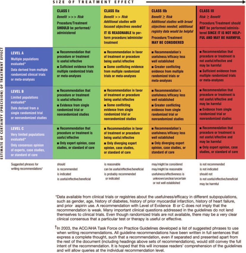
All guideline recommendations underwent a formal, blinded schema for classification of recommendations and level of
writing committee vote. Writing committee members were evidence is summarized in Table 1, which also illustrates how
required to recuse themselves if they had a significant the grading system provides an estimate of the size of the
relevant relationship with industry. The guideline recommen- treatment effect and an estimate of the certainty of the
dations were unanimously approved by all members of the treatment effect.
writing committee who were eligible to vote. The section The focus of these guidelines is the appropriate use of heart
“Pacing in Children and Adolescents” was reviewed by pacing devices (e.g., pacemakers for bradyarrhythmias and
additional reviewers with special expertise in pediatric elec- heart failure management, cardiac resynchronization, and
trophysiology. The committee thanks all the reviewers for implantable cardioverter-defibrillators [ICDs]), not the treat-
their comments. Many of their suggestions were incorporated ment of cardiac arrhythmias. The fact that the use of a device
into the final document. for treatment of a particular condition is listed as a Class I
indication (beneficial, useful, and effective) does not preclude
1.3. Methodology and Evidence the use of other therapeutic modalities that may be equally
The recommendations listed in this document are, whenever effective. As with all clinical practice guidelines, the recom-
possible, evidence based. An extensive literature survey was mendations in this document focus on treatment of an average
conducted that led to the incorporation of 527 references. patient with a specific disorder and may be modified by patient
Searches were limited to studies, reviews, and other evidence comorbidities, limitation of life expectancy because of coexist-
conducted in human subjects and published in English. Key ing diseases, and other situations that only the primary treating
search words included but were not limited to antiarrhythmic, physician may evaluate appropriately.
antibradycardia, atrial fibrillation, bradyarrhythmia, cardiac, These guidelines include sections on selection of pacemak-
CRT, defibrillator, device therapy, devices, dual chamber, ers and ICDs, optimization of technology, cost, and follow-up
heart, heart failure, ICD, implantable defibrillator, device of implanted devices. Although the section on follow-up is
implantation, long-QT syndrome, medical therapy, pace- relatively brief, its importance cannot be overemphasized:
maker, pacing, quality-of-life, resynchronization, rhythm, First, optimal results from an implanted device can be
sinus node dysfunction, sleep apnea, sudden cardiac death, obtained only if the device is adjusted to changing clinical
syncope, tachyarrhythmia, terminal care, and transplantation. conditions; second, recent advisories and recalls serve as
Additionally, the committee reviewed documents related to warnings that devices are not infallible, and failure of
the subject matter previously published by the ACC, AHA, electronics, batteries, and leads can occur.2,3
and HRS. References selected and published in this document The committee considered including a section on extrac-
are representative and not all-inclusive. tion of failed/unused leads, a topic of current interest, but
The committee reviewed and ranked evidence supporting elected not to do so in the absence of convincing evidence to
current recommendations, with the weight of evidence ranked support specific criteria for timing and methods of lead
as Level A if the data were derived from multiple randomized extraction. A policy statement on lead extraction from the
Downloaded from http://circ.ahajournals.org/ by guest on July 10, 2015e354 Circulation May 27, 2008
Table 1. Applying Classification of Recommendations and Level of Evidence
North American Society of Pacing and Electrophysiology instances, it includes a discussion of alternative acceptable
(now the HRS) provides information on this topic.4 Similarly, therapies. Many of the indications are modified by the term
the issue of when to discontinue long-term cardiac pacing or “potentially reversible.” This term is used to indicate abnor-
defibrillator therapy has not been studied sufficiently to allow mal pathophysiology (e.g., complete heart block) that may be
formulation of appropriate guidelines5; however, the question the result of reversible factors. Examples include complete
is of such importance that this topic is addressed to emphasize heart block due to drug toxicity (digitalis), electrolyte abnor-
the importance of patient-family-physician discussion and malities, diseases with periatrioventricular node inflammation
ethical principles. (Lyme disease), and transient injury to the conduction system
The text that accompanies the listed indications should be at the time of open heart surgery. When faced with a
read carefully, because it includes the rationale and support- potentially reversible situation, the treating physician must
ing evidence for many of the indications, and in several decide how long of a waiting period is justified before device
Downloaded from http://circ.ahajournals.org/ by guest on July 10, 2015Epstein et al ACC/AHA/HRS Guidelines for Device-Based Therapy e355
therapy is begun. The committee recognizes that this state- use all-cause mortality as the most appropriate end point of
ment does not address the issue of length of hospital stay clinical trials.8,9
vis-à-vis managed-care regulations. It is emphasized that These guidelines are not designed to specify training or
these guidelines are not intended to address this issue, which credentials required for physicians to use device therapy.
falls strictly within the purview of the treating physician. Nevertheless, in view of the complexity of both the cognitive
The term “symptomatic bradycardia” is used in this docu- and technical aspects of device therapy, only appropriately
ment. Symptomatic bradycardia is defined as a documented trained physicians should use device therapy. Appropriate
bradyarrhythmia that is directly responsible for development training guidelines for physicians have been published previ-
of the clinical manifestations of syncope or near syncope, ously.10 –13
transient dizziness or lightheadedness, or confusional states The 2008 revision reflects what the committee believes are
resulting from cerebral hypoperfusion attributable to slow the most relevant and significant advances in pacemaker/ICD
heart rate. Fatigue, exercise intolerance, and congestive heart therapy since the publication of these guidelines in the
failure may also result from bradycardia. These symptoms Journal of the American College of Cardiology and Circula-
may occur at rest or with exertion. Definite correlation of tion in 2002.14,15
symptoms with a bradyarrhythmia is required to fulfill the All recommendations assume that patients are treated with
criteria that define symptomatic bradycardia. Caution should optimal medical therapy according to published guidelines, as
be exercised not to confuse physiological sinus bradycardia had been required in all the randomized controlled clinical
(as occurs in highly trained athletes) with pathological bra- trials on which these guidelines are based, and that human
dyarrhythmias. Occasionally, symptoms may become appar- issues related to individual patients are addressed. The
ent only in retrospect after antibradycardia pacing. Neverthe- committee believes that comorbidities, life expectancy, and
less, the universal application of pacing therapy to treat a quality-of-life (QOL) issues must be addressed forthrightly
specific heart rate cannot be recommended except in specific with patients and their families. We have repeatedly used the
circumstances, as detailed subsequently. phrase “reasonable expectation of survival with a good
In these guidelines, the terms “persistent,” “transient,” and functional status for more than 1 year” to emphasize this
“not expected to resolve” are used but not specifically defined integration of factors in decision-making. Even when physi-
because the time element varies in different clinical condi- cians believe that the anticipated benefits warrant device
tions. The treating physician must use appropriate clinical implantation, patients have the option to decline intervention
judgment and available data in deciding when a condition is after having been provided with a full explanation of the
persistent or when it can be expected to be transient. Section potential risks and benefits of device therapy. Finally, the
2.1.4, “Pacing for Atrioventricular Block Associated With committee is aware that other guideline/expert groups have
Acute Myocardial Infarction,” overlaps with the “ACC/AHA interpreted the same data differently.16 –19
Guidelines for the Management of Patients With ST- In preparing this revision, the committee was guided by the
Elevation Myocardial Infarction”6 and includes expanded following principles:
indications and stylistic changes. The statement “incidental
finding at electrophysiological study” is used several times in 1. Changes in recommendations and levels of evidence were
this document and does not mean that such a study is made either because of new randomized trials or because
indicated. Appropriate indications for electrophysiological of the accumulation of new clinical evidence and the
studies have been published.7 development of clinical consensus.
The section on indications for ICDs has been updated to 2. The committee was cognizant of the health care, logistic,
reflect the numerous new developments in this field and the and financial implications of recent trials and factored in
voluminous literature related to the efficacy of these devices these considerations to arrive at the classification of
in the treatment and prophylaxis of sudden cardiac death certain recommendations.
(SCD) and malignant ventricular arrhythmias. As previously 3. For recommendations taken from other guidelines, word-
noted, indications for ICDs, cardiac resynchronization ther- ing changes were made to render some of the original
apy (CRT) devices, and combined ICDs and CRT devices recommendations more precise.
(hereafter called CRT-Ds) are continuously changing and can 4. The committee would like to reemphasize that the recom-
be expected to change further as new trials are reported. mendations in this guideline apply to most patients but
Indeed, it is inevitable that the indications for device therapy may require modification because of existing situations
will be refined with respect to both expanded use and the that only the primary treating physician can evaluate
identification of patients expected to benefit the most from properly.
these therapies. Furthermore, it is emphasized that when a 5. All of the listed recommendations for implantation of a
patient has an indication for both a pacemaker (whether it be device presume the absence of inciting causes that may be
single-chamber, dual-chamber, or biventricular) and an ICD, eliminated without detriment to the patient (e.g., nones-
a combined device with appropriate programming is indi- sential drug therapy).
cated. 6. The committee endeavored to maintain consistency of
In this document, the term “mortality” is used to indicate recommendations in this and other previously published
all-cause mortality unless otherwise specified. The committee guidelines. In the section on atrioventricular (AV) block
elected to use all-cause mortality because of the variable associated with acute myocardial infarction (AMI), the
definition of sudden death and the developing consensus to recommendations follow closely those in the “ACC/AHA
Downloaded from http://circ.ahajournals.org/ by guest on July 10, 2015e356 Circulation May 27, 2008
Guidelines for the Management of Patients With ST- response to activities of daily living that can be difficult to
Elevation Myocardial Infarction.”6 However, because of diagnose.25 The term “chronotropic incompetence” is used to
the rapid evolution of pacemaker/ICD science, it has not denote an inadequate heart rate response to physical activity.
always been possible to maintain consistency with other Although many experienced clinicians claim to recognize
published guidelines. chronotropic incompetence in individual patients, no single
metric has been established as a diagnostic standard upon
2. Indications for Pacing which therapeutic decisions can be based. The most obvious
example of chronotropic incompetence is a monotonic daily
2.1. Pacing for Bradycardia Due to Sinus and heart rate profile in an ambulatory patient. Various protocols
Atrioventricular Node Dysfunction have been proposed to quantify subphysiological heart rate
In some patients, bradycardia is the consequence of essential responses to exercise,26,27 and many clinicians would con-
long-term drug therapy of a type and dose for which there is sider failure to achieve 80% of the maximum predicted heart
no acceptable alternative. In these patients, pacing therapy is rate (220 minus age) at peak exercise as evidence of a blunted
necessary to allow maintenance of ongoing medical treat- heart rate response.28,29 However, none of these approaches
ment. have been validated clinically, and it is likely that the
appropriate heart rate response to exercise in individual
2.1.1. Sinus Node Dysfunction patients is too idiosyncratic for standardized testing.
Sinus node dysfunction (SND) was first described as a The natural history of untreated SND may be highly
clinical entity in 1968,20 although Wenckebach reported the variable. The majority of patients who have experienced
electrocardiographic (ECG) manifestation of SND in 1923. syncope because of a sinus pause or marked sinus bradycar-
SND refers to a broad array of abnormalities in sinus node dia will have recurrent syncope.30 Not uncommonly, the
and atrial impulse formation and propagation. These include natural history of SND is interrupted by other necessary
persistent sinus bradycardia and chronotropic incompetence medical therapies that aggravate the underlying tendency to
without identifiable causes, paroxysmal or persistent sinus bradycardia.24 MOST (Mode Selection Trial) included symp-
arrest with replacement by subsidiary escape rhythms in the tomatic pauses greater than or equal to 3 seconds or sinus
atrium, AV junction, or ventricular myocardium. The fre- bradycardia with rates greater than 50 bpm, which restricted
quent association of paroxysmal atrial fibrillation (AF) and the use of indicated long-term medical therapy. Supraventric-
sinus bradycardia or sinus bradyarrhythmias, which may ular tachycardia (SVT) including AF was present in 47% and
oscillate suddenly from one to the other, usually accompanied 53% of patients, respectively, enrolled in a large randomized
by symptoms, is termed “tachy-brady syndrome.”
clinical trial of pacing mode selection in SND.22,31 The
SND is primarily a disease of the elderly and is presumed
incidence of sudden death is extremely low, and SND does
to be due to senescence of the sinus node and atrial muscle.
not appear to affect survival whether untreated30 or treated
Collected data from 28 different studies on atrial pacing for
with pacemaker therapy.32,33
SND showed a median annual incidence of complete AV
The only effective treatment for symptomatic bradycardia
block of 0.6% (range 0% to 4.5%) with a total prevalence of
is permanent cardiac pacing. The decision to implant a
2.1% (range 0% to 11.9%).21 This suggests that the degen-
pacemaker for SND is often accompanied by uncertainty that
erative process also affects the specialized conduction system,
arises from incomplete linkage between sporadic symptoms
although the rate of progression is slow and does not
and ECG evidence of coexisting bradycardia. It is crucial to
dominate the clinical course of disease.21 SND is typically
diagnosed in the seventh and eighth decades of life, which is distinguish between physiological bradycardia due to auto-
also the average age at enrollment in clinical trials of nomic conditions or training effects and circumstantially
pacemaker therapy for SND.22,23 Identical clinical manifes- inappropriate bradycardia that requires permanent cardiac
tations may occur at any age as a secondary phenomenon of pacing. For example, sinus bradycardia is accepted as a
any condition that results in destruction of sinus node cells, such physiological finding that does not require cardiac pacing in
as ischemia or infarction, infiltrative disease, collagen vascular trained athletes. Such individuals may have heart rates of 40
disease, surgical trauma, endocrinologic abnormalities, auto- to 50 bpm while at rest and awake and may have a sleeping
nomic insufficiency, and others.24 rate as slow as 30 bpm, with sinus pauses or progressive sinus
The clinical manifestations of SND are diverse, reflecting slowing accompanied by AV conduction delay (PR prolon-
the range of typical sinoatrial rhythm disturbances. The most gation), sometimes culminating in type I second-degree AV
dramatic presentation is syncope. The mechanism of syncope block.34,35 The basis of the distinction between physiological
is a sudden pause in sinus impulse formation or sinus exit and pathological bradycardia, which may overlap in ECG
block, either spontaneously or after the termination of an presentation, therefore pivots on correlation of episodic
atrial tachyarrhythmia, that causes cerebral hypoperfusion. bradycardia with symptoms compatible with cerebral hypo-
The pause in sinus node activity is frequently accompanied perfusion. Intermittent ECG monitoring with Holter monitors
by an inadequate, delayed, or absent response of subsidiary and event recorders may be helpful,36,37 although the duration
escape pacemakers in the AV junction or ventricular myo- of monitoring required to capture such evidence may be very
cardium, which aggravates the hemodynamic consequences. long.38 The use of insertable loop recorders offers the advan-
However, in many patients, the clinical manifestations of tages of compliance and convenience during very long-term
SND are more insidious and relate to an inadequate heart rate monitoring efforts.39
Downloaded from http://circ.ahajournals.org/ by guest on July 10, 2015Epstein et al ACC/AHA/HRS Guidelines for Device-Based Therapy e357
The optimal pacing system for prevention of symptomatic 3. Permanent pacemaker implantation is not indicated for
bradycardia in SND is unknown. Recent evidence suggests SND with symptomatic bradycardia due to nonessential
that ventricular desynchronization due to right ventricular drug therapy. (Level of Evidence: C)
apical (RVA) pacing may have adverse effects on left
ventricular (LV) and left atrial structure and function.40 – 47 2.1.2. Acquired Atrioventricular Block in Adults
These adverse effects likely explain the association of RVA AV block is classified as first-, second-, or third-degree
pacing, independent of AV synchrony, with increased risks of (complete) block; anatomically, it is defined as supra-, intra-,
AF and heart failure in randomized clinical trials of pace- or infra-His. First-degree AV block is defined as abnormal
maker therapy45,48,49 and, additionally, ventricular arrhyth- prolongation of the PR interval (greater than 0.20 seconds).
mias and death during ICD therapy.50,51 Likewise, although Second-degree AV block is subclassified as type I and type II.
simulation of the normal sinus node response to exercise in Type I second-degree AV block is characterized by progres-
bradycardia patients with pacemaker sensors seems logical, a sive prolongation of the interval between the onset of atrial (P
clinical benefit on a population scale has not been demon- wave) and ventricular (R wave) conduction (PR) before a
strated in large randomized controlled trials of pacemaker nonconducted beat and is usually seen in conjunction with
therapy.52 These rapidly evolving areas of clinical investiga- QRS. Type I second-degree AV block is characterized by
tion should inform the choice of pacing system in SND (see progressive prolongation of the PR interval before a noncon-
Section 2.6, “Selection of Pacemaker Device”). ducted beat and a shorter PR interval after the blocked beat.
Recommendations for Permanent Pacing in Sinus Node Type II second-degree AV block is characterized by fixed PR
Dysfunction intervals before and after blocked beats and is usually
associated with a wide QRS complex. When AV conduction
occurs in a 2:1 pattern, block cannot be classified unequivo-
Class I
cally as type I or type II, although the width of the QRS can
1. Permanent pacemaker implantation is indicated for SND be suggestive, as just described. Advanced second-degree AV
with documented symptomatic bradycardia, including fre- block refers to the blocking of 2 or more consecutive P waves
quent sinus pauses that produce symptoms. (Level of with some conducted beats, which indicates some preserva-
Evidence: C)53–55 tion of AV conduction. In the setting of AF, a prolonged
2. Permanent pacemaker implantation is indicated for symp- pause (e.g., greater than 5 seconds) should be considered to
tomatic chronotropic incompetence. (Level of Evidence: be due to advanced second-degree AV block. Third-degree
C)53–57 AV block (complete heart block) is defined as absence of AV
3. Permanent pacemaker implantation is indicated for symp- conduction.
tomatic sinus bradycardia that results from required drug Patients with abnormalities of AV conduction may be
therapy for medical conditions. (Level of Evidence: C) asymptomatic or may experience serious symptoms related to
bradycardia, ventricular arrhythmias, or both. Decisions re-
Class IIa garding the need for a pacemaker are importantly influenced
by the presence or absence of symptoms directly attributable
1. Permanent pacemaker implantation is reasonable for SND to bradycardia. Furthermore, many of the indications for
with heart rate less than 40 bpm when a clear association pacing have evolved over the past 40 years on the basis of
between significant symptoms consistent with bradycardia experience without the benefit of comparative randomized
and the actual presence of bradycardia has not been clinical trials, in part because no acceptable alternative
documented. (Level of Evidence: C)53–55,58 – 60 options exist to treat most bradycardias.
2. Permanent pacemaker implantation is reasonable for syn- Nonrandomized studies strongly suggest that permanent
cope of unexplained origin when clinically significant pacing does improve survival in patients with third-degree
abnormalities of sinus node function are discovered or AV block, especially if syncope has occurred.63– 68 Although
provoked in electrophysiological studies. (Level of Evi- there is little evidence to suggest that pacemakers improve
dence: C)61,62 survival in patients with isolated first-degree AV block,69 it is
now recognized that marked (PR more than 300 milliseconds)
Class IIb first-degree AV block can lead to symptoms even in the
1. Permanent pacemaker implantation may be considered in absence of higher degrees of AV block.70 When marked
minimally symptomatic patients with chronic heart rate less first-degree AV block for any reason causes atrial systole in
than 40 bpm while awake. (Level of Evidence: C)53,55,56,58 – 60 close proximity to the preceding ventricular systole and
produces hemodynamic consequences usually associated
with retrograde (ventriculoatrial) conduction, signs and
Class III
symptoms similar to the pacemaker syndrome may occur.71
1. Permanent pacemaker implantation is not indicated for With marked first-degree AV block, atrial contraction occurs
SND in asymptomatic patients. (Level of Evidence: C) before complete atrial filling, ventricular filling is compro-
2. Permanent pacemaker implantation is not indicated for mised, and an increase in pulmonary capillary wedge pressure
SND in patients for whom the symptoms suggestive of and a decrease in cardiac output follow. Small uncontrolled
bradycardia have been clearly documented to occur in the trials have suggested some symptomatic and functional im-
absence of bradycardia. (Level of Evidence: C) provement by pacing of patients with PR intervals more than
Downloaded from http://circ.ahajournals.org/ by guest on July 10, 2015e358 Circulation May 27, 2008
0.30 seconds by decreasing the time for AV conduction.70 stitute an indication for pacemaker implantation except as
Finally, a long PR interval may identify a subgroup of specifically defined in the recommendations that follow.
patients with LV dysfunction, some of whom may benefit In general, the decision regarding implantation of a pace-
from dual-chamber pacing with a short(er) AV delay.72 These maker must be considered with respect to whether AV block will
same principles also may be applied to patients with type I be permanent. Reversible causes of AV block, such as electro-
second-degree AV block who experience hemodynamic com- lyte abnormalities, should be corrected first. Some diseases may
promise due to loss of AV synchrony, even without brady- follow a natural history to resolution (e.g., Lyme disease), and
cardia. Although echocardiographic or invasive techniques some AV block can be expected to reverse (e.g., hypervagotonia
may be used to assess hemodynamic improvement before due to recognizable and avoidable physiological factors, periop-
permanent pacemaker implantation, such studies are not erative AV block due to hypothermia, or inflammation near the
required. AV conduction system after surgery in this region). Conversely,
Type I second-degree AV block is usually due to delay in some conditions may warrant pacemaker implantation because
the AV node irrespective of QRS width. Because progression of the possibility of disease progression even if the AV block
to advanced AV block in this situation is uncommon,73–75 reverses transiently (e.g., sarcoidosis, amyloidosis, and neuro-
pacing is usually not indicated unless the patient is symptom- muscular diseases). Finally, permanent pacing for AV block
atic. Although controversy exists, pacemaker implantation is after valve surgery follows a variable natural history; therefore,
supported for this finding.76 –78 Type II second-degree AV the decision for permanent pacing is at the physician’s
block is usually infranodal (either intra- or infra-His), espe- discretion.84
cially when the QRS is wide. In these patients, symptoms are Recommendations for Acquired Atrioventricular Block
frequent, prognosis is compromised, and progression to in Adults
third-degree AV block is common and sudden.73,75,79 Thus,
type II second-degree AV block with a wide QRS typically Class I
indicates diffuse conduction system disease and constitutes an
indication for pacing even in the absence of symptoms. 1. Permanent pacemaker implantation is indicated for third-
However, it is not always possible to determine the site of AV degree and advanced second-degree AV block at any ana-
block without electrophysiological evaluation, because type I tomic level associated with bradycardia with symptoms
second-degree AV block can be infranodal even when the (including heart failure) or ventricular arrhythmias presumed
QRS is narrow.80 If type I second-degree AV block with a to be due to AV block. (Level of Evidence: C)59,63,76,85
narrow or wide QRS is found to be intra- or infra-Hisian at 2. Permanent pacemaker implantation is indicated for third-
electrophysiological study, pacing should be considered. degree and advanced second-degree AV block at any
anatomic level associated with arrhythmias and other
Because it may be difficult for both patients and their
medical conditions that require drug therapy that results in
physicians to attribute ambiguous symptoms such as fatigue
symptomatic bradycardia. (Level of Evidence: C)59,63,76,85
to bradycardia, special vigilance must be exercised to ac-
3. Permanent pacemaker implantation is indicated for third-
knowledge the patient’s concerns about symptoms that may
degree and advanced second-degree AV block at any
be caused by a slow heart rate. In a patient with third-degree
anatomic level in awake, symptom-free patients in sinus
AV block, permanent pacing should be strongly considered
rhythm, with documented periods of asystole greater than
even when the ventricular rate is more than 40 bpm, because
or equal to 3.0 seconds86 or any escape rate less than 40
the choice of a 40 bpm cutoff in these guidelines was not
bpm, or with an escape rhythm that is below the AV node.
determined from clinical trial data. Indeed, it is not the escape (Level of Evidence: C)53,58
rate that is necessarily critical for safety but rather the site of 4. Permanent pacemaker implantation is indicated for third-
origin of the escape rhythm (i.e., in the AV node, the His degree and advanced second-degree AV block at any ana-
bundle, or infra-His). tomic level in awake, symptom-free patients with AF and
AV block can sometimes be provoked by exercise. If not bradycardia with 1 or more pauses of at least 5 seconds or
secondary to myocardial ischemia, AV block in this circum- longer. (Level of Evidence: C)
stance usually is due to disease in the His-Purkinje system 5. Permanent pacemaker implantation is indicated for third-
and is associated with a poor prognosis; thus, pacing is degree and advanced second-degree AV block at any ana-
indicated.81,82 Long sinus pauses and AV block can also tomic level after catheter ablation of the AV junction. (Level
occur during sleep apnea. In the absence of symptoms, of Evidence: C)87,88
these abnormalities are reversible and do not require 6. Permanent pacemaker implantation is indicated for third-
pacing.83 If symptoms are present, pacing is indicated as in degree and advanced second-degree AV block at any ana-
other conditions. tomic level associated with postoperative AV block that is
Recommendations for permanent pacemaker implantation not expected to resolve after cardiac surgery. (Level of
in patients with AV block in AMI, congenital AV block, and Evidence: C)84,85,89,90
AV block associated with enhanced vagal tone are discussed 7. Permanent pacemaker implantation is indicated for third-
in separate sections. Neurocardiogenic causes in young pa- degree and advanced second-degree AV block at any ana-
tients with AV block should be assessed before proceeding tomic level associated with neuromuscular diseases with AV
with permanent pacing. Physiological AV block in the block, such as myotonic muscular dystrophy, Kearns-Sayre
presence of supraventricular tachyarrhythmias does not con- syndrome, Erb dystrophy (limb-girdle muscular dystrophy),
Downloaded from http://circ.ahajournals.org/ by guest on July 10, 2015Epstein et al ACC/AHA/HRS Guidelines for Device-Based Therapy e359
and peroneal muscular atrophy, with or without symptoms. 3. Permanent pacemaker implantation is not indicated for
(Level of Evidence: B)91–97 AV block that is expected to resolve and is unlikely to
8. Permanent pacemaker implantation is indicated for recur100 (e.g., drug toxicity, Lyme disease, or transient
second-degree AV block with associated symptomatic increases in vagal tone or during hypoxia in sleep apnea
bradycardia regardless of type or site of block. (Level of syndrome in the absence of symptoms). (Level of Evi-
Evidence: B)74 dence: B)99,100
9. Permanent pacemaker implantation is indicated for
asymptomatic persistent third-degree AV block at any 2.1.3. Chronic Bifascicular Block
anatomic site with average awake ventricular rates of 40 Bifascicular block refers to ECG evidence of impaired conduc-
bpm or faster if cardiomegaly or LV dysfunction is present tion below the AV node in the right and left bundles. Alternating
or if the site of block is below the AV node. (Level of bundle-branch block (also known as bilateral bundle-branch
Evidence: B)76,78 block) refers to situations in which clear ECG evidence for block
10. Permanent pacemaker implantation is indicated for second- in all 3 fascicles is manifested on successive ECGs. Examples
or third-degree AV block during exercise in the absence of are right bundle-branch block and left bundle-branch block on
myocardial ischemia. (Level of Evidence: C)81,82 successive ECGs or right bundle-branch block with associated
left anterior fascicular block on 1 ECG and associated left
Class IIa posterior fascicular block on another ECG. Patients with first-
1. Permanent pacemaker implantation is reasonable for persis- degree AV block in association with bifascicular block and
tent third-degree AV block with an escape rate greater than symptomatic, advanced AV block have a high mortality rate and
40 bpm in asymptomatic adult patients without cardiomeg- a substantial incidence of sudden death.64,101 Although third-
aly. (Level of Evidence: C)59,63,64,76,82,85 degree AV block is most often preceded by bifascicular block,
2. Permanent pacemaker implantation is reasonable for there is evidence that the rate of progression of bifascicular block
asymptomatic second-degree AV block at intra- or infra- to third-degree AV block is slow.102 Furthermore, no single
His levels found at electrophysiological study. (Level of clinical or laboratory variable, including bifascicular block,
Evidence: B)74,76,78 identifies patients at high risk of death due to a future brady-
3. Permanent pacemaker implantation is reasonable for first- arrhythmia caused by bundle-branch block.103
or second-degree AV block with symptoms similar to Syncope is common in patients with bifascicular block.
those of pacemaker syndrome or hemodynamic compro- Although syncope may be recurrent, it is not associated with an
mise. (Level of Evidence: B)70,71 increased incidence of sudden death.73,102–112 Even though
4. Permanent pacemaker implantation is reasonable for pacing relieves the neurological symptoms, it does not reduce
asymptomatic type II second-degree AV block with a the occurrence of sudden death.108 An electrophysiological
narrow QRS. When type II second-degree AV block study may be helpful to evaluate and direct the treatment of
occurs with a wide QRS, including isolated right bundle- inducible ventricular arrhythmias113,114 that are common in
branch block, pacing becomes a Class I recommendation. patients with bifascicular block. There is convincing evidence
(See Section 2.1.3, “Chronic Bifascicular Block.”) (Level that in the presence of permanent or transient third-degree AV
of Evidence: B)70,76,80,85 block, syncope is associated with an increased incidence of
sudden death regardless of the results of the electrophysiological
Class IIb study.64,114,115 Finally, if the cause of syncope in the presence of
bifascicular block cannot be determined with certainty, or
1. Permanent pacemaker implantation may be considered for if treatments used (such as drugs) may exacerbate AV
neuromuscular diseases such as myotonic muscular dys- block, prophylactic permanent pacing is indicated, espe-
trophy, Erb dystrophy (limb-girdle muscular dystrophy), cially if syncope may have been due to transient third-
and peroneal muscular atrophy with any degree of AV degree AV block.102–112,116
block (including first-degree AV block), with or without Of the many laboratory variables, the PR and HV intervals
symptoms, because there may be unpredictable progres- have been identified as possible predictors of third-degree AV
sion of AV conduction disease. (Level of Evidence: B)91–97 block and sudden death. Although PR-interval prolongation is
2. Permanent pacemaker implantation may be considered for common in patients with bifascicular block, the delay is often at
AV block in the setting of drug use and/or drug toxicity the level of the AV node. There is no correlation between the PR
when the block is expected to recur even after the drug is and HV intervals or between the length of the PR interval,
withdrawn. (Level of Evidence: B)98,99 progression to third-degree AV block, and sudden
death.107,109,116 Although most patients with chronic or intermit-
Class III
tent third-degree AV block demonstrate prolongation of the HV
1. Permanent pacemaker implantation is not indicated for interval during anterograde conduction, some investigators110,111
asymptomatic first-degree AV block. (Level of Evidence: have suggested that asymptomatic patients with bifascicular
B)69 (See Section 2.1.3, “Chronic Bifascicular Block.”) block and a prolonged HV interval should be considered for
2. Permanent pacemaker implantation is not indicated for permanent pacing, especially if the HV interval is greater than or
asymptomatic type I second-degree AV block at the equal to 100 milliseconds.109 Although the prevalence of HV-
supra-His (AV node) level or that which is not known to interval prolongation is high, the incidence of progression to
be intra- or infra-Hisian. (Level of Evidence: C)74 third-degree AV block is low. Because HV prolongation accom-
Downloaded from http://circ.ahajournals.org/ by guest on July 10, 2015You can also read