2019 Focused Update of the Guidelines of the Taiwan Society of Cardiology for the Diagnosis and Treatment of Heart Failure
←
→
Page content transcription
If your browser does not render page correctly, please read the page content below
Acta Cardiol Sin 2019;35:244-283
Review Article doi: 10.6515/ACS.201905_35(3).20190422A
Guidelines
2019 Focused Update of the Guidelines of the
Taiwan Society of Cardiology for the Diagnosis
and Treatment of Heart Failure
Chun-Chieh Wang,1 Cho-Kai Wu,2 Ming-Lung Tsai,1 Chii-Ming Lee,2 Wei-Chun Huang,3 Hsin-Hua Chou,4
Jin-Long Huang,5 Nai-Hsin Chi,6 Hsueh-Wei Yen,7 Bing-Hsiean Tzeng,8 Wei-Ting Chang,9 Hung-Yu Chang,10
Chao-Hung Wang,11 Yen-Yu Lu,12 Jui-Peng Tsai,13 Chun-Hung Su,14 Wen-Jin Cherng,1 Wei-Hsian Yin,10 Chia-Ti Tsai,2
Yen-Wen Wu,8 Jiunn-Lee Lin2,15 and Juey-Jen Hwang2,16
Heart failure is a growing epidemic, especially in Taiwan because of the aging population. The 2016 Taiwan Society
of Cardiology – Heart Failure with reduced Ejection Fraction (TSOC-HFrEF) registry showed that the guideline-
recommended therapies were prescribed suboptimally both at the time of hospital discharge and during follow-
up. We, therefore, conducted this 2019 focused update of the guidelines of the Taiwan Society of Cardiology for the
diagnosis and treatment of heart failure to reinforce the importance of new diagnostic and therapeutic modalities
of heart failure.
The 2019 focused update discusses new diagnostic criteria, pharmacotherapy, non-pharmacological management,
and certain co-morbidities of heart failure. Angiotensin receptor neprilysin inhibitor and If channel inhibitor is
introduced as new and recommended medical therapies. Latest criteria of cardiac resynchronization therapy,
implantable cardioverter-defibrillator, heart transplantation, and ventricular assist device therapy are reviewed in
the non-pharmacological management chapter. Co-morbidities in heart failure are discussed including chronic
kidney disease, diabetes, chronic obstructive pulmonary disease, and sleep-disordered breathing. We also explain
the adequate use of oxygen therapy and non-invasive ventilation in heart failure management. A particular chapter
for chemotherapy-induced cardiac toxicity is incorporated in the focused update to emphasize the importance of
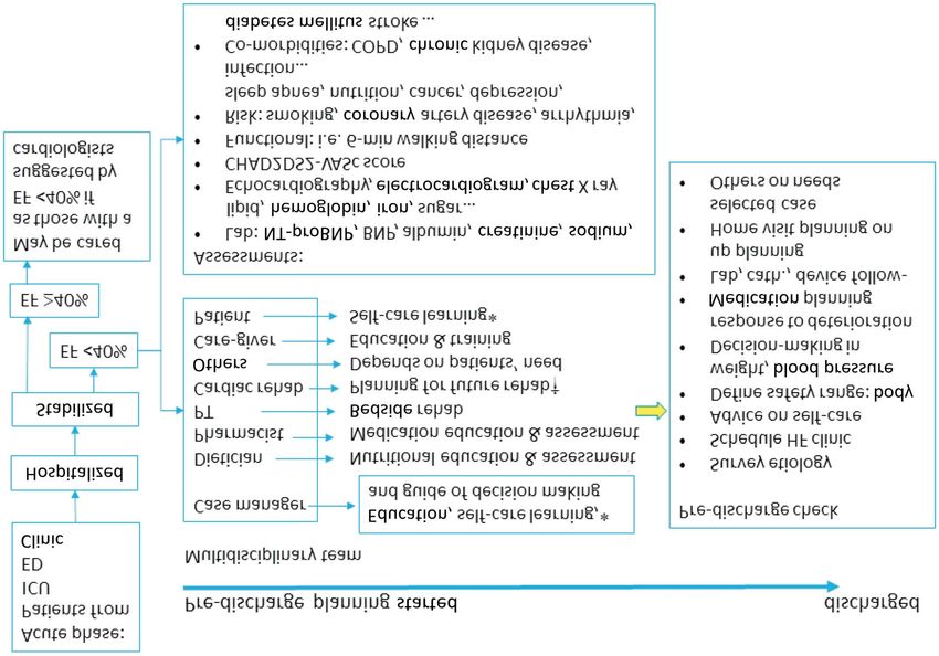
its recognition and management. Lastly, implications from the TSOC-HFrEF registry and post-acute care of heart
failure are discussed to highlight the importance of guideline-directed medical therapy and the benefits of
multidisciplinary disease management programs.
With guideline recommendations, we hope that the management of heart failure can be improved in our society.
Key Words: Biomarkers · Cardiac resynchronization therapy · Cardio-oncology · Co-morbidities · Guidelines ·
Heart failure · Pharmacotherapy · Post-acute care · Transplantation · Ventricular assist device
Received: April 16, 2019 Accepted: April 22, 2019
1
Division of Cardiology, Department of Internal Medicine, Linkou Chang Gung Memorial Hospital and Chang Gung University, Taoyuan;
2
Division of Cardiology, Department of Internal Medicine, National Taiwan University College of Medicine and Hospital, Taipei; 3Division of
Cardiology, Department of Internal Medicine, Kaohsiung Veterans General Hospital, Kaohsiung and School of Medicine, National Yang Ming
University, Taipei; 4Division of Cardiology, Department of Internal Medicine, Taipei Tzu Chi Hospital, Buddhist Tzu Chi Medical Foundation,
Taipei and School of Medicine, Tzu Chi University, Hualien; 5Cardiovascular Center, Taichung Veterans General Hospital, Taichung and School
of Medicine, National Yang-Ming University; 6Department of Surgery, National Taiwan University College of Medicine and Hospital, Taipei;
7
Division of Cardiology, Department of Internal Medicine, Kaohsiung Medical University Hospital, Kaohsiung; 8Division of Cardiology,
Cardiovascular Medical Center, Far Eastern Memorial Hospital, New Taipei City; 9Division of Cardiovascular Medicine, Chi-Mei Medical Center,
Tainan; 10Heart Center, Cheng Hsin General Hospital and Faculty of Medicine, School of Medicine, National Yang Ming University, Taipei;
11
Division of Cardiology, Department of Internal Medicine, Chang Gung Memorial Hospital, Keelung; 12Division of Cardiology, Department of
Internal Medicine, Sijhih Cathay General Hospital, New Taipei City; 13Division of Cardiology, Department of Internal Medicine, Mackay
Memorial Hospital, Taipei; 14Division of Cardiology, Department of Internal Medicine, Chung Shan Medical University Hospital and School of
Medicine, Chung Shan Medical University, Taichung; 15Division of Cardiovascular Medicine, Department of Internal Medicine, Shuang Ho
Hospital and Taipei Medical University, New Taipei City; 16National Taiwan University Hospital Yunlin Branch, Douliu City, Taiwan.
Corresponding author: Dr. Chun-Chieh Wang, Department of Cardiology, Chang Gung University & Chang Gung Memorial Hospital, Taipei &
Linkou Branches, Taiwan. Tel: 886-3-328-1200 ext. 8117; Fax: 886-3-328-9134; E-mail: chcwang@ms17.hinet.net, chcwang@cgmh.org.tw
Acta Cardiol Sin 2019;35:244-283 244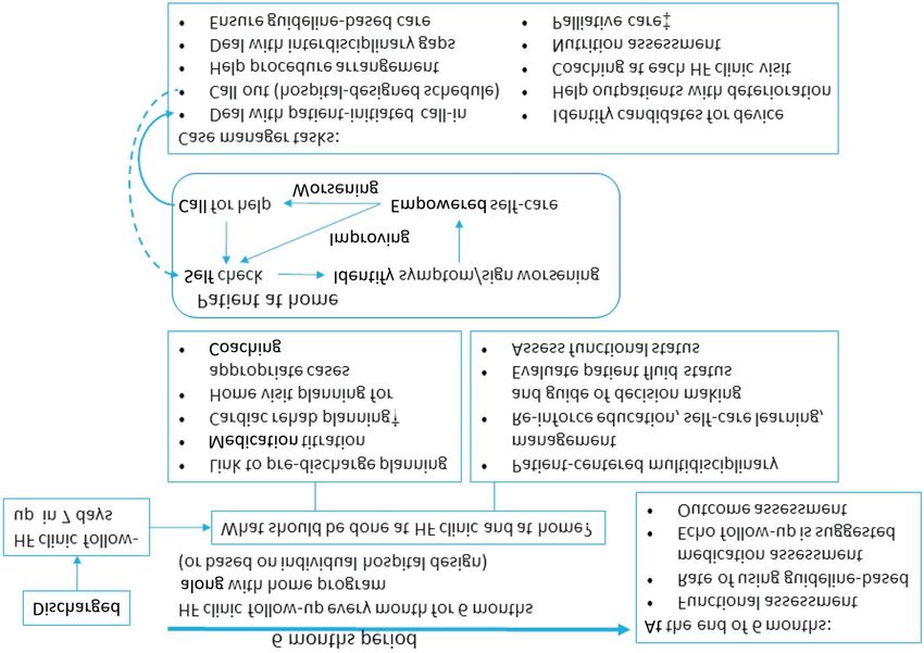
2019 TSOC Heart Failure Guideline

     The Taiwan Society of Cardiology (TSOC) Heart Fail-           sue Doppler parameters (such as S wave) and deforma-
ure Committee provides periodic reviews of new data to             tion imaging techniques (strain and strain rate) can be
produce focused updates that address clinically essen-             used to detect subtle, earlier changes in some HF pa-
tial advances in heart failure (HF) management. This               tients and they are suggested in selected cases.2,3 In a
2019 Focused Update deals with the following topics:               retrospective study enrolling 330 HFrEF Taiwanese pa-
(1) Diagnosis: echocardiography; (2) Diagnosis: bio-               tients, the authors assessed the predictive value of the
markers; (3) Pharmacotherapy: angiotensin converting               ratio of transmitral early filling velocity (E) to early dia-
enzyme inhibitors (ACEIs)/angiotensin receptor blockers            stolic tissue velocity (E¢) and the early diastolic strain
(ARBs)/angiotensin receptor neprilysin inhibitor (ARNI);           rate (E¢sr). They concluded that the E/E¢sr ratio was
(4) Pharmacotherapy: beta-blockers; (5) Pharmaco-                  better able to predict the prognosis of HFrEF than the
therapy: mineralocorticoid receptor antagonists; (6)               E/E¢ ratio. In addition, combined assessments of global
Pharmacotherapy: If channel inhibitors; (7) Non-phar-              longitudinal strain and E/E¢sr by speckle-tracking longi-
macological management: cardiac resynchronization                  tudinal strain could facilitate risk stratification of these
therapy and implantable cardioverter-defibrillators; (8)           patients.4
Non-pharmacological management: surgery; (9) Co-                        In patients with clinical HF, the definition of HF with
morbidities in HF: chronic kidney disease, diabetes, ch-           preserved ejection fraction (HFpEF) varies widely in pre-
ronic obstructive pulmonary disease, sleep-disordered              vious studies.5-7 In most patients, abnormalities of sys-
breathing; (10) Oxygen therapy in acute HF; (11) Che-              tolic and diastolic dysfunction coexist. Because ejection
motherapy-induced cardiac toxicity; (12) Implications              fraction (EF) is the most common selection criteria in
from the Taiwan Society of Cardiology – Heart Failure              clinical trials, echocardiographic EF is considered neces-
with reduced Ejection Fraction (TSOC-HFrEF) registry;              sary to classify patients with HF. In the 2013 American
and (13) Post-acute care of HF.                                    College of Cardiology (ACC)/American Heart Association
                                                                   (AHA) HF guidelines, HF was classified as HFrEF, HFpEF,
                                                                   and borderline HFpEF according to an EF £ 40%, 41~49%
DIAGNOSIS – ECHOCARDIOGRAPHY                                       and ³ 50%, respectively, with one subcategory of “HFpEF,
                                                                   improved” to describe a subset of HFrEF patients with
     Echocardiography is a term encompassing all cardiac           improvement or recovery in EF above 40% after treat-
ultrasound imaging techniques. We will focus on the use            ment.8 In the 2016 European Society of Cardiology (ESC)
of three-dimensional (3D) echocardiography, tissue Dop-            HF guidelines, “gray zone” HF (EF between 40~49%) was
pler imaging (TDI), deformation imaging (strain and strain         defined as HF with mid-range ejection fraction (HFmrEF).9
rate) and transthoracic echocardiography in the current            HfmrEF has been suggested to be a transitional zone for
guidelines to carefully assess the myocardial systolic and         HFpEF and HFrEF in some recent studies.10,11 In the cur-
diastolic function of both left and right ventricles.              rent guidelines, we also define patients with HF as
                                                                   HFpEF, HFmrEF, and HFrEF according to LVEF < 40%, 40%
Assessment of systolic function, classification of                 to 49%, and LVEF ³ 50% (Table 1).
heart failure
     To assess systolic function, we recommend the mo-             Evaluation of diastolic function
dified biplane Simpson’s rule. Left ventricular ejection                After an initial clinical diagnosis of HFpEF, further
fraction (LVEF) should be obtained from apical four- and           objective evidence of echocardiographic cardiac dys-
two-chamber views. Contrast agents can also add to the             function is required to validate the diagnosis. Patients
diagnostic accuracy for patients with poor quality im-             with suspected HFpEF or HFmrEF should have the fol-
ages.1 In contrast, the Teichholz and Quinones methods             lowing objective structural and/or functional alterations
of calculating LVEF from linear dimensions are not re-             of the heart:
commended in the setting of HF, especially for those               · Key structural alterations including left atrial volume
with regional wall motion abnormalities. In recent years,            index (LAVI) > 34 mL/m2 or a left ventricular (LV) mass
some studies have shown that 3D echocardiography, tis-               index ³ 115 g/m2 for males and ³ 95 g/m2 for females.9

                                                             245                                  Acta Cardiol Sin 2019;35:244-283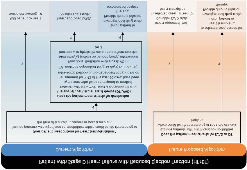
Chun-Chieh Wang et al.

Table 1. Types of heart failure
Types of heart failure                            HFpEF                                 HFmrEF                        HFrEF
Clinical expression                       Symptoms and/or signs                  Symptoms and/or signs        Symptoms and/or signs
Echocardiographic ejection fraction             LVEF ³ 50%                      LVEF between 40 and 49%            LVEF < 40%
Objective evidence                    Elevated natriuretic peptides*         Elevated natriuretic peptides*
                                      and echocardiographic cardiac          and echocardiographic cardiac
                                      structural change or diastolic         structural change or diastolic
                                                  #                                      #
                                      dysfunction                            dysfunction
* B-type natriuretic peptide > 100 pg/mL and/or N-terminal pro-B type natriuretic peptide > 300 pg/mL. # Refer to Figure 1 for
structural and function change and diastolic dysfunction grading.
HFmrEF, HF with mid-range ejection fraction; HFpEF, HF with preserved ejection fraction; HFrEF, HF with reduced EF; LVEF, left
ventricular ejection fraction.

· Key functional alterations including an average E/e’ >                    A diagnostic algorithm for HFpEF and diastolic car-
  14, a septal e’ velocity < 7 cm/s or a lateral e’ velocity <          diac dysfunction is shown in Figure 1A, and the grade of
  10 cm/s.12                                                            diastolic dysfunction is shown in Figure 1B. A recent ret-
· Other indirect echocardiographically derived measure-                 rospective study including 451 Taiwanese HFpEF pa-
  ments including longitudinal strain or tricuspid regurgi-             tients evaluated their risks of outcomes based on the
  tation velocity (TRV; TRV > 2.8 m/s).12                               2016 and 2009 diastolic dysfunction grading algorithm.
· A recent recommendation from the American Society                     After a follow-up period of 2,976 days, the net reclassifi-
  of Echocardiography and the European Association of                   cation index increased significantly after grading with
  Cardiovascular Imaging has focused on the assessment                  the 2016 algorithm (10.6%, p < 0.001). Therefore, the
  of diastolic dysfunction in HFpEF.12 There are three ty-              2016 diastolic dysfunction grading algorithm appears to
  pes of abnormal filling patterns recognized convention-               improve the prognostic value in Taiwanese patients with
  ally in patients in sinus rhythm.                                     HFpEF.
· When the mitral inflow pattern shows an E/A ratio £
  0.8 as well as a peak E velocity of £ 50 cm/s, then mean              Evaluation of right ventricular function and
  left atrial pressure is considered low. The correspond-               pulmonary artery pressure
  ing grade of diastolic dysfunction is grade I.                             Echocardiography should also address right ventric-
· When the mitral inflow pattern shows an E/A ratio ³ 2,                ular (RV) size and function, as well as right atrial size and
  mean left atrial pressure is elevated and is considered               dimensions.8 RV function is a useful parameter to pre-
  to be grade III diastolic dysfunction.                                dict mortality and morbidity in patients with HF.13,14 To
· When mitral inflow shows an E/A £ 0.8 and a peak E ve-                measure RV function, the following parameters are es-
  locity > 50 cm/s, or if the E/A ratio is > 0.8 but < 2, other         pecially useful:
  criteria should be evaluated including peak TRV > 2.8                 · Tricuspid annular plane systolic excursion (TAPSE; ab-
  m/s, average E/e’ > 14 or LAVI > 34 mL/m2. In patients                  normal TAPSE < 17 mm indicates RV systolic dysfunc-
  in whom one of the three main criteria is not available,                tion).
  the ratio of pulmonary vein peak systolic to peak dia-                · Tissue Doppler-derived tricuspid lateral annular systolic
  stolic velocity or systolic time velocity integral to dia-              velocity (s¢) (s¢ velocity < 9.5 cm/s indicates RV systolic
  stolic time-velocity integral < 1 supports the presence                 dysfunction).1,15,16
  of elevated LV filling pressure. If these three para-                 · RV fractional area change, which is expressed as a per-
  meters are available and none or only one exceeds the                   centage change in the RV chamber area from end-dias-
  cutoff value, the patient is considered to have grade I                 tole to end-systole, rather than changes in volume.16
  diastolic dysfunction. If two of the three or all three               · Systolic pulmonary artery pressure derived from an op-
  parameters exceed the cutoff values, then the patient                   timal recording of the maximal systolic tricuspid pres-
  is considered to have grade II diastolic dysfunction.                   sure gradient.
  Otherwise, the diastolic dysfunction grade cannot be                  · Estimation of right atrial or central venous pressure
  evaluated and should not be reported.                                   (CVP) based on inferior vena cava size and its breath-

Acta Cardiol Sin 2019;35:244-283                                  246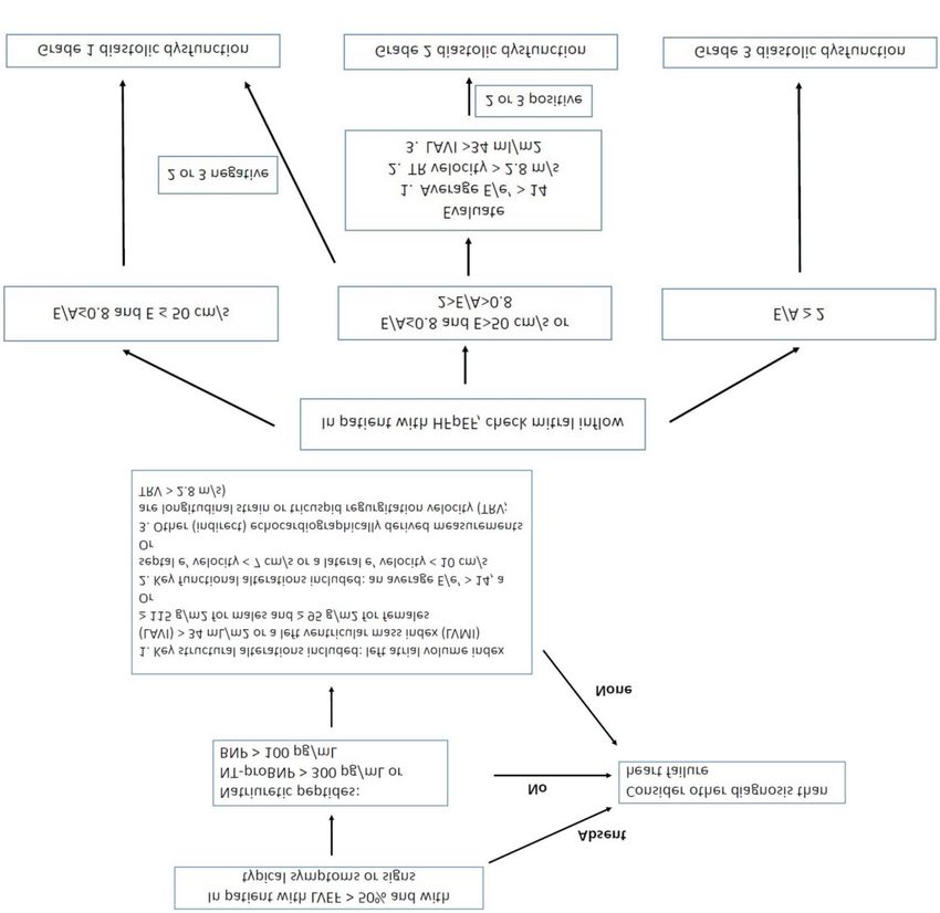
2019 TSOC Heart Failure Guideline

                A

B
Figure 1. (A) Diagnosis of heart failure with preserved ejection fraction. (B) Grading of diastolic dysfunction. BNP, B-type natriuretic peptide; HF,
heart failure; HFpEF, heart failure with preserved ejection fraction; LVEF, left ventricular ejection fraction; NT-proBNP, N-terminal pro B-type
natriuretic peptide.

    ing-related collapse.                                                     Transesophageal echocardiography and stress
                                                                              echocardiography
    For patients with severe HF and cardiologists with                            Transesophageal echocardiography is recommended
experience in 3D echocardiography, 3D measurements                            in patients with an inadequate thoracic echo window, in
of RV volume may be more accurate and clinically rele-                        patients with complicated valvular disease which cannot
vant.3 Newer techniques to assess RV function include                         be distinguished from transthoracic echo or does not
3D speckle-tracking echocardiography, pulsed-wave TDI,                        match the patients’ symptoms using transthoracic echo
color TDI, and strain imaging.16,17                                           alone, in suspected aortic dissection, suspected endo-

                                                                        247                                       Acta Cardiol Sin 2019;35:244-283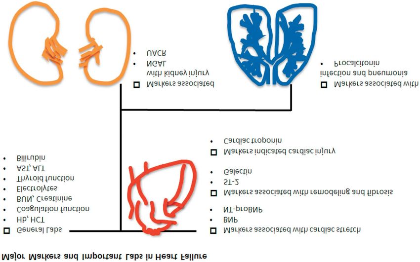
Chun-Chieh Wang et al.

carditis or congenital heart disease, and to rule out in-            specific NP reduction as a treatment goal are still un-
tracavitary thrombi in patients with atrial fibrillation             clear. Although some smaller studies have shown an im-
(AF) requiring cardioversion. Stress echocardiography,               provement in clinical outcomes,25,26 further studies are
on the other hand, can be used to assess the severity of             required to elucidate the benefits.
ischemic heart disease and myocardial viability.18 Stress                 Besides HF, ischemic heart disease, uncontrolled hy-
echocardiography can also detect exercise diastolic dys-             pertension, increasing age, renal dysfunction, anemia,
function for HFpEF patients with an inconclusive diagno-             pulmonary diseases, and sepsis can also increase NP lev-
sis at rest.19                                                       els. The grey zone of BNP as a diagnostic tool for HF is
                                                                     100-400 pg/mL, and 300-450 pg/mL for NT-proBNP. In
                                                                     elderly patients (age > 75 years), the grey zone of NT-
DIAGNOSIS – BIOMARKERS                                               proBNP can be extended to 300-1800 pg/mL.27 Because
                                                                     the level can also be elevated in various conditions other
      Routine diagnostic evaluations for HF should include           than HF, NPs are preferred as an exclusion tool in first-
laboratory tests, including biomarkers for HF, which can             line screening. A normal concentration in an untreated
be used to assist in the diagnosis and as prognostic pre-            patient has a high negative predictive value for the diag-
dictors. The use of biomarkers to diagnose HF is more                nosis of HF. Moreover, the level of NPs can be lower in
convenient than echocardiography as a first line tool at             patients with a higher body mass index (BMI) (> 35 kg/
outpatient service or emergency departments. However,                m2) due to increased clearance receptors in adipocytes,28
these biomarkers can also be elevated in conditions other            thus the use of NPs in these groups of patients should
than HF. Therefore, biomarkers should be used cau-                   be applied with caution. For patients receiving treat-
tiously and be limited to exclusion, especially in patients          ment with ARNI, NP-proBNP is preferred to evaluate the
with atypical presentations.                                         patient’s prognosis because the mechanism of action of
      We recommend measuring B-type natriuretic pep-                 ARNI elevates the level of BNP.
tide (BNP) or N-terminal proB-type natriuretic peptide                    Cardiac troponins should also be sampled in pa-
(NT-proBNP) to assist in confirming or excluding the di-             tients with suspected or newly diagnosed HF. Cardiac
agnosis of HF (Figure 2). Natriuretic peptides (NPs) are             troponins are an established marker of cardiac injury.
biomarkers associated with stretched myocardial myo-                 Several factors are associated with elevated troponins,
cytes20 which can counteract stress by inducing vasodi-              including subendocardial ischemia, cardiomyocyte ne-
lation, natriuresis, diuresis, and inhibition of cardiac and         crosis, cardiomyocyte damage from inflammatory cyto-
vascular myocyte growth. Evidence from some large co-                kines, oxidative stress, apoptosis, and leakage of tro-
hort studies supports the use of NPs, especially BNP and             ponins from the cytosolic pool due to increased mem-
NT-proBNP, 21 to predict and diagnose new-onset HF.                  brane permeability.29 For patients with newly diagnosed
Other studies also support the potential for predicting              HF, troponins can be measured to evaluate the possible
the prognosis of HF including hospitalization and overall            etiology and also to predict the prognosis. Patients with
mortality. Recent studies from Taiwan with regards to                acute coronary syndrome-induced HF should consider
patients with acute decompensated HF have reported                   revascularization. Nevertheless, cardiac troponins can
that a higher BNP was associated with worse function                 also be elevated in patients with myocarditis and severe
class and a two-fold increased risk of in-hospital mortal-           HF. An elevated cardiac troponin level in HFrEF patients
ity. 22 In patients with HFpEF, elevated NP levels have              is significantly associated with mortality and cardiovas-
also been shown to be a marker associated with a poor                cular (CV) events.30-32 However, data on the prognostic
prognosis, including mortality and HF-related hospital-              value in patients with HFpEF are limited.33,34
ization.23,24                                                             In addition to NPs and cardiac troponins, other
      We also suggest the use of NPs as a prognostic pre-            markers are also associated with HF (Figure 3). Markers
dictor to monitor the effectiveness of HF therapy before             of cardiomyocyte remodeling such as ST-2 and Ga-
hospital discharge. However, the effectiveness and ben-              lectin-3 have been shown to be predictors and markers
efits of serial follow-up measurements or targeting a                of HF. An elevated level of soluble ST-2 suggests de-

Acta Cardiol Sin 2019;35:244-283                               248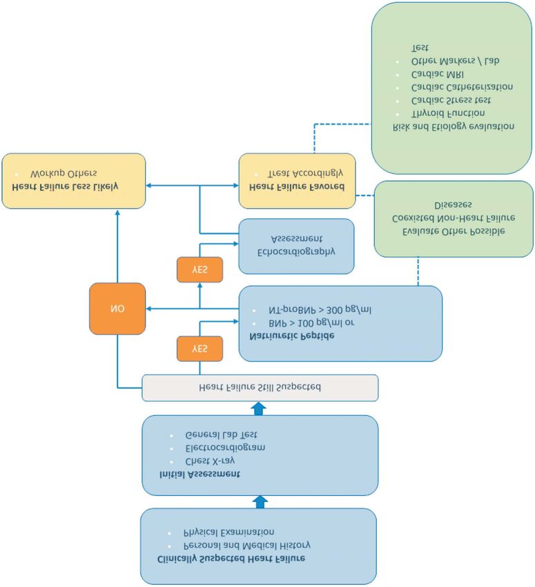
2019 TSOC Heart Failure Guideline

Figure 2. Algorithm of B-type natriuretic peptide (BNP) or N-terminal proB-type natriuretic peptide (NT-proBNP) to assist in the differential
diagnosis of HF. HF, heart failure; MRI, magnetic resonance imaging.

creased cardiac protection in cardiac injuries. Studies                   These kidney biomarkers can reflect nephrotoxic injury
have also shown that ST-2 can be an independent mar-                      and systemic endothelial dysfunction. Although the di-
ker to predict HF hospitalizations and mortality.35 Among                 rect mechanism in HF is unclear, these markers may be
patients with acute myocardial infarction (AMI), a high                   an early indicator of kidney injury in HF.37 Furthermore,
serum ST-2 level has also been shown to be a predictor                    the occurrence of pneumonia in patients with acute HF
of HF.36 Macrophages secrete Galectin-3, and this is as-                  is a commonly discussed issue, and initiating appropri-
sociated with cardiac fibrosis. Accordingly, studies have                 ate antibiotic therapy is essential. Procalcitonin is a
suggested that elevated Galectin-3 could be a prognos-                    valuable diagnostic marker for infection in the setting
tic indicator of HF. Urinary albumin to creatinine ratio                  of acute exacerbations of HF. The combination of mul-
(UACR) and neutrophil gelatinase-associated lipocalin                     tiple biomarkers may have potential benefits for the di-
(NGAL) have been used as markers for kidney injury.                       agnosis and prognostic prediction for patients with HF.
Recent studies have also suggested that UACR and                          However, further validation for clinical cohorts is re-
NGAL can be markers to assess the prognosis of HF.33,36                   quired.

                                                                    249                                     Acta Cardiol Sin 2019;35:244-283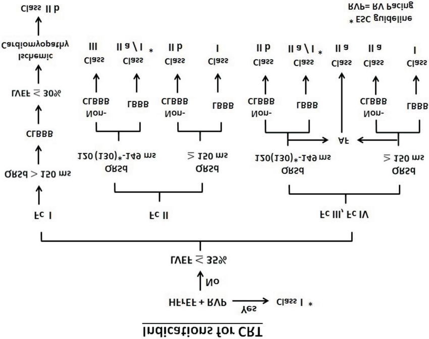
Chun-Chieh Wang et al.

Figure 3. Key biomarkers and necessary laboratory parameters in the differential diagnosis of HF. ALT, alanine aminotransaminase; AST, aspartate
aminotransaminase; BNP, B-type natriuretic peptide; BUN, blood urea nitrogen; Hb, hemoglobin; HCT, hematocrit; HF, heart failure; NGAL, neutrophil
gelatinase-associated lipocalin; NT-proBNP, N-terminal proB-type natriuretic peptide; UACR, urinary albumin to creatinine ratio.

PHARMACOTHERAPY – ANGIOTENSIN                                                up-titrated to the target doses used in randomized con-
CONVERTING ENZYME INHIBITORS/ANGIOTENSIN                                     trolled trials (RCTs) if tolerable.40
RECEPTOR BLOCKERS/ANGIOTENSIN RECEPTOR                                            The development of ARNI has caused a paradigm
NEPRILYSIN INHIBITOR                                                         shift from “add-on” to “replacement” in RAA axis bloc-
                                                                             kers. Although pivotal trials for sacubitril/valsartan are
     HF is the final common pathway of various cardiac                       still lacking, the overwhelming superiority of ARNI to
diseases and is characterized by high morbidity and                          ACEIs in the Prospective Comparison of ARNI with ACEI
mortality. A major issue in the treatment of HF is cardiac                   to Determine Impact on Global Mortality and Morbidity
remodeling after either acute or chronic myocardial in-                      in Heart Failure (PARADIGM-HF) trial41 led to the incor-
jury.38 The renin-angiotensin-aldosterone (RAA) system                       poration of ARNI into the revised ESC guidelines for HF
is deeply involved in cardiac remodeling. Numerous                           in 2016.42
large clinical trials have demonstrated that successfully
blocking the RAA axis both reduces morbidity and also                        Angiotensin converting enzyme inhibitors
improves the survival of HF patients.                                            Multiple large-scale RCTs have clearly established
     Although ACEIs and ARBs have been incorporated                          the benefits of angiotensin converting enzyme inhibi-
into guidelines of international cardiology societies, in-                   tion in patients with mild, moderate, or severe symp-
cluding the ACC/AHA and ESC, for the management of                           toms of HF and in patients with or without coronary ar-
HF, the prescription rate of RAA system blockers is rela-                    tery disease (CAD) [Cooperative North Scandinavian
tively low. According to the TSOC registry,39 the prescrip-                  Enalapril Survival Study (CONSENSUS): enalapril, 1987;
tion rates of ACEIs and ARBs are 27.5% and 34.6%, re-                        Studies of Left Ventricular Dysfunction (SOLVD): en-
spectively. The combined prescription rate of 62.1% for                      alapril, 1991; Survival And Ventricular Enlargement trial
either an ACEI or ARB is lower than that in Western                          (SAVE): captopril, 1992; Acute Infarction Ramipril Effi-
countries. The doses of both ACEIs and ARBs should be                        cacy study (AIRE): ramipril, 1993; Trandolapril Cardiac

Acta Cardiol Sin 2019;35:244-283                                       250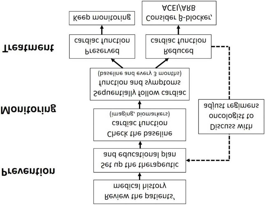
2019 TSOC Heart Failure Guideline

Evaluation study (TRACE): trandolapril 1995; Assess-                ARBs have no beneficial effects on mortality when com-
ment of Treatment with Lisinopril and Survival trial (AT-           bined with ACEIs, and may increase the risk of hypo-
LAS): lisinopril, 1999].                                            tension or hyperkalemia (VAL-HeFT: valsartan/ACEI,
     No significant differences among the available ACEIs           ACEI, 2001;43 CHARM add-on: candesartan/ACEI, ACEI,
have been reported with regards to their effects on                 2003;48 VALsartan In Acute myocardial InfarctioN Trial
symptoms or survival. (ACC/AHA 2017)                                (VALIANT): valsartan, captopril, or both, 2003; ONgoing
· ACEIs reduce morbidity and mortality in HFrEF.                    Telmisartan Alone and in combination with Ramipril
· ACEIs should be started at low doses and titrated up-             Global Endpoint Trial (ONTARGET): telmisartan, ramipril,
  ward to doses shown to reduce the risk of CV events in            or both, 200849).
  clinical trials.                                                  · An ARB is recommended to reduce the risk of HF hospital-
· ACEIs can produce angioedema and should be given                    ization and CV death in symptomatic patients intolerant
  with caution to patients with low systemic blood pres-              to ACEIs (because of cough or angioedema); (Class A,
  sure, renal insufficiency, or elevated serum potassium              Level I for ACC/AHA 2017; Class A, Level B for ESC 2016).
  ([K] > 5.0 mEq/L). The dose of ACEIs should be reduced            · An ARB may be considered to reduce the risk of HF hos-
  or held temporarily if serum K > 5.5 mEq/L and be dis-              pitalization and death in patients who are symptomatic
  continued if K > 6.0 mEq/L.                                         despite treatment with a beta-blocker who are unable
· If maximal doses are not tolerable, moderate doses                  to tolerate a mineralocorticoid receptor antagonist
  should be tried; abrupt withdrawal of ACE inhibition                (MRA) (Class IIb, Level C for ESC 2016).
  can lead to clinical deterioration and should be avoided.         · Patients already tolerating ARBs for other indications may
  (ACC/AHA 2017)                                                      be continued on ARBs if they subsequently develop HF.
· Although the use of an ARNI instead of an ACEI for                · ARBs should be started at low doses and titrated up-
  HFrEF is superior, for the patients for whom ARNI is not            ward, with an attempt to use doses shown to reduce
  appropriate, the continued use of an ACEI for all classes           the risk of CV events in clinical trials.
  of HFrEF remains strongly advised. (ACC/AHA 2017)                 · ARBs should be given with caution to patients with low
                                                                      systemic blood pressure, renal insufficiency, or ele-
Angiotensin receptor blockers                                         vated serum potassium (> 5.0 mEq/L). The dose of ARBs
     ACEIs are associated with side effects including cough           should be reduced or held temporarily if serum K > 5.5
and angioedema, which may compromise its clinical im-                 mEq/L and be discontinued if K > 6.0 mEq/L.
plication. Moreover, escape phenomenon with an eleva-
tion in angiotensin II levels may be detected 3 to 6 months         Angiotensin receptor neprilysin inhibitor
after the initiation of ACEI treatment. ARBs were devel-                 The benefits of ACEIs regarding decreased morbidity
oped with the rationale that angiotensin II production              and mortality have been shown consistently for HF pa-
continues in the presence of ACE inhibition, driven th-             tients across the clinical spectrum, from asymptomatic
rough alternative enzyme pathways. ARBs do not inhibit              to severely symptomatic. Similar benefits have been
kininase and are associated with a much lower inci-                 shown for ARBs in populations with mild-to-moderate
dence of cough and angioedema than ACEIs.                           HF who are unable to tolerate ACEIs.
     The findings of multiple large-scale RCTs have shown                In ARNI, a single molecule with dual action, the ARB
that long-term therapy with ARBs reduces mortality and              valsartan, blocks the action of angiotensin II at AT1 recep-
morbidity, especially in ACEI-intolerant patients. [Evalua-         tors, thus inhibiting activation of the RAA system and pre-
tion of Losartan in The Elderly II study (ELITE II): losar-         venting vasoconstriction, renal sodium and fluid retention
tan, captopril, 2000; Valsartan in Heart Failure trial              and cardiac remodeling. On the other hand, the active me-
(Val-HeFT): valsartan, 2001;43,44 Candesartan in Heart              tabolite in sacubitril LBQ657 inhibits neprilysin and there-
failure: Assessment of Reduction in Mortality and mor-              by increases NPs, which in turn leads to vasodilation.
bidity trial (CHARM): candesartan, 2003;45,46 Heart fail-                In the PARADIGM-HF study, patients with mild-to-
ure Endpoint evaluation of AII-Antagonist Losartan study            moderate HF characterized by either (1) a mildly elevated
(HEAAL): losartan, high vs. low dose 200947]. However,              BNP (> 150 pg/mL) or NT-proBNP (³ 600 pg/mL), or (2)

                                                              251                                  Acta Cardiol Sin 2019;35:244-283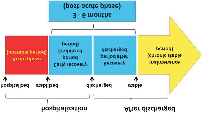
Chun-Chieh Wang et al.

BNP ³ 100 pg/mL or NT-proBNP ³ 400 pg/mL with a prior                 for HF. The mortality and morbidity in patients with HF
hospitalization in the preceding 12 months who were                   resulting from LV systolic dysfunction have been shown
able to tolerate both a target dose of enalapril (10 mg               to be reduced by three beta-blockers (bisoprolol, car-
twice daily) and then subsequently an ARNI (sacubitril/               vedilol, and metoprolol succinate).51-54 Nebivolol, a beta-
valsartan; 200 mg twice daily), were randomized. Com-                 blocker with vasodilating properties, has been shown to
pared with the enalapril group, sacubitril/valsartan signif-          be effective and well-tolerated in older patients with
icantly reduced the combined risk of the primary end-                 HF.55 Beta-blockers have also been shown to improve LV
point (death from a CV cause or first hospitalization for             function and outcomes in Taiwanese studies, 56,57 and
HF) [21.8% vs. 26.5%; hazard ratio (HR) 0.80, 95% confi-              also in long-term hemodialysis patients with HF in a Na-
dence interval (CI) 0.73-0.87; p < 0.001]. In particular, the         tional Health Insurance Research Database study.58
risk of CV death was reduced by 20%, death due to wors-                    The TSOC-HFrEF multicenter registry collected data
ening HF by 21%, and sudden cardiac death (SCD) by 20%.               from 21 medical centers or teaching hospitals in Taiwan,
      Sacubitril/valsartan therapy is recommended to re-              and showed that only 59.6% of patients with HF received
place ACEI therapy to further reduce the risk of HF hospi-            beta-blocker therapy at discharge,39 which is lower than
talization and mortality in ambulatory HFrEF patients who             in Northern America Organized Program To Initiate life-
remain symptomatic despite optimal therapy with an ACEI,              saving treatMent In HospitaliZEd Patients with Heart Fail-
a beta-blocker, and an MRA, and who fit trial criteria.               ure registry (OPTIMIZE-HF)59 and Europe ESC Heart Fail-
· The use of an ARNI is associated with hypotension and               ure Pilot survey (ESC-HF Pilot)60 studies. At 12 months of
   a low-frequency incidence of angioedema.                           follow-up, the prescription rate increased to 66.3%,61 and
· The target dose is 97/103 mg twice daily. Clinical expe-            the percentage of patients receiving > 50% of the target
   rience will provide further information about the opti-            dose of beta-blockers increased from 20.6% at discharge
   mal titration and tolerability of ARNI, particularly re-           to 26.3% at 1-year follow-up.51 However, these results
   garding blood pressure, adjustments in concomitant HF              were lower than in the QUALIFY global survey, which re-
   medications, and the rare complication of angioedema.              ported that the percentages of patients receiving the tar-
· ARNI should not be administered concomitantly with                  get dose and > 50% of the target dose of beta-blockers
   an ACEI or within 36 hours of the last dose of an ACEI.            were 14.8% and 51.8%, respectively.62 The mean heart
· ARNI should not be administered to patients with a his-             rate in the TSOC-HFrEF registry at 1-year follow-up was
   tory of angioedema.                                                80.7 ± 16.0 bpm, indicating that there was still a need for
                                                                      further drug up-titration or medications. 51,61 A multi-
     Recently, the comParIson Of sacubitril/valsartaN ver-            disciplinary disease management program reported an
sus Enalapril on Effect on nt-pRo-bnp in patients stabilized          increase in beta-blocker prescription rate to 77% at dis-
from an acute Heart Failure episode trial (PIONEER-HF)50              charge in a Taiwan single-center study.63
showed promising results in HFrEF patients who were hos-                   The prevalence of chronic obstructive pulmonary dis-
pitalized for acute decompensated HF. The initiation of               ease (COPD) and/or asthma in the TSOC-HFrEF registry
sacubitril/valsartan therapy after hemodynamic stabiliza-             was 11%,62 which is lower than the 31% in the Acute De-
tion resulted in a significantly greater reduction in NT-             compensated HEart failure national REgistry (ADHERE) and
proBNP concentration than enalapril therapy, with no sig-             19% in the EuroHeart Failure Survey II (EHFS-II).64,65
nificant difference in the rate of renal dysfunction, symp-           Cardioselective b-blockers, including bisoprolol, carvedilol,
tomatic hypotension, hyperkalemia, or angioedema. How-                and metoprolol, were recommended for patients with co-
ever, the role of ARNI in the setting of acute HF should be           existing HF and COPD in the 2015 Taiwan cardiologist-
confirmed in a more extensive prospective study.                      pulmonologist consensus handbook and previous stud-
                                                                      ies.66-71 In the Val-HeFT study, cardioselective b-blockers
                                                                      were shown to have a better 23-month mortality rate than
PHARMACOTHERAPY – BETA-BLOCKERS                                       non-selective b-blockers66,70 in patients with coexisting HF
                                                                      and COPD. In a Taiwan nationwide study, beta-blockers
    Beta-blockers are recommended as first-line therapy               were shown to reduce mortality, HF exacerbations, and

Acta Cardiol Sin 2019;35:244-283                                252
2019 TSOC Heart Failure Guideline

the need for hospitalization in patients with coexisting HF         as well as a 24% reduction in all-cause mortality.
and COPD.72 Moreover, beta-blockers were not shown to                   The effects of MRAs on morbidity and mortality
be associated with COPD exacerbations.72 However, the               among patients with AMI complicated by LV dysfunction
suboptimal use of beta-blockers has also been shown in              and HF were evaluated in the Eplerenone post-acute
patients with concurrent HF and COPD in Taiwan.72,73                myocardial infarction Heart failure Efficacy and SUrvival
                                                                    Study (EPHESUS).76 Patients with LV dysfunction (LVEF £
                                                                    40%) following AMI who developed symptoms of HF or
PHARMACOTHERAPY – MINERALOCORTICOID                                 had a history of diabetes mellitus were randomly as-
RECEPTOR ANTAGONISTS                                                signed to receive eplerenone (25 mg per day initially, ti-
                                                                    trated to a maximum of 50 mg daily) or placebo in addi-
· MRAs are recommended in patients with chronic symp-               tion to optimal medical therapy (ACEIs, ARBs, diuretics
  tomatic HFrEF and New York Heart Association (NYHA)               and beta-blockers). After a mean follow-up duration of
  functional class II-IV who are already receiving ACEIs or         16 months, the rates of all-cause mortality, CV death,
  ARBs and beta blockers to reduce mortality and HF hos-            and hospitalizations for CV events were significantly
  pitalization.                                                     lower in the eplerenone group.
· In patients following an AMI who have reduced LV func-                Of note, patients with hyperkalemia (defined as a
  tion and develop symptoms of HF or have a history of              serum potassium level > 5.0 mEq/L) or advanced CKD
  diabetes, treatment with MRAs in addition to optimal              (defined as a serum creatinine concentration > 2.5
  medical therapy is recommended to reduce mortality                mg/dL or eGFR < 30 mL/min/1.73 m2) were all excluded
  and hospitalizations from a CV cause.                             from these randomized trials to avoid life-threatening
· MRAs should be avoided in patients with advanced                  hyperkalemia in patients with HFrEF.
  chronic kidney disease (CKD) (creatinine > 2.5 mg/dL or
  estimated glomerular filtration rate [eGFR] < 30 mL/min/          Dosages of mineralocorticoid receptor antagonists
  1.73 m2) or hyperkalemia (potassium level > 5.0 mEq/L).           and laboratory monitoring
                                                                         Spironolactone and eplerenone should be initiated
     The benefits of MRA treatment in patients with                 at a dose of 25 mg daily and up-titrated to 50 mg daily
HFrEF were investigated in two landmark studies: the                after 4~8 weeks. In patients at risk of hyperkalemia or
Randomized Aldactone Evaluation study (RALES)74 and                 worsening renal function (patients aged ³ 75 years, with
the Eplerenone in Mild Patients Hospitalization And Sur-            diabetes mellitus, or eGFR < 60 mL/min/1.73 m2),77 an
vIval Study in Heart Failure trial (EMPHASIS-HF).75                 initial regimen of spironolactone 25 mg or eplerenone
     The RALES trial randomly assigned patients with NYHA           25 mg every other day is advised.
functional class III or IV and an LVEF of no more than 35%               The most significant risk related to MRA treatment
who had been treated with an ACEI or loop diuretic to re-           is hyperkalemia (defined as a potassium level more than
ceive spironolactone (25 mg daily) or placebo. After a              5.5 mEq/L), which occurred in 19.0% of the spirono-
mean follow-up of 24 months, patients in the spirono-               lactone group in the RALES trial and 11.8% of the ep-
lactone group showed a 30% reduction in all-cause mortal-           lerenone group in the EMPHASIS-HF trial. The develop-
ity compared with the placebo group, as well as 29% re-             ment of hyperkalemia is associated with morbidity and
duction in SCD and a 35% reduction in the frequency of              mortality.78 However, the treatment benefits of spirono-
hospitalizations for worsening HF. The EMPHASIS-HF trial            lactone were maintained at least until the potassium
enrolled patients with mild symptoms (NYHA functional               level exceeded 5.5 mEq/L, and this benefit lost statistical
class II) and an LVEF of no more than 35% to receive                significance as the potassium level approached 6.0
eplerenone (a selective MRA) or placebo, in addition to             mEq/L.79 Routine follow-up of potassium level and renal
recommended optimal medical therapy (an ACEI, an ARB,               function is recommended 1 week and 1 month after
or both and a beta-blocker). After a mean follow-up period          starting or increasing the dose of MRAs. Subsequent
of 21 months, the eplerenone group showed a 37% reduc-              monitoring should occur at least monthly for the first 3
tion in the composite of CV death or hospitalization for HF         months and every 3-6 months thereafter according to

                                                              253                                 Acta Cardiol Sin 2019;35:244-283
Chun-Chieh Wang et al.
the baseline renal function. Patients should be educated risk of CV death and HF hospitalization in HFrEF patients
to avoid foods high in potassium once potassium levels (LVEF £ 35%) with NYHA functional class II to IV in sinus
are higher than 5.0 mEq/L. The dose of MRAs should be rhythm and a resting heart rate ³ 70 bpm who are re-
reduced if potassium levels rise above 5.5 mEq/L. If po- ceiving a maximal dose of beta-blockers or cannot toler-
tassium levels rise above 6.0 mEq/L, MRAs should be ate or have contraindications for a beta-blocker after re-
withheld. The potassium level should be rechecked ceiving an ACEI (or ARB) and an MRA. A high resting
within 3-7 days, and MRAs should only be restarted if heart rate is not only a well-validated risk marker but
the follow-up potassium level is less than 5.0 mEq/L. also a modifiable risk factor in HF.81,86 The magnitude of
heart rate reduction with a beta-blocker plus ivabradine,
rather than background beta-blocker dose, primarily de-
PHARMACOTHERAPY – If CHANNEL INHIBITORS termines the subsequent effect on outcomes,87 since a
substantial proportion of patients with HF cannot toler-
Ivabradine is a new therapeutic agent that explicitly ate the doses of beta-blockers used in large clinical tri-
inhibits ion movement through the f-channel, thereby als. The most common reasons for patients not receiving
inhibiting the If current in the sinoatrial node slowing di- target doses include hypotension and fatigue, and con-
astolic depolarization, the sole effect being heart rate traindications to beta-blockers such as asthma, frequent
reduction, without altering other cardiac functions.80 hypoglycemic episodes or others. Therefore, patients
The Systolic Heart failure treatment with the IF inhib- who cannot tolerate optimal beta-blocker doses may
itor ivabradine Trial (SHIFT) demonstrated the efficacy of benefit from the addition of ivabradine.81,88
ivabradine in reducing the composite endpoint of CV Although both beta-blockers and ivabradine are
death or HF hospitalization. Ivabradine reduced the com- known to reduce resting heart rate, beta-blockers likely
posite endpoint for HF in patients with symptomatic reduce ventricular arrhythmias by blocking beta-1 re-
HFrEF and LVEF £ 35%, in sinus rhythm and with a heart ceptors throughout the myocardium. It is therefore li-
rate ³ 70 beats bpm who had been hospitalized for HF kely that beta-blockers have a more pronounced benefit
within the previous 12 months, receiving treatment with by reducing sudden death,52,89 whereas ivabradine has
an evidence-based dose of beta-blockers (or maximum an isolated effect on sinoatrial nodal tissue and increases
tolerated dose), an ACEI (or ARB), and an MRA.81 Heart diastolic time without affecting blood pressure,90 result-
rate reduction with ivabradine has been shown to be safe ing in improvements in myocardial perfusion and stroke
in severe HF and to improve clinical outcomes independ- volume and maintaining cardiac output. 91 Ivabradine
ently of disease severity.82 Patients receiving ivabradine has been shown to have a significant effect on pump
have been shown to spend fewer days in the hospital as failure death with no effect on SCD; these differences in
they benefit from a reduction in recurrent hospitaliza- effect indicate that combining the two may result in fur-
tions,83 which is an essential marker of prognosis and re- ther benefits and cancel unwanted effects.92 However,
mains a primary objective to reduce healthcare costs. The bradycardia has been reported to found more common
initiation of ivabradine before discharge has been shown in ivabradine-treated patients.93 Moreover, in a meta-
to reduce the risk of rehospitalization during the vulnera- analysis study, patients receiving ivabradine were shown
ble phase after hospitalization for HF.83 to have more AF than controls.94 Close follow-up is there-
Ivabradine treatment is associated with a marked re- fore suggested to monitor these effects.
duction in LV volume and a significant improvement in LVEF,
therefore suggesting that it modifies disease progression in
patients with HF.84 A great deal of clinical evidence has NON-PHARMACOLOGICAL MANAGEMENT –
shown that the use of ivabradine can address unmet needs CARDIAC RESYNCHRONIZATION THERAPY &
in the management of systolic HF, as it improves symptoms, IMPLANTABLE CARDIOVERTER-DEFIBRILLATORS
increases exercise capacity, improves the quality of life, pre-
vents re-hospitalization, and prolongs survival.85 Cardiac resynchronization therapy for HF
Ivabradine should be considered to attenuate the Cardiac resynchronization therapy (CRT) has been
Acta Cardiol Sin 2019;35:244-283 2542019 TSOC Heart Failure Guideline
shown to improve cardiac performance in appropriately CRT104,105 trials specified an LVEF < 30%, while the RE-
selected patients and to improve symptoms and well-be- VERSE106-108 trial specified < 40% and the BLOCK-HF109
ing95-97 and reduce morbidity and mortality.98 Of the im- trial < 50%. Relatively few patients with an LVEF of 35-
provements in quality-adjusted life years with CRT among 40% have been randomized. However an individual par-
patients with moderate to severe HF, two-thirds may be ticipant data meta-analysis suggested no reduction in
attributed to improved quality of life and one-third to in- the effect of CRT in this group. The results of CRT trials
creased longevity.99 The indications are listed in Figure 4. about remodeling and HF events support a standard th-
reshold of 35% to achieve benefits from CRT in patients
Left ventricular dysfunction with NYHA functional class II through IV HF symptoms.110
Only the Comparison of Medical Therapy, Pacing,
and Defibrillation in Heart Failure (COMPANION)100 and QRS morphology and duration
the CArdiac REsynchronization in Heart Failure (CARE- The prevalence of mechanical dyssynchrony has
HF)101,102 trials have compared the effect of CRT to guide- been documented in 40% of patients with dilated car-
line-directed medical therapy (GDMT). Most other trials diomyopathy and QRS duration ³ 120 ms, and up to 70%
have compared CRT therapy with defibrillation backup of patients with QRS duration ³ 150 ms and intraven-
(CRT-D) to implantable cardioverter defibrillators (ICDs), tricular mechanical delay, as identified by several echo-
and a few have compared CRT-pacemaker (CRT-P) to cardiographic techniques.111,112 The COMPANION100 and
backup pacing. Most studies of CRT have specified that CARE-HF trials101,102 included patient with a QRS dura-
the LVEF should be < 35%, but the RAFT103 and MADIT- tion ³ 120 ms, and LVEF £ 35% and compared GDMT to
Figure 4. Indications for cardiac resynchronization therapy. AF, atrial fibrillation; CLBBB, complete left bundle branch block; CRT, cardiac
resynchronization therapy; Fc, functional class; HFrEF, heart failure with reduced ejection fraction; LBBB, left bundle branch block; LVEF, left ventricu-
lar ejection fraction; QRSd, QRS duration; RVP, right ventricular pacing.
255 Acta Cardiol Sin 2019;35:244-283Chun-Chieh Wang et al.
CRT pacing therapy without backup defibrillation (CRT- ated with more obvious CRT benefits. Optimal CRT bene-
P) and to CRT-D. Both CRT-P and CRT-D reduced the risk fits have been observed with a biventricular pacing per-
of the primary composite endpoint by approximately centage as close to 100% as possible.120-123
20% compared with GDMT alone. The CARE-HF trial en-
rolled subjects with a QRS duration ³ 150 ms (89% of The roles of imaging tests
the patients) or a QRS duration 120 to 150 ms with Not all patients respond favorably to CRT.95 Several
echocardiographic evidence of dyssynchrony (11% of characteristics can predict improvements in morbidity and
the patients) and was the first study to show a signifi- mortality, and the extent of reverse remodeling is one of
cant (36%) reduction in death rate. The prospective Ec- the most important mechanisms of action of CRT. Patients
hoCRT trial113,114 suggested possible harm from CRT in with an ischemic etiology have been shown to have less
patients with a QRS duration < 130 ms, and therefore improvement in LV function due to myocardial scar tissue,
CRT is not recommended if the QRS duration is < 130 ms which is less likely to undergo favorable remodeling.124 Im-
in the ESC guidelines.98,113,114 However, randomization in aging tests with echocardiography for dyssynchrony have
the Echo-CRT trial was not stratified by QRS duration not yet been shown to be of value in selecting patients for
and only in subgroup analysis so that unmeasured re- CRT.125 Patients with extensive myocardial scarring have
sidual confounding was possible.114 been shown to have less improvement in LV function with
QRS morphology has also been associated with a ben- CRT.126-128 Optimizing the site of the LV lead can be achi-
eficial response to CRT. Several studies have shown that eved using imaging studies.128,129 Pacing thresholds are
patients with left bundle branch block (LBBB) morphology higher in scarred myocardium and, if possible, placing
are more likely to respond favorably to CRT, whereas there the pacing lead in such regions should be avoided.130,131
is less certainty about patients with non-LBBB morphol-
ogy.98,115 Therefore, the Taiwan National Health Insurance Recommendations
Administration only reimburses CRT for patients with a · CRT is indicated for patients with LV dysfunction (LVEF
QRS duration ³ 120 ms, LBBB, and LV dysfunction. £ 35%), LBBB (QRS ³ 120 ms) and HF NYHA functional
class II-IV.
HFrEF with ventricular pacing dependent · CRT is indicated for patients with HFrEF and RV pacing
When LVEF is reduced, RV pacing may exacerbate dependent regardless of functional class.
cardiac dyssynchrony. LV dyssynchrony can be prevented · High biventricular pacing percentage (³ 98%) is benefi-
by CRT, which might improve patient outcomes.109,116-118 cial in patients with CRT and AF.
CRT rather than RV pacing is recommended for patients · Imaging studies can provide information regarding opti-
with HFrEF regardless of NYHA functional class who are mal sites for the LV lead.
indicated for ventricular pacing in order to reduce mor-
bidity.109 Upgrading to CRT should be considered in pa- Implantable cardioverter-defibrillators
tients with HF and a high proportion of RV pacing de- A high proportion of deaths among patients with HF,
spite optimal medical therapy. especially those with milder symptoms, occur suddenly
and unexpectedly. Many of these are due to electrical
Cardiac resynchronization in patients with atrial disturbances, including ventricular arrhythmias, brady-
fibrillation cardia, and asystole, although some are due to coronary,
A subgroup analysis of patients with AF from the RAFT cerebral or aortic vascular events. Treatments that im-
study found no benefit from CRT-D compared with ICD, prove or delay the progression of cardiovascular disease
although less than half of the patients had > 90% bi- (CVD) will reduce the annual rate of sudden death. ICDs
ventricular capture.119 Observational studies have reported are effective in preventing bradycardia and correcting
that when biventricular capture is < 98%, the prognosis of potentially lethal ventricular arrhythmias. Some anti-
patients with CRT declines.116 Large observational studies arrhythmic drugs may also reduce the rates of tachy-
have investigated the optimal level of biventricular pacing arrhythmias and sudden death. However they do not re-
percentage and found that a higher percentage is associ- duce overall mortality and may actually increase it. Indi-
Acta Cardiol Sin 2019;35:244-283 2562019 TSOC Heart Failure Guideline
cations are listed in the algorithm in Figure 5. studies conducted since the widespread introduction of
beta-blockers suggest that it does not reduce mortality
Secondary prevention of sudden cardiac death in patients with HFrEF. 137-139 Dronedarone 140,141 and
Compared with amiodarone treatment, ICDs reduce class I antiarrhythmic agents140,142 should not be used to
mortality in survivors of cardiac arrest and in patients who prevent arrhythmias in this population. Some guide-
have experienced sustained symptomatic ventricular ar- line-recommended therapies including beta-blockers,
rhythmias. Therefore, the Taiwan National Health Insur- MRAs, sacubitril/valsartan, and CRT-Ps have been shown
ance Administration reimburses indications for secondary to reduce the risk of sudden death.143
prevention. An ICD is recommended in such patients when Sudden death has been shown to be strongly reduced
the intent is to increase survival; the decision to implant by beta-blockers (41-65%).54,144,145 However, ACEIs and
should take into account the patient’s wishes and their ARBs do not fully suppress aldosterone synthesis and do
quality of life, the LVEF (survival benefit is uncertain when not provide significant benefits with regards to a decrease
the LVEF is > 35%) and the absence of other diseases likely in SCD. MRAs prevent SCD by controlling potassium loss,
to cause death within the following year.132-134 blocking the effect of aldosterone on the formation of col-
lagen, and by increasing the myocardial uptake of nore-
Primary prevention of sudden cardiac death pinephrine, which decreases sympathetic activation.146
Although amiodarone may have been shown to re- Spironolactone treatment has been shown to result in a
duce mortality in older trials of HF,135,136 contemporary 31% reduction in cardiac death, and eplerenone treatment
Figure 5. ICD class I indications. CABG, coronary artery bypass graft; CAD, coronary artery disease; CLBBB, complete left bundle branch block; CRT,
cardiac resynchronization therapy; CRT-D, cardiac resynchronization therapy defibrillator; EPS, electrophysiologic study; ICD, implantable
cardioverter-defibrillator; LVEF left ventricular ejection fraction; MI, myocardial infarction; NIDCM, non-ischemic dilated cardiomyopathy; NSVT,
non-sustained ventricular tachycardia; NYHA, New York heart association; PTCA, percutaneous transluminal coronary angioplasty; tx, treatment; VF,
ventricular fibrillation; VT, ventricular tachycardia.
257 Acta Cardiol Sin 2019;35:244-283Chun-Chieh Wang et al.
has been shown to result in a reduction in death from CV thmic deaths were reduced, this was offset by an in-
causes or hospitalization for CV events (relative risk, 0.83; crease in non-arrhythmic deaths. Accordingly, an ICD is
95% CI, 0.72-0.94; p = 0.005). A reduction in sudden death contraindicated during this period. A wearable defibril-
from cardiac causes (relative risk, 0.79; 95% CI 0.64-0.97; p lator may be considered if the patient is deemed to be at
= 0.03) has also been reported.147,148 high risk of ventricular fibrillation, although evidence
An ICD can reduce the rate of SCD in patients with from randomized trials is lacking.162-164
symptomatic ventricular arrhythmia. 149,150 In patients
with moderate or severe HF, a reduction in sudden death Recommendations
may be partially or wholly offset by an increase in death · Secondary prevention is indicated and reimbursed by
due to worsening HF.137 In patients with mild HF (NYHA the Taiwan National Health Insurance Administration.
functional class II-III), an ICD will prevent about two · SCD is an important issue for patients with LV dysfunc-
deaths per year for every 100 devices implanted.137 On tion, especially for those after an MI. An ICD is recom-
average, patients with ischemic heart disease are at a mended.
greater risk of sudden death than patients with dilated · SCD can be reduced with MRAs, beta-blockers, and
cardiomyopathy, and, therefore, although the relative ARNI rather than ACEIs/ARBs.
benefits are similar, the absolute benefit is greater in pa- · Anti-arrhythmic agents (amiodarone, dronedarone) can-
tients with ischemic heart disease. 150 Patients with a not decrease the incidence of SCD in HF.
longer QRS duration may also benefit more from an ICD.
However, these patients should often receive a CRT de-
vice.137,151 ICD therapy is not recommended in patients NON-PHARMACOLOGICAL MANAGEMENT –
with NYHA functional class IV with severe symptoms re- SURGERY
fractory to pharmacological therapy who are not candi-
dates for CRT, a ventricular assist device or cardiac trans- Guidelines for heart transplantation listing were es-
plantation, because such patients have a very limited tablished in 2006 and modified in 2016; the two ver-
life expectancy and are likely to die from pump failure. sions of the guidelines are compared with the indica-
Patients with serious co-morbidities who are unlikely to tions in Taiwanin Table 2. More recent studies have
survive for more than 1 year are unlikely to obtain sub- adopted stricter cardiopulmonary stress tests and em-
stantial benefits from an ICD.152-156 phasized the importance of anaerobic threshold to en-
Compared with traditional pharmacological therapy, sure the accuracy of the test results.
several large RCTs including the Multicenter Automatic The current trend is more toward durable mechani-
Defibrillator Implantation Trial (MADIT), MADIT II, and cal support. The suggested indications and contraindica-
Sudden Cardiac Death in Heart Failure Trial (SCD-HeFT) all tions for mechanical support are shown in Table 3.
showed significant benefits and cost-effectiveness in the The suggested timing of mechanical circulatory
primary prevention of SCD by ICD implantation in pa- (MCS) support is based on the INTEragency Registry for
tients with HFrEF.157 Subgroup analysis of the MADIT and Mechanically Assisted Circulatory Support (INTERMACS)
MADIT II trials also showed the same outcome of primary patient profiles as shown in Table 4.
prevention of SCD with ICDs in an Asian population.158,159 The current algorithm for stage D HF and HFrEF, in
Of 313 Taiwanese patients without ICD implantation who which transplantation is the first consideration. In the
satisfied the MADIT II criteria, 152 (49%) died after 4.60 ± future, the shortage of organs and improvements in du-
4.31 years of follow-up. Of these patients, 68 (45%) died rable LV assist device (LVAD) may change the algorithm
of SCD, similar to the conventional group in the MADIT II (Figure 6).167,168
study (51%), and survival during the first 2 years in this
cohort was inferior to the conventional group in the Surgery for heart failure
MADIT II study.158 Two other RCTs showed no benefits in The standard surgery for stage D HF is still heart
patients who had an ICD implanted within 40 days after transplantation. For stage D HF and HFrEF, temporary or
myocardial infarction (MI).160,161 Although sudden arrhy- permanent mechanical support can also be considered.
Acta Cardiol Sin 2019;35:244-283 258You can also read