A Case of Eosinophilic Pustular Folliculitis since Birth - MDPI
←
→
Page content transcription
If your browser does not render page correctly, please read the page content below
children
Case Report
A Case of Eosinophilic Pustular Folliculitis since Birth
Satoshi Yoshida 1 , Kazuki Yatsuzuka 1, * , Kenji Chigyo 2 , Yuta Kuroo 1 , Koji Takemoto 2 and Koji Sayama 1

                                           1   Department of Dermatology, Ehime University Graduate School of Medicine, Ehime 791-0295, Japan;
                                               ehimeehime101@gmail.com (S.Y.); y.kuroo@gmail.com (Y.K.); sayama@m.ehime-u.ac.jp (K.S.)
                                           2   Department of Pediatrics, Ehime Prefectural Niihama Hospital, Ehime 792-0042, Japan;
                                               kenji1991911@gmail.com (K.C.); takemoto.koji.1970@gmail.com (K.T.)
                                           *   Correspondence: doctorkynt@gmail.com; Tel.: +81-89-9605350

                                           Abstract: A newborn male infant presented with multiple pustules and erosions with erythema
                                           involving his scalp and forehead at birth. One week after birth, new pustules continued to appear,
                                           forming crusted, ring-shaped plaques with pigmentation. Tests for possible pathogens were negative.
                                           Tzanck smear and skin biopsy revealed pustules beneath the stratum corneum at sites correspond-
                                           ing to hair follicles, which contained eosinophils and neutrophils. Taken together, a diagnosis of
                                           eosinophilic pustular folliculitis (EPF) was made. The pustules on the head disappeared rapidly with
                                           topical corticosteroid treatment, although new eruptions were still observed on the trunk about one
                                           month after birth. To our knowledge, only two cases of EPF since birth have been reported to date.
                                           Here, we also discuss the differential diagnosis of noninfectious pustular diseases at birth, including
                                           erythema toxicum neonatorum and transient neonatal pustular melanosis. These diseases, and EPF,
                                           may present with very similar clinical symptoms at birth, and the Tzanck test or biopsy may be
                                           required for differential diagnosis.

                                           Keywords: eosinophilic pustular folliculitis; transient neonatal pustular melanosis; erythema toxicum
                                           neonatorum; neonatal eosinophilic pustulosis


Citation: Yoshida, S.; Yatsuzuka, K.;
Chigyo, K.; Kuroo, Y.; Takemoto, K.;       1. Introduction
Sayama, K. A Case of Eosinophilic                Pustules are rarely present at birth. Regardless of the time of presentation, pustular
Pustular Folliculitis since Birth.         diseases can be infectious or noninfectious. Infectious pustular diseases may be life-
Children 2021, 8, 30. https://doi.org/
                                           threatening and require a particularly early diagnosis. These diseases can be differentiated
10.3390/children8010030
                                           through the identification of bacteria, fungi, and viruses [1].
                                                 Noninfectious pustular diseases include erythema toxicum neonatorum (ETN), tran-
Received: 22 December 2020
                                           sient neonatal pustular melanosis (TNPM), and eosinophilic pustular folliculitis (EPF).
Accepted: 5 January 2021
                                           ETN and TNPM usually resolve spontaneously, whereas EPF often requires treatment.
Published: 7 January 2021
                                           These diseases may present with very similar clinical symptoms at birth and must be
Publisher’s Note: MDPI stays neu-
                                           differentiated for effective, targeted treatment. Here, we describe a rare case of EPF since
tral with regard to jurisdictional clai-
                                           birth, along with differential diagnosis from other diseases associated with pustules seen
ms in published maps and institutio-       at birth.
nal affiliations.
                                           2. Case Presentation
                                                A male baby was born with numerous pustules on his scalp and forehead. His 35-
                                           year-old mother had a history of Graves’ disease and Moyamoya disease, which were
Copyright: © 2021 by the authors. Li-      treated with propylthiouracil and aspirin, and he was born by cesarean section at term. The
censee MDPI, Basel, Switzerland.
                                           perinatal period was uneventful, and the infant had a birth weight of 2700 g and Apgar
This article is an open access article
                                           scores of eight and nine at 1 and 5 min, respectively.
distributed under the terms and con-
                                                At birth, multiple pustules measuring 1–5 mm and a few erosions with erythema were
ditions of the Creative Commons At-
                                           observed on his scalp and forehead (Figure 1). There were no systemic symptoms, such
tribution (CC BY) license (https://
                                           as fever or dyspnea. His blood showed a normal C-reactive protein level (0.01 mg/dL;
creativecommons.org/licenses/by/
                                           reference range: 0–0.4 mg/dL), elevated white blood cell count (12,500/µL; reference
4.0/).

Children 2021, 8, 30. https://doi.org/10.3390/children8010030                                              https://www.mdpi.com/journal/children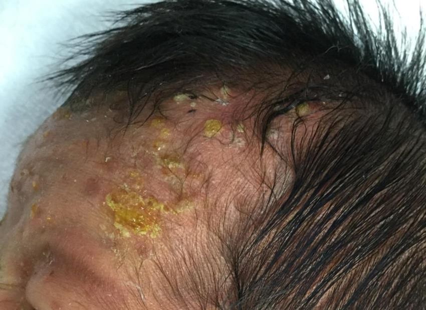
Children 2021, 8, x FOR PEER REVIEW                                                                                                 2 of 6
Children 2021, 8, 30                                                                                                                2 of 6

                                  reference range: 0–0.4 mg/dL), elevated white blood cell count (12,500/μL; reference range:
                                 range:   3400–9400/µL),
                                  3400–9400/μL),             normal eosinophil
                                                    normal eosinophil               count reference
                                                                         count (500/μL;     (500/µL;range:
                                                                                                       reference range: normal
                                                                                                             0–658/μL),   0–658/µL),
                                                                                                                                  β-D-
                                 normal   β-D-glucan
                                  glucan level          levelreference
                                                (9.9 pg/mL;    (9.9 pg/mL;    reference
                                                                         range:           range: elevated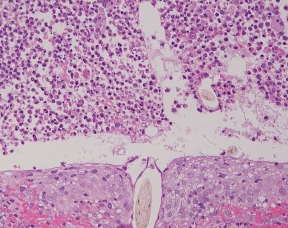
Children 2021, 8, 30 3 of 6
Children 2021, 8, x FOR PEER REVIEW 3 of 6
Children 2021, 8, x FOR PEER REVIEW 3 of 6
Figure2.2.One
Figure Oneweek
week after
after birth,
birth, the
the patient
patient developed
developed ring-shaped
ring-shaped plaques
plaquesconsisting
consistingofofpustules,
pustules,
crusts,
Figureand erythema
2. One on his
week after scalp
birth, and forehead.
the patient Pigmentation
developed was seen
ring-shaped plaques on the forehead,
consisting of pustules,consistent
crusts, and erythema on his scalp and forehead. Pigmentation was seen on the forehead, consistent
with previous
crusts, erythema
and erythema andscalp
on his pustules.
and forehead. Pigmentation was seen on the forehead, consistent
with previous
with previouserythema
erythemaand
and pustules.
pustules.
(a)
(a)
(b) (c)
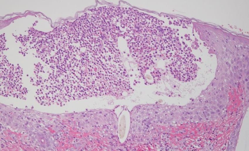
FigureFigure 3. Hematoxylin
3. Hematoxylin andand eosinstaining.
eosin staining. (a)
(a)AAskin biopsy
skin revealed
biopsy pustules
revealed beneathbeneath
pustules the stratum
the corneum,
stratum which coin-which
corneum,
cided with the epidermal (b)opening of hair follicles. Original magnification ×100. (b) A small amount (c) of inflammatory cell
coincided with the epidermal opening of hair follicles. Original magnification ×100. (b) A small amount of inflammatory
infiltration, such as eosinophils and lymphocytes, was also seen around the hair follicles in the dermis, and between the
cell infiltration,
Figuredermal such fibers.
3. Hematoxylin
collagen as eosinophils
andOriginal and lymphocytes,
eosin staining. (a) A×200.
magnification was
skin (c) Thealso
biopsy seen contained
revealed
pustules around the
pustules hair follicles
beneath the
eosinophils, in the dermis,
stratum
neutrophils, corneum, and
and hairs, between
which
sug- coin-
cided
the withcollagen
dermal
gesting the
the epidermal opening
fibers. of
destruction the hairof
Original Original×
hair follicles.
magnification
follicles. Original
200. (c) magnification
The pustules
magnification ×200. ×100. (b) Aeosinophils,
contained small amount of inflammatory
neutrophils, cell
and hairs,
infiltration,the
suggesting such as eosinophils
destruction of the and
hair lymphocytes, wasmagnification
follicles. Original also seen around
×200.the hair follicles in the dermis, and between the
dermal collagen fibers. Original magnification ×200. (c) The pustules contained eosinophils, neutrophils, and hairs, sug-
gesting the destruction of the hair follicles. Original magnification ×200.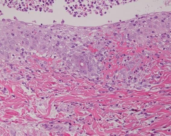
the right side (Figure 4). On the 10th day after birth, the patient’s blood test results showed
an elevated white blood cell count (12,400/μL; reference range: 3400–9400/μL) and ele-
vated eosinophil count (1240/μL; reference range: 0–658/μL), and pustules with erythema
were found on the seborrheic areas of the trunk, such as the axillae and inguinal regions.
Children 2021, 8, 30 About one month after birth, no new pustules or erythema were seen at the sites where 4 of 6
topical corticosteroid had been applied, although new eruptions were still observed on
the trunk.
(a) (b)
Figure
Figure 4.
4. (a)
(a)New
Newpustules
pustulescontinued
continued totoappear
appear on
onthe
theright
rightforehead
forehead after
after topical
topical ketoconazole
ketoconazole treatment.
treatment. (b)
(b) The
The pustules
pustules
and
and erythema
erythema disappeared
disappeared on
on the
the left forehead after
left forehead after topical
topical corticosteroid
corticosteroid treatment.
treatment.
3. Discussion
3. Discussion
To diagnose noninfectious
To noninfectious pustular
pustular diseases,
diseases, infectious
infectious pustular
pustular diseases
diseases must
must be
ruled out. Infectious pustular diseases
ruled out. Infectious pustular diseases include bullousinclude bullous impetigo, congenital cutaneous
candidiasis, herpes
candidiasis, herpes simplex virus infection, varicella, etc. [1]. These Thesediseases
diseasesare aredifferenti-
differen-
ated based
tiated basedon onaacombination
combinationof ofsystemic
systemic symptoms,
symptoms, bacterial
bacterial and fungal cultures, direct
microscopy, Tzanck
microscopy, Tzanck test,
test, and
and PCR
PCR analyses.
analyses.
ETN is a transient physiological
ETN is a transient physiological rash rash found
found in about
in about 40%40% of newborn
of newborn infants infants [2].
[2]. Such
Such rashes typically develop within 72 h after birth but are rarely
rashes typically develop within 72 h after birth but are rarely seen at the time of delivery seen at the time of
delivery [3,4]. ETN is characterized by erythema and papules
[3,4]. ETN is characterized by erythema and papules or pustules, 1–3 mm in diameter, on or pustules, 1–3 mm in
diameter, on the face, trunk, and limbs. Since it does not affect the
the face, trunk, and limbs. Since it does not affect the palms of the hands or soles of thepalms of the hands or
solesitofisthe
feet, feet, itthat
thought is thought
an immunethat an immunetoresponse
response microbial to colonies
microbialincolonies in hairisfollicles
hair follicles an im-
portant etiologic factor [5]. The individual lesions commonly disappear within a fewa
is an important etiologic factor [5]. The individual lesions commonly disappear within
few hours,
hours, without
without pigmentation
pigmentation [2,6].[2,6].
ETNETN typically
typically resolves
resolves withinwithin a week
a week and and is not
is not ac-
accompanied by systemic symptoms; therefore, no special
companied by systemic symptoms; therefore, no special treatments are required. treatments are required.
TNPM is
TNPM is common
common in in black
black neonates,
neonates, characterized
characterized by by flaccid
flaccid 1–3
1–3 mm
mm pustules
pustules on on the
the
chin, neck, trunk, and thighs beginning at birth [7]. These pustules quickly
chin, neck, trunk, and thighs beginning at birth [7]. These pustules quickly transition into transition into
pigmented macules,
pigmented macules, whichwhich usually
usually fade
fade within
within aa month
month [6,8].
[6,8]. No
No specific
specific therapies
therapies forfor
TNPM are required as it resolves spontaneously, and no systemic symptoms are associated
TNPM are required as it resolves spontaneously, and no systemic symptoms are associ-
with the lesions [6].
ated with the lesions [6].
Although a previous report proposed that TNPM and ETN may represent different
Although a previous report proposed that TNPM and ETN may represent different
points on the spectrum of the same disease [9], the pathogenic mechanisms underlying
points on the spectrum of the same disease [9], the pathogenic mechanisms underlying
TNPM remain unclear.
TNPM remain unclear.
EPF in infancy is a rare disease first reported by Lucky et al. in 1984 [10]. EPF is
a disease that causes pruritic erythema, blisters, and pustules centered on hair follicles
without systemic symptoms. The predominant site of EPF in infancy is the scalp, although
seborrheic areas of the face, trunk, and limbs may also be affected. The skin manifestations
of EPF are characterized by pustules measuring 1–3 mm accompanied by erythema, leading
to the formation of plaques and pigmented spots. Unlike adult EPF, the lesions of EPF in
infancy exhibit secondary crusting [6,11]. These eruptions follow a cyclical course from
three months to five years [11]. The best way to quickly diagnose EPF without assessment
of the clinical course is to perform a Tzanck smear of a pustule or skin biopsy. The pustules
in EPF contain large numbers of eosinophils and are accompanied by dermal infiltration
of eosinophil-dominated inflammatory cells around the hair follicles. If the lesions do
not show folliculitis on biopsy, the condition is preferentially referred to as neonatal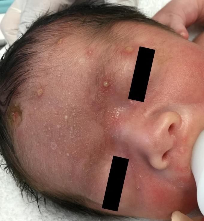
Children 2021, 8, 30                                                                                                                   5 of 6

                                   eosinophilic pustulosis (NEP) [12]. As EPF and NEP present with similar symptoms and
                                   a similar clinical course, it may not be necessary to distinguish between EPF and NEP.
                                   Topical corticosteroids were reported previously to show beneficial effects in both EPF and
                                   NEP [12].
                                         Our patient presented with many pustules but relatively minimal crusting at birth
                                   compared to typical infant cases of EPF. This may be because our case was bathed in amni-
                                   otic fluid without exposure to air at the onset of EPF. However, one week after birth, our
                                   case exhibited typical crusting pustules. According to the clinical course over one month,
                                   the negative results of pathogen tests, the findings of the skin biopsy, and the response to
                                   treatment, we could differentiate EPF from the other infectious and noninfectious pustular
                                   diseases described above.
                                         Although the cause of EPF in infancy has not been clarified, the increased number
                                   of eosinophils in the peripheral blood during attacks is suggestive of an immunological
                                   pathomechanism [10]. A clinical study of human polyomavirus 6 as a potential etiological
                                   agent for EPF in adults suggested a hypothetical association between persistent antigenic
                                   stimuli from infectious agents and tissue eosinophilia through activation of T helper 2 cells [13].
                                         EPF develops within 24 h after birth in about 25% of affected infants under one year
                                   of age; however, the symptoms are rarely seen at birth [10,11,14]. To our knowledge, only
                                   two cases of EPF since birth have been reported to date [10,14]. Both cases exhibited the
                                   same clinical presentation and course as ordinary EPF in infancy; however, these previous
                                   cases were not accompanied by any maternal medical history or drug treatments. It has
                                   not been elucidated whether maternal immune disorders and oral medications influence
                                   the pathogenesis of EPF. The mother in our case had Graves’ disease and was taking
                                   propylthiouracil and aspirin. In our case, the eruption worsened with an increase in
                                   blood eosinophil count, which may have been related to the mother’s oral medication.
                                   Several adult cases of drug-induced EPF have been reported, leading to increased blood
                                   eosinophils [15,16]. In addition, a previous study reported that patients with Graves’
                                   disease have a genetic predisposition to Th2 predominance [17]. It is well known that
                                   patients suffering from Th2-dominant diseases often show increased levels of peripheral
                                   eosinophils. Therefore, the mother’s medical history may have affected the pathogenesis
                                   of our patient’s EPF. Further case studies are required to shed light on the involvement of
                                   these conditions in the onset of the disease.

                                   Author Contributions: Conceptualization, S.Y., K.Y., Y.K., K.C. and K.T.; writing-original draft
                                   preparation, S.Y. and K.Y.; writing-review and editing, S.Y., K.Y. and K.S. All authors have read and
                                   agreed to the published version of the manuscript.
                                   Funding: This research received no external funding.
                                   Informed Consent Statement: Informed consent was obtained from all subjects involved in the study.
                                   Data Availability Statement: The data presented in this study are available on request from the
                                   corresponding author. The data are not publicly available due to privacy restrictions.
                                   Conflicts of Interest: The authors declare no conflict of interest.

References
1.    Ghosh, S. Neonatal Pustular Dermatosis: An Overview. Indian J. Dermatol. 2015, 60, 211. [PubMed]
2.    Hidano, A.; Purwoko, R.; Jitsukawa, K. Statistical Survey of Skin Changes in Japanese Neonates. Pediatr. Dermatol. 1986, 3,
      140–144. [CrossRef] [PubMed]
3.    Reginatto, F.P.; de Villa, D.; Muller, F.M.; Peruzzo, J.; Peres, L.P.; Steglich, R.B.; Cestari, T.F. Prevalence and characterization of
      neonatal skin disorders in the first 72h of life. J Pediatr. 2017, 93, 238–245. [CrossRef] [PubMed]
4.    Leung, A.K. Erythema toxicum neonatorum present at birth. J. Singap. Paediatr. Soc. 1986, 28, 163–166.
5.    Marchini, G.; Nelson, A.; Edner, J.; Lonne-Rahm, S.; Stavreus-Evers, A.; Hultenby, K.; Stavr, A. Erythema Toxicum Neonatorum
      Is an Innate Immune Response to Commensal Microbes Penetrated into the Skin of the Newborn Infant. Pediatr. Res. 2005,
      58, 613–616. [CrossRef] [PubMed]
6.    van Praag, M.C.; van Rooij, R.W.; Folkers, E.; Spritzer, R.; Menke, H.E.; Oranje, A.P. Diagnosis and treatment of pustular disorders
      in the neonate. Pediatr Dermatol. 1997, 14, 131–143. [CrossRef] [PubMed]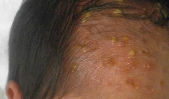
Children 2021, 8, 30                                                                                                                   6 of 6

7.    Wyre, H.W.; Murphy, M.O. Transient neonatal pustular melanosis. Arch. Dermatol. 1979, 115, 458. [CrossRef] [PubMed]
8.    Merlob, P.; Metzker, A.; Reisner, S.H. Transient Neonatal Pustular Melanosis. Arch. Pediatr. Adolesc. Med. 1982, 136, 521. [CrossRef]
      [PubMed]
9.    Ferrándiz, C.; Coroleu, W.; Ribera, M.; Lorenzo, J.; Natal, A. Sterile Transient Neonatal Pustulosis Is a Precocious Form of
      Erythema toxicum neonatorum. Dermatology 1992, 185, 18–22. [CrossRef] [PubMed]
10.   Lucky, A.W.; Esterly, N.B.; Heskel, N.; Krafchik, B.R.; Solomon, L.M. Eosinophilic pustular folliculitis in infancy. Pediatr. Dermatol.
      1984, 1, 202–206. [CrossRef] [PubMed]
11.   Buckley, D.A.; Munn, S.E.; Higgins, E.M. Neonatal eosinophilic pustular folliculitis. Clin. Exp. Dermatol. 2001, 26, 251–255.
      [CrossRef] [PubMed]
12.   Asgari, M.; Leiferman, K.M.; Piepkorn, M.; Kuechle, M.K. Neonatal eosinophilic pustulosis*. Int. J. Dermatol. 2004, 45, 131–134.
      [CrossRef] [PubMed]
13.   Hashida, Y.; Higuchi, T.; Nakajima, S.; Nakajima, K.; Ujihara, T.; Kabashima, K.; Sano, S.; Daibata, M. Human Polymavirus 6
      Detected in Cases of Eosinophilic Pustular Folliculitis. J. Infect. Dis. 2020. [CrossRef] [PubMed]
14.   Lee, J.H.; Kang, J.H.; Cho, B.K.; Park, H.J. Generalized Eosinophilic Pustular Folliculitis of Infancy Responding to Hy-droxyzine.
      Ann. Dermatol. 2015, 27, 458–460. [CrossRef] [PubMed]
15.   Ooi, C.G.; Walker, P.; Sidhu, S.K.; Gordon, L.A.; Marshman, G. Allopurinol induced generalized eosinophilic pustular fol-liculitis.
      Australas. J. Dermatol. 2006, 47, 270–273. [CrossRef] [PubMed]
16.   Kimura, K.; Ezoe, K.; Yokozeki, H.; Katayama, I.; Nishioka, K. A case of eosinophilic pustular folliculitis (Ofuji’s disease) in-duced
      by patch and challenge tests with indeloxazine hydrochloride. J. Dermatol. 1996, 23, 479–483. [CrossRef] [PubMed]
17.   Izumi, Y.; Hidaka, Y.; Tada, H.; Takano, T.; Kashiwai, T.; Tatsumi, K.-I.; Ichihara, K.; Amino, N. Simple and practical parameters
      for differentiation between destruction-induced thyrotoxicosis and Graves’ thyrotoxicosis. Clin. Endocrinol. 2002, 57, 51–58.
      [CrossRef] [PubMed]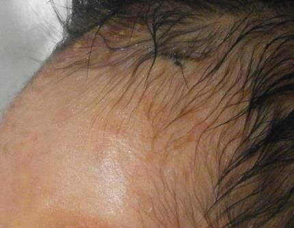
You can also read