ARROCase: Thymoma Jacob Miller, MD; Alexander Chin, MD, MBA - Stanford Hospital and Clinics
←
→
Page content transcription
If your browser does not render page correctly, please read the page content below
ARROCase: Thymoma

                Jacob Miller, MD; Alexander Chin, MD, MBA
                       Stanford Hospital and Clinics
                               Stanford, CA

June 19, 2020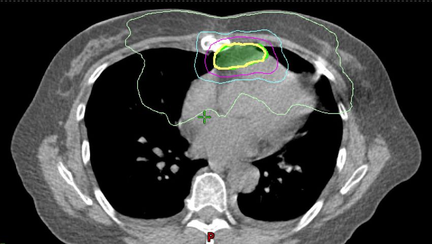
Clinical Presentation
•    58 year-old woman presents to an ophthalmologist with ptosis, diplopia,
     malaise, and muscle weakness with chewing.
      – PMH/PSH: None.
      – FH: No family history of malignancy or neuromuscular disease.
      – SH: Married, works as insurance agent. Never smoker, no alcohol/drug
         use.
      – Medications/allergies: Non-contributory.
      – Physical Examination: +Diplopia, bilateral ptosis. Mild proximal muscle
         weakness.
      – Suspecting myasthenia gravis, she is referred to a neurologist.

June 19, 2020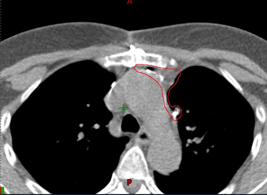
Workup
•    Neurology consultation
      – AChR modulating, blocking, and binding antibodies were each
        elevated.
      – A clinical diagnosis of myasthenia gravis was made, and she was
        started on prednisone and pyridostigmine with symptomatic
        improvement.
      – Prednisone was later tapered and mycophenolate was started.
•    Imaging was ordered to rule out thymoma
      – Chest x-ray demonstrated a left anterior mediastinal mass.
      – Chest CT+C demonstrated an anterior mediastinal mass measuring
        6.6x5.3x2.4cm without gross invasion of the lung or heart and without
        nodal, pleural, or intrathoracic metastases.

June 19, 2020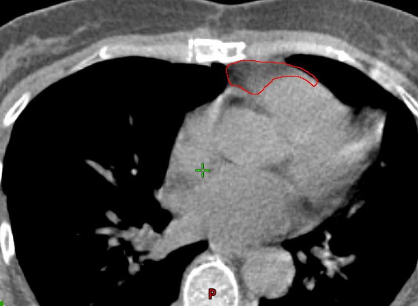
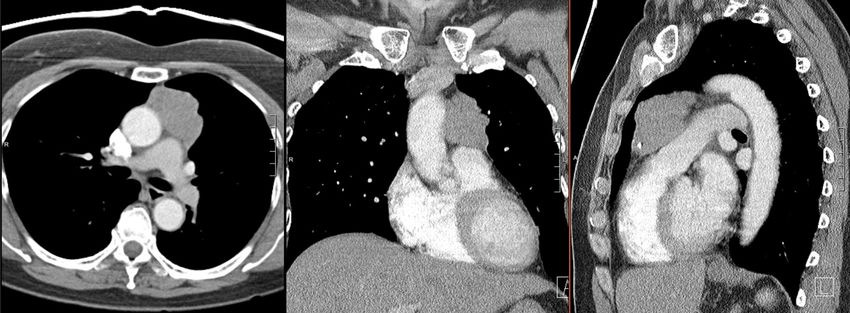
Workup
•    Chest X-Ray demonstrated a left-sided mass obscuring the aortic contour in the
     anterior mediastinum.

June 19, 2020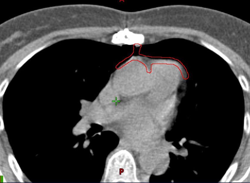
Workup
•    Chest CT demonstrated a left-sided anterior mediastinal mass measuring
     6.6x5.3x2.4cm without gross invasion of the lung, pleura, or heart, and without
     nodal, pleural, or intrathroracic metastases.

June 19, 2020
Workup
• Further workup:
– CBC/CMP unremarkable
– Pulmonary function testing demonstrated FVC 3.56L (85% predicted), FEV1
2.68 (81% predicted), suggesting possible restrictive process.
– AFP, beta-HCG were not elevated.
• Consultation with thoracic surgery:
– Myasthenic symptoms had resolved with pyridostigmine/prednisone.
– The anterior mediastinal tumor was clinically consistent with a resectable
thymoma, and initial biopsy was not indicated.
– Recommendation was for a median sternotomy and total thymectomy.
• Differential diagnosis (anterior mediastinal mass)
– Thymoma, thymic carcinoma, thymic cyst, carcinoid, thymic lipoma,
seminoma, germ cell tumor, teratoma, lymphoma, enlarged/ectopic thyroid
June 19, 2020Resection
• Resection:
– After median sternotomy, the mass encased the left phrenic nerve and was adherent to
the left upper lobe and anterolateral (left) pericardium
– Total thymectomy, en bloc resection of the left phrenic nerve, partial pericardiectomy,
and en bloc left upper lobe wedge resection were performed for gross total resection.
– Uncomplicated postoperative course
• Surgical Pathology:
– Histology: thymoma, WHO Type B2, 5.5cm. Lymphocytes admixed with lesional cells. [1]
– Extent: transcapsular invasion with involvement of the visceral pleura, parietal pleura,
and pericardium
– Margins: microscopic positive posterior margin (R1), 0.1cm to right+left lateral margins
– Modified Masaoka stage: stage IIIA (+pericardial/pleural invasion, no great vessel
invasion) [2]
– AJCC 8th edition stage: pT2 cN0M0 (II)
• Post-discharge follow-up: no dyspnea despite left phrenic nerve sacrifice.
June 19, 2020Adjuvant Therapy
• Radiotherapy: [2,3]
– Given extent (Masaoka stage IIIA) and microscopic positive margin, adjuvant
radiotherapy was recommended.
– Simulation:
• Supine, arms over head, vac bag, IV contrast
• 4DCT, inspiration breath hold, expiration breath hold CT with 2mm slice
thickness
– Technique: volumetric-modulated arc radiotherapy
June 19, 2020Adjuvant Therapy
• Radiotherapy: [2,3]
– Target volumes:
• Fuse pre-resection imaging and contour pre-resection GTV
• Postoperative CTV encompasses entire surgical bed, clips, anterior
mediastinum, and areas of pericardial/pleural contact with the preoperative
GTV at risk for microscopic disease. Discussion with surgeon encouraged.
• Motion-inclusive ITV vs. inspiration breath hold
• Elective nodal irradiation not indicated
• CTV-to-PTV margin dictated by image-guidance and LINAC tolerance (5mm)
– Prescription dose:
• 54 Gy in 30 fractions prescribed to cover 95% of PTV
• Motion management/IGRT: daily CBCT with respiratory gating during
treatment to exclude extreme breaths vs. inspiration breath hold
• Chemotherapy: not indicated
June 19, 2020Postoperative Radiotherapy: CTV June 19, 2020
Postoperative Radiotherapy: CTV

June 19, 2020
Postoperative Radiotherapy: CTV

June 19, 2020
Postoperative Radiotherapy: Plan
    PTV
    CTV

    100% IDL – 54 Gy
    75% IDL – 40.5 Gy
    50% IDL – 27 Gy
    20% IDL – 10.8 Gy

June 19, 2020
Postoperative Radiotherapy: Plan
PTV
CTV
100% IDL – 54 Gy
75% IDL – 40.5 Gy
50% IDL – 27 Gy
20% IDL – 10.8 Gy
June 19, 2020Postoperative Radiotherapy: Plan
PTV
CTV
100% IDL – 54 Gy
75% IDL – 40.5 Gy
50% IDL – 27 Gy
20% IDL – 10.8 Gy
June 19, 2020Postoperative Radiotherapy: Plan
PTV
CTV
100% IDL – 54 Gy
75% IDL – 40.5 Gy
50% IDL – 27 Gy
20% IDL – 10.8 Gy
June 19, 2020Postoperative Radiotherapy: Plan
PTV
CTV
100% IDL – 54 Gy
75% IDL – 40.5 Gy
50% IDL – 27 Gy
20% IDL – 10.8 Gy
June 19, 2020Postoperative Radiotherapy: Plan
• VMAT plan:
– Fields: Two arcs, 6 MeV, 54 Gy in 30 fractions prescribed to 95% PTV coverage
– Lungs: Mean 9.7 Gy; V30=6%; V20=12%; V5=61%
– Heart: Mean 8.9 Gy; V30=9%; V10=25%
– Esophagus: Dmax 32 Gy; mean 12 Gy
– Great vessels: D0.03ccRadiotherapy Course and Follow-Up
• She experienced grade 1 fatigue during radiotherapy, with no dyspnea,
esophagitis, or weight loss.
• A surveillance chest CT with contrast was completed six months after radiotherapy
per NCCN guidelines, with no evidence of intrathoracic recurrence.
– Recurrence patterns defined in Gomez et al. [3]
• NCCN guidelines for follow-up: [2]
– Chest CT with contrast every 6 months for the first two years, then annually
for 5 years (thymic carcinoma) or 10 years (thymoma)
June 19, 2020Thymic Masses: Presentation
• Symptoms:
– Myasthenic symptoms: ptosis, diplopia, dysphagia, difficulty chewing, dysarthria,
hypophonia, facial weakness, dyspnea. 30-50% of patients with thymomas have
myasthenic symptoms. 10-20% of patients with myasthenia gravis have a thymoma.
– Mass effect: dyspnea, chest pain, cough, odynophagia, SVC syndrome,
pleural/pericardial effusions, restrictive lung physiology
– Phrenic nerve involvement: dyspnea, diaphragmatic paralysis
– Other paraneoplastic syndromes: variety of other common and uncommon
autoimmune diseases have been associated with thymomas, including pure red cell
aplasia, immunodeficiencies, and thymoma-associated multiorgan autoimmunity.
• Management: short- and long-term immunosuppression, thymectomy,
pyridostigmine/IVIG (MG), supportive transfusions (PRCA). Thymectomy alone may
not reverse these syndromes.
• Epidemiology: [2]
– Incidence: 1.5 cases per million, similar incidence between men and women
– Most common adult primary thymic neoplasm
– Highest incidence between 40-60 years of age
June 19, 2020Thymic Masses: Work-Up
• NCCN 1.2020 recommended work-up: [2]
– Required: Chest CT with contrast, beta-HCG+AFP (rule out germ cell tumors),
CBC/CMP, AChR antibodies
– Optional: PFTs, PET/CT, MRI (for equivocal CT, may help distinguish thymoma
vs. thymic carcinoma vs. thymic cyst vs. other histologies)
– Biopsy:
• Upfront resection without biopsy can be pursued if a primary thymic neoplasm is felt to be
likely (well-defined anterior mediastinal mass, negative beta-HCG/AFP, absence of adenopathy,
absence of continuity with thyroid).
• For unresectable tumors or if there is uncertainty regarding histology, core biopsy should be
performed (CT-guided, open, or thoracoscopic). Thoracentesis and cytology can also be
pursued to establish diagnosis.
June 19, 2020Thymic Neoplasms: Classification
WHO Type Muller-Hermelink Levine and Rosai Distribution
Type A Medullary type thymoma Encapsulated 4-7% (17% MG*)
Type AB Mixed type thymoma Encapsulated 28-34% (16% MG)
Type B1 Predominantly cortical Malignant type I 9-20% (57% MG)
Type B2 Cortical type Malignant type I 20-36% (71% MG)
Type B3 Well-differentiated Malignant type I 10-14% (46% MG)
carcinoma
Type C Thymic carcinoma Malignant type II 5-10% (Thymic Neoplasms: Staging
Masaoka-Koga Staging [2] 5-Year OS [7]
I Macroscopically encapsulated, no microscopic transcapsular invasion 96%
IIA Microscopic transcapsular invasion
IIB Macroscopic invasion into surrounding fatty tissue, or grossly adherent to but not 86%
breaking through mediastinal pleura or pericardium
IIIA Macroscopic invasion of neighboring organ (e.g., pericardium or lung) without great
vessel invasion
69%
IIIB Macroscopic invasion of neighboring organ (e.g., pericardium or lung) with great
vessel invasion
IVA Pleural or pericardial dissemination
50%
IVB Lymphogenous or hematogenous metastasis
June 19, 2020Thymic Neoplasms: Staging
AJCC 8th Edition Staging
T category TX: primary tumor cannot be assessed
T0: no evidence of primary tumor
T1: tumor encapsulated or extending into mediastinal fat
T2: direct invasion of the pericardium (partial or full-thickness)
T3: direct invasion into any of the following: lung, brachiocephalic vein, superior vena cava,
phrenic nerve, chest wall, or extrapericardial pulmonary artery or veins
T4: invasion into any of the following: aorta, arch vessels, intrapericardial pulmonary
artery, myocardium, trachea, esophagus
N category NX: regional nodes cannot be assessed
N0: no regional nodal metastases
N1: metastasis in anterior (perithymic) lymph nodes
N2: metastasis in deep intrathoracic or cervical lymph nodes
M category M0: no pleural, pericardial, or distant metastases
M1a: separate pleural or pericardial nodule(s)
M1b: pulmonary intraparenchymal nodule or distant organ metastasis
Group Stage I: T1N0M0
II: T2N0M0
IIIA: T3N0M0
IIIB: T4N0M0
IVA: N1 or M1a
IVB: N2 or M1b
June 19, 2020Management
• Given rarity, there is no randomized evidence to guide management.
• Resectable tumors (with or without initial biopsy) should proceed to initial resection by a
team with experience in the management of thymic neoplasms. [2]
– Myasthenic symptoms should be managed and optimized prior to resection with
immunosuppression, pyridostigmine, and/or IVIG.
• Initially unresectable tumors should first be treated with chemotherapy +/- radiotherapy. [2]
– Potentially-resectable tumors: chemotherapy → restaging
• Resectable after restaging: resection +/- PORT
• Unresectable after restaging: definitive RT +/- chemotherapy
– Unresectable tumors: concurrent chemoradiotherapy
• Systemic therapy: [2]
– First-line thymoma: CAP q3 weeks (cisplatin, doxorubicin, cyclophosphamide)
– First-line thymic carcinoma: Carboplatin/paclitaxel q3 weeks
– Second-line thymoma: everolimus, octreotide, pemetrexed, gemcitabine
– Second-line thymic carcinoma: sunitinib, pemetrexed, everolimus, pembrolizumab
– Concurrent chemotherapy: cisplatin+etoposide or carboplatin+paclitaxel
June 19, 2020Management: PORT
• Controversial. Given rarity, there is no randomized evidence to guide management. [2-4]
– Masaoka stage I, R0: no PORT
– Masaoka stage II, R0: consider PORT for high-risk features (e.g., large size, WHO type B3/C)
– Masaoka stage III: PORT
– Masaoka stage IV: individualized based on resectability, symptoms
– R1-2 resection: PORT +/- chemotherapy (e.g., for R2 resection or thymic carcinoma)
– Thymic carcinoma: PORT (even if stage I-II)
• Conflicting evidence for LC, DFS, and OS benefit in different subgroups
– NCDB (PMID: 28126540): PORT improved OS for Masaoka stage IIB, III, and positive margins. No
SS benefit for OS among stage I-IIA [5]
– Japanese Consortium (PMID: 25565590): PORT improved RFS but not OS for stage II-III thymic
carcinoma, and did not improve RFS or OS for stage II-III thymoma. [6]
– ITMIG (PMID: 27346413): PORT improved OS in stage II-III R0 thymoma. [7]
– Meta-analysis (PMID: 27026316): PORT improved OS in stage III/IV but not stage II thymoma. [8]
– All observational series are subject to selection biases in PORT vs. no PORT cohorts.
June 19, 2020Management: PORT
• Treatment planning: consensus atlas is not available, but reporting guidelines exist [3], with lower inter-
rater agreement in postoperative cases relative to definitive cases [9]
• Radiotherapy:
– Target volumes: [3]
• Fuse pre-resection imaging and contour pre-resection GTV
• Postoperative CTV encompasses entire surgical bed, clips, anterior mediastinum, and areas of
pericardial/pleural contact with the preoperative GTV at risk for microscopic disease.
Discussion with surgeon encouraged.
• Motion-inclusive ITV vs. breath hold
• Elective nodal irradiation not recommended by NCCN
• CTV-to-PTV margin dictated by image-guidance and LINAC
– Prescription dose:
• R0: 45-50 Gy at 1.8-2 Gy per fraction
• R1: 54 Gy at 1.8-2 Gy per fraction
• R2: 60-70 Gy at 1.8-2 Gy per fraction, similar to unresectable disease
• Hemithoracic RT with boost to high-risk areas is rarely used [3]
– Motion management: inspiration breath hold vs. respiratory gating during treatment to exclude
extreme breaths with daily CBCT.
• Postoperative chemotherapy: can be considered for thymic carcinoma or R2 resection
June 19, 2020Management: Unresectable Disease
• Potentially-resectable tumors: induction chemotherapy → resection (if feasible) → risk-
adapted PORT [10]
• Unresectable tumors / R2 resection: Definitive concurrent chemoradiotherapy [2]
– Target volumes (definitive):
• Fuse pre-radiotherapy CT/MRI/PET and contour GTV
• No routine GTV-to-CTV expansion, but CTV should include areas of
pericardial/pleural contact with the GTV at risk for microscopic disease. If
chemotherapy precedes radiotherapy, CTV should include pre-chemotherapy
extent of disease adapted to anatomy at time of simulation.
• Motion-inclusive ITV
• Elective nodal irradiation not recommended by NCCN
• CTV-to-PTV margin dictated by image-guidance and LINAC
– Prescription dose: 60-70 Gy at 1.8-2 Gy per fraction
– Motion management: inspiration breath hold vs. respiratory gating during treatment to
exclude extreme breaths with daily CBCT
June 19, 2020Management: Unresectable Disease
• Potentially-resectable tumors: induction chemotherapy → resection (if feasible) → risk-
adapted PORT [10]
• Unresectable tumors / R2 resection: Definitive concurrent chemoradiotherapy [2]
– Evidence:
• Kim et al: phase II trial of 22 patients with unresectable thymoma treated with CAP
q 3-4 weeks x 3 → surgical resection (76% R0) → PORT → CAP q 3-4 weeks x 3 [11]
• Loehrer: phase II trial of 26 patients with unresectable thymoma treated with CAP q
3 weeks x 2-4 → definitive radiotherapy (54 Gy) [12]
• Fan et al: phase II trial of 56 patients with unresectable thymoma/thymic carcinoma
treated with definitive chemoradiotherapy (60 Gy) with concurrent EP q 4 weeks x
2 → adjuvant EP q 4 weeks x 2 [13]
• Concurrent chemotherapy:
– Generally indicated for suitable candidates in the definitive setting
– Cisplatin+etoposide or carboplatin+paclitaxel
June 19, 2020Surveillance
• NCCN 1.2020: Chest CT+contrast every 6 months for 2 years, and then annually for
5 years for thymic carcinomas and 10 years for thymomas.
• Late toxicity: given long life expectancy in most cases, late toxicities can include
pneumonitis, dyspnea, cardiac toxicity, and secondary malignancies.
• Recurrence: [2]
– Most common site of recurrence is along the pleura/pericardium.
– Nodal recurrence is uncommon, but thymic carcinomas may metastasize to
the bone, liver, kidneys, and lymph nodes.
– Resection of limited pleural/pericardial metastases can lead to long-term
disease control, with prognosis associated with WHO Grade. [14]
June 19, 2020References
• [1] Tumours of the lung, pleura, thymus and heart. In: World Health Organization Classification of tumours, Travis WD,
Brambilla E, Muller-Hermelink HK, Harris CC (Eds), IARC Press, Lyon, France 2004.
• [2] NCCN Clinical Practice Guidelines in Oncology. Thymomas and Thymic Carcinomas. Version 1.2020. November 2019.
• [3] Gomez et al. J Thorac Oncol . 2011 Jul;6(7 Suppl 3):S1743-8. PMID: 21847057.
• PORT:
– [4] Ahmad et al. J Thorac Cardiovasc Surg. 2015;149(1):95. Epub 2014 Oct 5. PMID: 25524678.
– [5] Jackson et al. J Thorac Oncol. 2017;12(4):734. Epub 2017 Jan 25. PMID: 28126540.
– [6] Omasa et al. Cancer. 2015;121(7):1008. Epub 2015 Jan 6. PMID: 25565590.
– [7] Rimner et al. J Thorac Oncol. 2016;11(10):1785. Epub 2016 Jun 23. PMID: 27346413.
– [8] Lim et al. Int J Radiat Oncol Biol Phys . 2016 Apr 1;94(5):1129-36. PMID: 27026316.
– [9] Holliday et al. J Radiat Oncol. 2016 Mar; 5(1): 55–61. PMID: 27570583.
• Unresectable Disease:
– [10] Hamaji et al. Ann Thorac Surg. 2015;99(5):1848. PMID: 25770014.
– [11] Kim et al. Lung Cancer 2004 Jun;44(3):369-79. PMID: 15140551.
– [12] Loehrer et al. J Clin Oncol. 1997 Sep;15(9):3093-9. PMID: 9294472.
– [13] Fan et al. Int J Radiat Oncol Biol Phys. 2020 May 1;107(1):98-105. PMID: 31987968.
• Recurrent Disease:
– [14] Okumura et al. J Surg Oncol. 2007;95(1):40. PMID: 17192865.
• ESMO Thymic Guidelines: [15] Ann Oncol . 2015 Sep;26 Suppl 5:v40-55. PMID: 26314779
Please provide feedback regarding this case or other ARROcases to arrocase@gmail.com
June 19, 2020You can also read