Assessment of Canine Elbow joint for osteoarthritis - and treatment with Synovetin OA
←
→
Page content transcription
If your browser does not render page correctly, please read the page content below
Assessment of
Canine Elbow joint
 for osteoarthritis
and treatment with Synovetin OA®
              Steven M. Fox, MS, DVM, MBA, PhD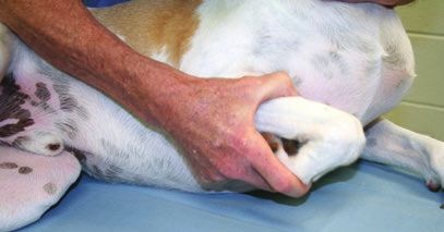
PAIN
Pain is the clinical sign most frequently associated with osteoarthritis (OA).1 The clinical manifestation
of this pain is lameness. When an animal presents with clinical lameness, a determination must be made
whether the animal is unable to use the limb or is unwilling to use the limb. Inability to use the limb may be
attributable to musculoskeletal changes, such as joint contracture or muscle atrophy. These anomalies are
best addressed with physical rehabilitation. On the other hand, unwillingness to use a limb is most often
attributable to pain. Herein, lameness is an avoidance behavior.
Ironically, articular cartilage is frequently the focus of studies regarding OA. However, clinical treatment
of the OA patient is most often focused on the alleviation of pain. Appreciating that articular cartilage is
aneural, the focus of OA pain management resides in the periarticular structures. No pain is elicited by
stimulation of cartilage, and stimulation of normal synovial tissue rarely evokes pain.2
OA pain is the result of a complex interplay between structural change, biochemical alterations,
peripheral and central pain-processing mechanisms, and individual cognitive processing of nociception.
The source of pain in the joint ‘organ’ is multifocal: direct stimulation of the joint capsule and bone
receptors by cytokines/ligands of inflammatory and degradative processes, physical stimulation of the
joint capsule from distension (effusion) and stretch (laxity, subluxation, abnormal articulation), physical
stimulation of subchondral bone from abnormal loading, and (likely) physical stimulation of muscle,
tendon, and ligaments. Bony changes at the joint margins and beneath areas of damaged cartilage can
be major sources of OA pain. Subchondral bone contains unmyelinated nerve fibers, which increase in
number with OA.3 Increased pressure on subchondral bone (associated with OA) results in stimulation
of these nociceptors. This is thought to contribute to the vague but consistent pain frequently
associated with OA. In humans, OA is believed to be responsible for increased intraosseous pressure,
which may contribute to chronic pain, particularly nocturnal pain. Human OA patients report pain, even
at rest, associated with raised intraosseous pressure.4

LAMENESS
Most often lameness in pets is identified by the owner, who subsequently seeks further consultation
and advice from their veterinarian, or is identified by the veterinarian during routine examination.
Most simply, dogs (and cats) are lame because they cannot or will not use one or more limbs in a normal
fashion. Pain associated with OA is recognized to become more persistent and intense as the disease
progresses. The condition may be asymptomatic in the early stages. With progression of the disease,
discomfort may be continuous or exacerbated by motion and weight bearing. In the later stages of OA,
pain can become pervasive and affect nearly all activities and behaviors.

DIAGNOSIS OF OA
A proper diagnosis depends on a complete history and full assessment of the patient, possibly including:
• A complete physical, orthopedic, and neurologic examination
• Radiographs of affected area(s)
• Advanced imaging, such as computed tomography, magnetic resonance imaging, nuclear scintigraphy
• Advanced gait analysis, such as force plate (kinetic) analysis of gait and motion (kinematic) analysis
• Clinicopathologic examination, including hematology and serum chemistries, especially creatine
  kinase and electrolytes, and synovial fluid analysis

                                                                                                                 2
• Electrodiagnostic testing: ultrasound, electromyography, nerve conduction velocity measurements,
  and potential recordings with repetitive nerve stimulation
• Muscle biopsy examination, including histopathology and histochemical analysis
• Special tests: muscle percussion, serology for pathogens (e.g. Neospora, Toxoplasma), measurement
  of acetylcholine receptor antibody, immunohistochemistry, and molecular diagnostic techniques

ANAMNESIS
The medical history, signalment, and owner’s complaint(s) comprise the process of anamnesis. Most
canine patients do not vocalize from their pain of OA, and many pet owners do not believe their pet
is in pain if it does not vocalize. Nevertheless, signs suggesting animal discomfort include lameness,
muscle atrophy, reluctance to exercise, general malaise, lethargy, inappetence or anorexia, change
in temperament, licking or biting an affected joint, restlessness, insomnia, seeking warmth, seeking
comfortable bedding, and difficulty posturing to toilet. Supraspinal influences are known to alter the
behavior of humans with OA1, and it is reasonable to presume the same occurs in dogs.
Pet owners often recognize lameness only when there is gait asymmetry; however, dogs with bilateral
OA, such as with hip or elbow dysplasia, have a symmetrically abnormal gait and may not favor a single
limb. These patients shift weight from hind to forelimbs or vice versa with resultant muscle atrophy
of the affected limbs and increased development in compensating limbs. Rarely are dogs nonweight
bearing simply due to OA. Pet owners do often report that their dog is stiff after resting, particularly
following strenuous exercise, but they report that the pet will ‘warm out of the stiffness’. The amount
of time required to warm out of this stiffness gradually increases with progression of the disease. Pet
owners also frequently report a shortened stride and stiff gait. This is associated with a decreased
range of motion (ROM) in the joint, often due to joint capsule fibrosis and osteophyte formation.

EXAMINATION
For many years degenerative joint disease (DJD) (often used interchangeably with the term OA) was
considered a disease of the cartilage. DJD is most appropriately considered a disease of the entire
joint, with the influence of multiple structures including articular cartilage. Pain is a hallmark of DJD,
provoked by instability; therefore a comprehensive physical examination is the essential diagnostic tool.
An orthopedic examination should be part of every routine examination and should be conducted in
conjunction with a neurologic examination (when appropriate) to identify neurologic causes for pain
or lameness, such as a nerve root signature sign secondary to a laterally herniated intervertebral
disc (IVD) or brachial plexus pathology.
A consistent ‘routine’ for examining a patient is advised, and it is also recommended that the ‘lame’ limb
be examined last. A consistent examination pattern (e.g., distal limb to proximal limb, and left side to
right side or vice versa) is helpful to avoid missing a structure during the examination, and leaving the
most painful limb for last in the examination avoids the early elicitation of pain which may render the
patient noncompliant for further examination. A thorough examination also requires the aid of an assis-
tant who is adequately trained to hold and restrain the animal. The assistant is also important for identify-
ing the animal’s painful response to examination, such as body shifts and change of facial expression.

                                                                                                                3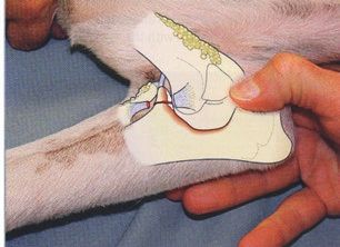
Animal restraint
Appropriate animal restraint by the assistant (with the patient standing on the examination table) is with
one arm over or under the patient’s trunk, while the other arm is placed under and around the patient’s
neck (Figure 1A). This constraint allows the assistant to quickly tighten his/her grip to control the animal
and avoid the patient from harming anyone, should it become confrontational. In lateral recumbency,
the assistant should be at the animal’s dorsum, ‘lightly leaning’ on the animal with his/her forearms while
holding the hind and forelimbs (Figure 1B). One forearm should be placed on the animal’s neck, with
that hand grasping the forelimb that is closest to the table, or the ‘down limb’. The other arm is placed
over the top of the abdomen and the hand grasps the ‘down’ hindlimb. With this restraint, the assistant
can rapidly increase the amount of weight on his/her forearms, thereby controlling the animal’s
movements. Regarding restraint, large dogs are analogous to horses: if you control their head, you
control their body.

                                                                  Figure 1A and 1B: Restraint for examination.
                                                                  Standing restraint (A) of large dogs is done with
                                                                  the neck cradled close to the assistant’s chest with
                                                                  one arm, while the other arm controls the patient’s
                                                                  trunk by placement either under or over the trunk.
                                                                  If the patient struggles or becomes aggressive,
                                                                  the assistant holds the dog as tightly as possible.
                                                                  Lateral restraint (B) of large dogs is done with
                                                                  the assistant’s forearm over the dog’s neck. If
                                                                  the patient struggles, more weight is applied
                                                                  on his/her forearm.

THE ORTHOPEDIC EXAMINATION

Forelimb examination
In the growing dog, forelimb lameness differentials mostly reflect abnormal stressors on normal bone
or normal stressors on abnormal bone (excluding fractures and minor soft tissue injuries) and include:
• Osteochondritis dissecans (OCD): shoulder
• Luxation/subluxation: shoulder (congenital)
• Avulsion: supraglenoid tubercle
• OCD: elbow
• Ununited anconeal process (UAP)
• Fragmented coronoid process (FCP)
• Ununited medial epicondyle
• Elbow incongruity:
  - congenital
  - physical injury

                                                                                                                         4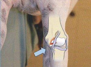
• Premature closure of growth plates, such as with radius curvus
• Retained cartilaginous core (distal ulna)
• Panosteitis* (a disease of diaphyseal bone)
• Hypertrophic osteodystrophy*

In the adult dog, forelimb lameness differentials mostly reflect abnormal stressors on normal bone
or normal stressors on abnormal bone (excluding fractures and minor soft tissue injuries) and include:
• Arthritis
• OCD: shoulder
• Luxation/subluxation: shoulder
• Fragmented Coronoid Process (FCP)
• Ununited medial epicondyle
• Elbow incongruity
• Angular limb deformity
• Hypertrophic osteopathy
• Bone/soft tissue neoplasia*
• Inflammatory arthritis
* Denote pathology/disease conditions which are not considered OA but often manifest similar clinical presentations.

For the purpose of examination, the forelimb can be anatomically segmented into the paw, antebrachium,
brachium, scapula, and interpositional joints. Although the entire limb should be examined in every
patient, the orthopedic examination can be focused more on areas prone to disease and signalment of
the individual patient.

Paw
The paw should be thoroughly examined with flexion and extension of each digit, as well as inspection
of each nail and nail bed. Findings incidental to those suggesting OA might include:
• Pad lacerations
• Foreign bodies
• Split nails
• Overgrown nails
• Nail bed tumors
• Phalangeal luxations/fractures
Some patients resist manipulation of the paws. Here, the assistant can be very helpful by talking to the
patient or scratching the patient to distract him/her from the examination.

                                                                                                                       5
Carpus
The carpus should be placed under stress in flexion, extension, valgus, and varus (Figures 2-5). The
normal carpus should flex comfortably until the palmar surface of the paw nearly touches the flexor
surface of the antebrachium. Findings from the carpal examination may include:
• Young dog ‘carpal laxity syndrome’
• Carpal flexural deformity of young dogs
• Degenerative joint disease
• Hyperextension
• Inflammatory arthritis
• Luxation
• Fracture (including an intra-articular fracture, possibly mistaken as OA)

Figure 2: Carpus flexion. The carpus should be comfortably flexed with the         Figure 3: The carpus should be stressed in
palmar surface nearly touching the flexor surface of the antebrachium.             extension, looking for signs of discomfort/pain.

Figure 4: Placing the carpus in valgus stress identifies integrity of the medial   Figure 5: Placing a varus stress on the carpus
radial collateral ligament.                                                        challenges integrity of the lateral ulnar collateral
                                                                                   ligament.

Joint capsule distension is easily palpated and suggests joint inflammation.

                                                                                                                                          6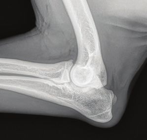
Antebrachium
Periosteum of bone is a sensitive tissue, well innervated with
nociceptive axons. Therefore, examination of both the radius
and ulna should focus on deep palpation for a response of
bone pain (Figure 6). Panosteitis is commonly revealed in this
manner. Osteosarcoma is another condition that results in
pain on palpation of the metaphyseal region of bones. Although
an orthopedic examination would include assessment of
the antebrachium, OA includes only diarthrodial joints.
Nevertheless, joint pain should be localized and differentiated   Figure 6: Digital palpation is made on the
from the pain of long bones and soft tissues.                     anteromedial aspect of the antebrachium,
                                                                  where there is minimal muscle cover. In the
Physeal disturbances are relatively common in the growing         normal dog, the elbow joint is parallel to the
dog, the severity of which depends on the amount of growth        carpal joint.
remaining following injury until physeal closure. Resultant
aberrant growth is expressed as angular limb deformities of
the carpus and/or the elbow. In general, the plane of the elbow
joint should be parallel to the plane of the carpal joint.
Sources of lameness within the radius/ulna include:
• Hypertrophic osteopathy
• Angular limb deformities
• Panosteitis

Elbow
The elbow is the most common forelimb joint responsible for
lameness, especially in growing dogs of predisposed breeds
(i.e., large breeds, sporting dogs, and Rottweilers). The elbow
should be manipulated through a complete ROM (Figure 7),
noting the abnormal presence of crepitus or painful response,
particularly in full extension. In a normal dog, hyperextension
of the elbow should elicit minimal to no discomfort.
Valgus and varus stresses placed upon the joint are performed     Figure 7: Examination of the elbow joint
to assess integrity of the joint capsule and collateral           includes manipulation through a full range of
                                                                  motion, as well as pronation and supination of
ligaments/tendons. Joint effusion accompanying disease            the antebrachium.
often distends the joint, palpable by placement of the thumb
and index finger in the normally concave depression caudal
to the distal humeral epicondyles.

                                                                                                                   7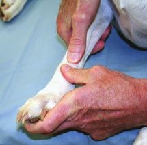
Common orthopedic diseases of the elbow joint include FCP, UAP, and OCD. Palpation of the medial
joint, in the area of the medial coronoid, often elicits a painful response in dogs suffering from any (or all)
of these conditions (Figures 8 and 9).

Figure 8: Fragmented medial coronoid, ununited anconeal       Figure 9: Osteochondritis dissecans of the elbow most
process, and osteochondritis are common diseases of the       commonly occurs on the distal, medial humeral trochlea
elbow, constituting elbow dysplasia. Patients with any of     (condyle).
these pathologies often resent deep digital pressure on the
medial aspect of the joint near the affected location.
Further, joint capsule distension is common with any of
these conditions and can best be identified with palpation,
as with the thumb placement in the figure.

Less common findings of the elbow, aside from OA, include:
• Subluxations or luxation (can be associated with OA)
• Fractures
• Radioulnar incongruities (can be associated with OA)
• Inflammatory arthropathies
• Neoplasia

Brachium
Osteosarcoma is a common tumor of the forelimb, frequently residing in the proximal humerus (and
distal radius/ulna). Deep palpation along the length of the humerus is conducted to reveal evidence of
pain and areas of inflammation or swelling. Other abnormal conditions of the brachium (not associated
with OA) include:
• Hypertrophic osteodystrophy
• Fractures
• Hypertrophic osteopathy
• Panosteitis

                                                                                                                       8
Shoulder
As with examination of all joints, the shoulder joint should be examined through a full ROM to include
flexion, extension, adduction, and abduction, as well as internal and external rotation (Figure 10). Of
particular note is examination of the shoulder joint in extension. The examiner should be mindful to
avoid placing the forelimb into extension with his/her hand placed caudal to the elbow joint (Figure 11).
Placing the hand behind the elbow when forcing the shoulder into extension also forces the elbow into
extension. A resultant painful response from the patient might actually be from elbow disease rather
than shoulder disease. The examiner’s hand placed above the elbow allows the elbow to be placed in
a neutral position and avoids this complication.
Figure 10: Examination of the shoulder in flexion. The shoulder Figure 11: Avoid placing the forelimb in extension with hand
joint should also be assessed in abduction and adduction. Note placement caudal to the elbow. This typically causes simultaneous
restraint of the patient with the assistant’s forearm over the hyperextension of the elbow and may give a false impression that
patient’s neck. the source of discomfort is in the shoulder joint when it may
reside in the elbow joint.
Painful conditions associated with the shoulder joint include:
• OCD (especially in young animals, which can lead to OA).
• Biceps tenosynovitis
• Mineralization of the supraspinatus
• Infraspinatus contracture
• DJD (of unknown etiology)
• Articular fractures
• Incomplete ossification of the caudal glenoid process
• Medial shoulder instability (leading to OA)
• Luxation, either congenital or acquired (leading to OA)
Stabilization of the shoulder joint is maintained by both medial and lateral glenohumeral ligaments, the
shape of the articular surfaces (humeral head and glenoid), and musculotendinous units of the rotator
cuff: the supraspinatus, infraspinatus, teres minor, and subscapularis. Abnormal excursion of the
shoulder joint, with or without pain, suggests involvement of several of these periarticular soft tissue
structures. Medial shoulder instability typically results in excessive shoulder abduction as well as pain
at the end of abduction.
9Scapula
The scapula is not a common source of forelimb pain.
However, atrophy of scapular muscles is frequently
associated with disuse of the forelimb as well as many
neurologic conditions. Tumors, acromion fractures, midbody
fractures, and scapular luxation from the thoracic wall are
commonly seen when pain is localized to the scapular area,
so deep palpation and manipulation of the scapula should
be performed when this anatomic structure is suspect.
The biceps tendon should be palpated from its origin on
the supraglenoid tubercle through its excursion within the Figure 12: Examination of the shoulder joint
with superimposed arthrology. A ‘drawer
intertubercular groove in the proximal humerus. Biceps manipulation’ of the shoulder joint should be
tenosynovitis frequently results in the patient’s painful part of the examination, as well as palpation of
the biceps tendon (red) from its origin on the
response to this deep palpation (Figure 12). Another supraglenoid tubercle of the scapula through
maneuver that may elicit pain is to flex the shoulder joint the intertubercular groove of the humerus.
while simultaneously extending the elbow joint. This places
maximal stretch on the biceps tendon and may exacerbate
a pain response.
DIAGNOSTIC IMAGING
The foundation for diagnosing OA is the physical examination. Diagnostic imaging is a logical next step
in a diagnostic sequence. The indications for diagnostic imaging include: to confirm or refute a clinically
suspected lesion, to suggest or document the site of a suspected lesion, to characterize the nature and
extent of a known or suspected lesion, to follow the progression of disease or healing, to aid in establishing
prognosis, to plan or evaluate surgical therapies, to suggest or guide additional diagnostic procedures,
and to screen for diseases with obscure clinical signs.5
Survey radiography
Evaluation on conventional radiographs of the osteoarthritic joint should include: narrowing or ablation
of the joint space with radiographs taken in standing position, increased density to the subchondral bone
(eburnation), new bone formation of joint margins (osteophytosis), joint deformity with preservation of
articular margins, proliferative and lytic changes at the attachment sites of the joint capsule and supporting
ligaments, meniscal calcification, and partial-to-complete ankylosis. Amongst these, osteophytosis,
subchondral bone sclerosis, remodeling, and joint space narrowing are the most common.
Osteophytes are characteristic of OA, develop in areas of the joint subject to low stress, and are usually
marginal (peripheral). The osteophyte is believed to form from metaplasia of synovium into cartilage with
the formation of chondroblasts and cartilage at the margin of the articular surface.6 Radiographically,
osteophytes appear as lips of new bone around the edges of the joint. They develop initially in the peri-
articular regions covered by the synovial membrane. Periosteal and synovial osteophytes may develop
10from the periosteum or synovial membrane and are termed buttressing, especially when located at the
medial aspect of the stifle joint. Osteophyte formation can develop at the site of bony attachment of the
joint capsule or adjacent ligament or tendon insertion, termed enthesophytes. Clinically, osteophytes of
the (human) knee are associated with pain and predict pain more accurately than the narrowing of knee
joint space in all radiological views.7
Joint space narrowing, which is considered more accurate on weight-bearing radiographs, has been an
accepted indicator of articular cartilage degeneration in human patients,8 although others question its
diagnostic value compared to other indirect indicators.9 The impracticality of obtaining weight-bearing
radiographs has limited their use for evaluating the joint space in dogs. Areas of the joint that are subject
to increased load bearing show subchondral bone changes that accompany OA, including eburnation, cyst
formation, flattening, and deformity.10
After localization of the lameness by means of a physical examination, survey radiographs can provide
morphologic information about the area of interest. Additional diagnostic imaging can be performed
based upon the type of information sought and the anatomic structure to be evaluated.
Osteophyte deposition:
• Distal humeral trochlea
• Anconeal notch
Subchondral sclerosis
• Radial head
of ulnar trochlear notch
Lateral view of the canine elbow: radiographic changes suggestive of osteoarthritis.
11Supplemental diagnostic imaging
Table 1 presents a comparison of various imaging modalities in the clinical setting.
Table 1: Comparison of imaging modalities
Modality Advantages/disadvantages
Conventional radiography • Can lead to definitive or differential diagnosis
• Can define the nature and extent of involvement and characterize
aggressiveness of the lesion
• Greater spatial resolution than either MRI or CT
• Two-dimensional display of three-dimensional object gives
superimposition of structures that may obscure important features
• Requires minimum of two views for interpretation
Ultrasonography • Real-time noninvasive evaluation of muscular and tendinous structures
• Does not use ionizing radiation
• Can directly image cartilage (user dependent) and synovium, evaluate
amount and nature of joint fluid, and localize periarticular mineralization
• Particularly well suited to evaluation of soft tissue structures
• Limited access to joint regions
Nuclear medicine • High sensitivity for detecting early disease, as well as disease progression
(scintigraphy) • Surveys all joints during a single examination
• Lacks spatial resolution
• Involves injection of radiolabeled phosphate compound (e.g. technetium-
99m-labeled methylene diphosphonate [99mTc-MDP])
• Nonspecific
• Expensive; requires specialty training, special equipment, special licensing
Computed tomography • Whereas conventional radiographs have five radiographic opacities
(CT) (metal, bone, soft tissues, fat, and air) CT systems can record thousands
of separate opacities
• Information is captured by several radiation sensors, converted into
a digital file, and viewed as a tomographic slice on a computer screen
• High contrast and resolution of osseous tissues are hallmarks
• CT imparts a perception of depth
• Various image display formats can enhance soft tissue or osseous
structures individually
• Reconstruction of slices can present data in a plane other than that
in which the information was obtained
• Anesthesia or profound sedation is required
Magnetic resonance • Does not use ionizing radiation
imaging (MRI) • Excellent tissue contrast
• Can generate images in any plane
• Patient must be motionless (general anesthesia)
• Excellent for imaging cruciate ligament damage, elbow dysplasia, IVD, and
early detection of articular cartilage destruction
• Costly; requires skilled technical support
12ARTHROSCOPY
A variety of joint disorders lend themselves well to the minimally invasive diagnostic and therapeutic
technique of arthroscopy (Table 2). Lesions are often diagnosed before degenerative changes are
radiographically apparent. This is due to the magnification of joint surfaces, joint capsule, and intra-
articular structures. Arthroscopy has been used in a variety of situations including: diagnostic evaluation
of joints, removal of loose fragments or foreign bodies, debridement with septic arthritis, osteophyte
excision, synovectomy with rheumatoid arthritis (RA), and arthrolysis of contractures. There are few
complications associated with arthroscopy, although equipment is expensive and technical expertise is
essential. Iatrogenic articular cartilage trauma is often a reflection of the arthroscopist’s experience and
instrument damage is costly. Knowledge of regional anatomy is essential to the arthroscopist. Swelling
following arthroscopy is normally absorbed within 24–48 hours after the procedure, and patient
recovery time is frequently reduced compared with arthrotomy.
Table 2: Applications of arthroscopy
Recommended as follow-up to Arthroscopic Arthroscopic therapeutic
physical/radiographic signs of: diagnoses include: interventions include:
• Joint capsular thickening • OCD • OCD of shoulder, elbow, stifle,
• Meniscal injuries and hock
• Increased synovial fluid
• Fragmented medial coronoid process
volume • Fragmented medial
coronoid process • Ruptured cranial cruciate ligament
• Periarticular swelling
• Osteophyte formation • DJD
• Bony sclerosis • Intra-articular fractures
• Narrowed joint space • Synovitis
• Cartilaginous or osseous • Bicipital tendonitis
defects or deformities • Bicipital tendon rupture
• Bone chips or fragments • Neoplasia
• Joint laxity
13Table 3: Progression for diagnosis and treatment
Method Features
History • Assess body conformation
Distant observation • Note decrease in weight bearing or altered limb motion
• Observe for trembling while standing
• Note asymmetric joint or soft tissue swelling
• Discern muscle atrophy
• Notice digit and joint alignment (dogs with tarsocrural OCD tend
to be straight legged)
Gait assessment • Chronic lameness often ‘disappears’ in the exam room
• Gait is observed at a walk and trot, with the dog moving towards and away
from the observer, as well as from the side
• Observe ambulation on various surfaces, as well as on inclines and stairs
‘Covert lameness’ may become apparent with tight circles or stair climbing
• Gait abnormalities may include:
– Shortened stride – Ataxia
– Toe-in/toe-out – Head bob
– Stumbling – Asymmetric pelvic motion
– Audible click – Weakness
– Leg crisscrossing – Hypermetria
– Dragging toenails – Vocalization
– Limb circumduction
Standing palpation • Examine the contralateral limb simultaneously, looking for asymmetry from:
– Trauma – Congenital defects
– Degenerative changes – Neoplasia
– Inflammation
• Palpate for:
– Swelling
– Heat
– Malalignment
– Crepitus
– Muscle atrophy
Recumbent examination • Arthroscopy
Diagnostic aids • Diagnostic imaging
– Radiography – Nuclear medicine
– Fluoroscopy – Computed tomography
– Ultrasonography – Magnetic resonance imaging
• Routine laboratory evaluation
– Hematology – Arthrocentesis11
– Biochemical profile – Microbiologic examination
– Urinalysis – Serology
Consider referral For additional diagnostics and assessment for treatment with Synovetin OA®
14REFERENCES
1. Hadler N. Why does the patient with osteoarthritis hurt? In: Brandt KD, Doherty M, Lohmander LS (eds). Osteoarthritis.
Oxford University Press, New York, 1998, pp. 255–61.
2. Kellgren JH, Samuel EP. The sensitivity and innervation of the articular capsule. J Bone Joint Surg. 1950;4:193–205.
3. Reimann I, Christensen SB. A histological demonstration of nerves in subchondral bone. Acta Orthop Scand. 1977;
48:345–52.
4. Arnoldi CC, Djurhuus JC, Heerfordt J, et al. Intraosseous phlebography, intraosseus pressure measurements, and 99mTc
polyphosphate scintigraphy in patients with painful conditions in the hip and knee. Acta Orthop Scand. 1980;51:19–28.
5. Suter PF. Normal radiographic anatomy and radiographic examination. In: Suter PF. Thoracic Radiography: Thoracic Disease
of the Dog and Cat. Wettswil, Switzerland, 1984, p. 2.
6. Moskowitz R. Bone remodeling in osteoarthritis: subchondral and osteophytic responses. Osteoarthr Cartilage.
1999;7:323–4.
7. Cicuttini FM, Baker J, Hart DJ, et al. Association of pain with radiological changes in different compartments and
views of the knee joint. Osteoarthr Cartilage. 1996;4:143–7.
8. Leach RE, Gregg T, Siber FJ. Weight-bearing radiograpy in osteoarthritis of the knee. Radiology. 1970;97:265–8.
9. Brandt KD, Fife RS, Braunstein EM, et al. Radiographic grading of the severity of knee osteoarthritis: relation of the
Killgren and Lawrence grade to a grade based on joint space narrowing, and correlation with arthroscopic evidence
of articular cartilage degeneration. Arthritis Rheumatol. 1991;34:1381–6.
10. Morgan JP. Radiological pathology and diagnosis of degenerative joint disease in the stifle joint of the dog. J Small Anim
Pract. 1969;10:541–4.
11. Lozier SM, Menard M. Arthrocentesis and synovial fluid analysis. In: Bojrab MJ (ed). Current Techniques in Small Animal
Surgery, edn 4. Williams & Wilkins, Baltimore, 1998, p. 1057.
© 2020 Exubrion Therapeutics, Inc. All Rights Reserved. Printed in USA. March 2020 EXN-SYN-059You can also read