Patients with inferior vena cava thrombosis frequently present with lower back pain and bilateral lower-extremity deep vein thrombosis

Page created by Wanda Franklin
 
CONTINUE READING
Patients with inferior vena cava thrombosis frequently present with lower back pain and bilateral lower-extremity deep vein thrombosis
Vasa 2013; 42: 275 – 283                         C. Kraft et al.: Clinical presentation of IVC thrombosis
                                                                                                                                © 2013 Hans Huber Publishers, Hogrefe AG, Bern   DOI 10.1024/0301-1526/a000288

                                                                                                                                                                                                                                             Original communication 275

                                                                                                                                Patients with inferior vena cava thrombosis
                                                                                                                                frequently present with lower back pain and bilateral
                                                                                                                                lower-extremity deep vein thrombosis
                                                                                                                                Christiane Kraft, Carola Hecking, Jan Schwonberg, Marc Schindewolf, Edelgard Lindhoff-Last,
                                                                                                                                and Birgit Linnemann
http://econtent.hogrefe.com/doi/pdf/10.1024/0301-1526/a000288 - Sunday, October 04, 2015 3:01:52 PM - IP Address:144.76.86.22

                                                                                                                                J. W. Goethe University Hospital Frankfurt/Main, Germany

                                                                                                                                  Summary                                                                        Zusammenfassung
                                                                                                                                  Background: Inferior vena cava (IVC) thrombosis is rare, and                   Klinische Charakteristika von Patienten mit Thrombose der
                                                                                                                                  data about the clinical presentation of patients are scarce.                   Vena cava inferior
                                                                                                                                  Therefore, we reviewed all cases of IVC thrombosis consecu-                    Hintergrund: Thrombosen mit Beteiligung der Vena cava in-
                                                                                                                                  tively registered in the MAISTHRO (MAin-ISar-THROm-                            ferior (VCI) sind extrem selten und es fehlen Daten, wie sich
                                                                                                                                  bosis) database and described patients’ characteristics in                     Patienten mit dieser Thromboseform klinisch präsentieren.
                                                                                                                                  terms of their clinical presentations in the acute setting of                  Anhand unseres prospektiven Thromboseregisters MAIS-
                                                                                                                                  IVC thrombosis.                                                                THRO haben wir ein Kollektiv von VCI-Thrombosepatienten
                                                                                                                                  Patients and methods: From the MAISTHRO registry, which                        zusammengestellt und stellen hier die klinischen Charakte-
                                                                                                                                  enrolled 1470 consecutive patients with documented histo-                      ristika dieser Patientengruppe dar.
                                                                                                                                  ries of venous thromboembolism, we identified 60 patients                      Patienten und Methoden: Aus dem MAISTHRO-Register,
                                                                                                                                  (0,4 %; females 60 %) with IVC thrombosis and 888 patients                     in dem 1470 konsekutive Patientenfälle mit venöser
                                                                                                                                  (60.4 %; females 55 %) with isolated lower-extremity deep vein                 Thromboembolie erfasst sind, konnten wir 60 Patienten
                                                                                                                                  thrombosis (LE-DVT).                                                           (0,4 %; Frauenanteil 60 %) mit VCI-Thrombose und 888 Pa-
                                                                                                                                  Results: The median age at the time of IVC thrombosis mani-                    tienten (60,4 %; Frauenanteil 55 %) mit isolierter tiefer Bein-
                                                                                                                                  festation was 36.5 years (9 to 83). IVC thrombosis was the                     venenthrombose identifizieren.
                                                                                                                                  initial VTE event in 47 patients (78 %). In the majority of cases,             Ergebnisse: Bei den VCI-Thrombosepatienten lag das Mani-
                                                                                                                                  IVC thrombosis extended to the lower-extremity veins, and                      festationsalter im Median bei 36.5 Jahren (Range 9–83). In
                                                                                                                                  both lower extremities were affected in 17 cases (28 %). The                   47 Fällen (78 %) war die VCI-Thrombose das erste Throm-
                                                                                                                                  initial clinical symptom of IVC thrombosis was lower back or                   boseereignis. In der Mehrzahl der Fälle dehnte sich die
                                                                                                                                  abdominal pain which preceded typical symptoms of LE-DVT                       VCI-Thrombose auf die Becken- und Beinvenen aus; eine
                                                                                                                                  in 29 (48 %) patients. Symptomatic pulmonary embolism was                      beidseitige Thrombose fand sich in 17 Fällen (28 %). Gefragt
                                                                                                                                  more frequently observed in IVC thrombosis patients when                       nach der Initialsymptomatik gaben 29 (48 %) der Patienten
                                                                                                                                  compared to a sex- and age-matched subgroup of LE-DVT                          Rücken- und Bauchschmerzen an, für die sich kein ande-
                                                                                                                                  patients, although the difference was not significant (27 % vs.                res pathologisches Korrelat finden ließ. Erst mit zeitlicher
                                                                                                                                  12 %; p = 0.064). Malignant disease was the only established                   Latenz trat die typische Klinik einer Beinvenenthrombose
                                                                                                                                  VTE risk factor with a higher prevalence among IVC throm-                      hinzu. Symptomatische Lungenembolien waren häufiger bei
                                                                                                                                  bosis patients than patients with isolated LE-DVT (27 % vs.                    VCI-Thrombose als bei isolierter Beinvenenthrombose, auch
                                                                                                                                  9 %; p = 0.015). Congenital IVC anomalies were identified in                   wenn der Unterschied nicht signifikant war (27 % vs. 12 %;
                                                                                                                                  another eight IVC thrombosis patients (13 %).                                  p = 0,064). Eine maligne Erkrankung war der einzige etab-
                                                                                                                                  Conclusions: IVC thrombosis should be considered a differen-                   lierte Risikofaktor, der häufiger bei VCI-Thrombosepatienten
                                                                                                                                  tial diagnosis for inexplicable lower back or abdominal pain                   als bei Patienten mit isolierter Beinvenenthrombose vorkam
                                                                                                                                  especially in young patients. Malignant disease and congenital                 (27 % vs. 9 %; p = 0,015). Angeborene Anomalien der Vena
                                                                                                                                  IVC anomalies seem to be predisposing factors for thrombosis                   cava inferior fanden sich hingegen bei 8 Patienten mit einer
                                                                                                                                  involving the inferior vena cava.                                              VCI-Thrombose (13 %).
                                                                                                                                                                                                                 Schlussfolgerungen: Eine Thrombose der Vena cava inferior
                                                                                                                                                                                                                 sollte als Differenzialdiagnose in Betracht gezogen werden bei
                                                                                                                                                                                                                 zunächst unerklärlichen Rücken- oder Bauchschmerzen, ins-
                                                                                                                                                                                                                 besondere wenn jüngere Patienten betroffen sind. Malignome
                                                                                                                                  Key words: Venous thrombosis, inferior vena cava, lower                        und angeborene VCI-Anomalien scheinen prädisponierende
                                                                                                                                  back pain, congenital anomaly, malignant disease                               Faktoren für die Entstehung einer VCI-Thrombose zu sein.
C. Kraft et al.: Clinical presentation of IVC thrombosis                              Vasa 2013; 42: 275 – 283
                                                                                                                                                                                                                                           © 2013 Hans Huber Publishers, Hogrefe AG, Bern

                                                                                                                                276 Original communication

                                                                                                                                   Introduction                              March 2000. Since then, 1470 con-                          bosis. All ultrasound studies were
                                                                                                                                                                             secutive patients with acute or previ-                     performed in the supine position for
                                                                                                                                   Deep vein thrombosis of the lower         ous venous thromboembolism (VTE)                           the examination of the inferior vena
http://econtent.hogrefe.com/doi/pdf/10.1024/0301-1526/a000288 - Sunday, October 04, 2015 3:01:52 PM - IP Address:144.76.86.22

                                                                                                                                   extremity (LE-DVT) is estimated           who were referred to our university                        cava, the iliac veins and the femoral
                                                                                                                                   to occur with an incidence of 1 per       hospital’s outpatient department were                      veins and in the sitting position for
                                                                                                                                   1000 per year [1]. Typically, LE-DVT      enrolled. We identified patients with                      the examination of the popliteal and
                                                                                                                                   is suspected in cases of unilateral leg   thrombosis involving the IVC and                           calf veins. From the common femoral
                                                                                                                                   swelling and pain. Less frequently, pa-   compared the patients’ character-                          vein to the distal calf veins, the diag-
                                                                                                                                   tients present with tenderness along      istics to the total cohort of patients                     nosis of venous thrombosis was based
                                                                                                                                   the course of the deep veins or with      with isolated lower-extremity DVT.                         on the absence of complete venous
                                                                                                                                   a prominence of superficial veins         Because of the difference in the distri-                   compressibility. The IVC and iliac
                                                                                                                                   functioning as collaterals. In con-       bution of manifestation age, further                       veins were screened by colour-coded
                                                                                                                                   trast, thrombosis involving the infe-     analysis was performed comparing                           ultrasound, and thrombosis was de-
                                                                                                                                   rior vena cava (IVC) is a rare event,     the IVC thrombosis cohort with a                           fined as the absence of spontaneous
                                                                                                                                   and data on IVC thrombosis and its        randomly selected LE-DVT subgroup                          or provoked blood flow within these
                                                                                                                                   clinical presentation are scarce. In      matched for sex and age (± 2 years)                        venous segments. If an examination
                                                                                                                                   the literature, case reports and stud-    in a 1 : 1 approach. In 3 adolescent                       was not conclusive or it was impos-
                                                                                                                                   ies dealing with this infrequent form     IVC thrombosis patients – 9, 12 and                        sible to determine the proximal ex-
                                                                                                                                   of thrombosis primarily involve small     14 years of age – no corresponding                         tension of thrombosis, an additional
                                                                                                                                   numbers of patients.                      match partners were available.                             imaging test was performed (Table I).
                                                                                                                                   Our interest in this special form of      Information about the clinical pre-                        During routine follow-up when at-
                                                                                                                                   DVT arose through observations of         sentation of VTE was provided by pa-                       tending our outpatient department,
                                                                                                                                   several young patients in our outpa-      tient interviews using a standardised                      all patients underwent at least one
                                                                                                                                   tient department that presented with      questionnaire and by medical reports                       complete ultrasound examination
                                                                                                                                   extensive LE-DVT involving the IVC.       sent by general practitioners or ad-                       after a standard protocol including
                                                                                                                                   Some of these patients suffered from      mitting hospitals. The interview was                       the iliac veins and inferior vena cava
                                                                                                                                   inexplicable lower back pain for sev-     performed after a median period of                         with the intention to screen for con-
                                                                                                                                   eral days to weeks before thrombosis      31 months (interquartile range 13                          genital anomalies.
                                                                                                                                   manifestation. Consultations with an      to 71) from the acute VTE event.                           Diagnostic procedures for pulmo-
                                                                                                                                   orthopaedic specialist and X-rays of      When the family histories were as-                         nary embolism (PE) in the acute
                                                                                                                                   their spines did not reveal the causes    sessed, only patients with VTE events                      DVT situation were only performed
                                                                                                                                   of the pain. Therefore, the symptoms      in first-degree relatives were consid-                     if patients presented with the typical
                                                                                                                                   of these patients were treated with an-   ered to have a positive family history.                    clinical symptoms that suggest PE.
                                                                                                                                   algesics until leg swelling occurred,     The study protocol was approved by                         For the diagnosis of acute PE, patients
                                                                                                                                   which led to the diagnosis of exten-      the local ethics committee and has                         underwent multi-detector spiral CT
                                                                                                                                   sive thrombosis involving the inferior    been registered in ClinicalTrials.                         scan or ventilation/perfusion lung
                                                                                                                                   vena cava.                                Gov (NCT00631423). All patients                            scintigraphy (Table I).
                                                                                                                                   With a retrospective case-control         enrolled in this study provided writ-
                                                                                                                                   study design, we aimed to describe        ten informed consent.                                      Clinical presentation
                                                                                                                                   clinical characteristics of patients                                                                 For detailed information about the
                                                                                                                                   with thrombosis involving the IVC         Diagnosis of VTE                                           clinical presentation of IVC throm-
                                                                                                                                   and compare them to a cohort of pa-       IVC thrombosis was diagnosed by                            bosis, we contacted all patients again
                                                                                                                                   tients with isolated LE-DVT matched       performing a colour-coded duplex                           and asked them to specify the symp-
                                                                                                                                   for sex and age.                          ultrasound examination, conven-                            toms at the time of the venous throm-
                                                                                                                                                                             tional venography, contrast-medi-                          bosis manifestation. We registered
                                                                                                                                                                             ated computed tomography (CT) or                           the first symptom and the symptoms
                                                                                                                                   Patients and methods                      magnetic resonance imaging studies                         at the time when thrombosis was con-
                                                                                                                                                                             (MRI). LE-DVT was diagnosed by                             firmed by an imaging technique. In
                                                                                                                                   Study population                          either compression ultrasonography                         addition, patients were asked about
                                                                                                                                   Patient data were obtained from the       or conventional venography. Ultraso-                       the time interval between the start
                                                                                                                                   MAISTHRO (MAin-ISar-THROm-                nography was the preferred method                          of symptoms and the time of diagno-
                                                                                                                                   bosis) registry [2], which began in       in cases of suspected venous throm-                        sis. Furthermore, we aimed to assess
Vasa 2013; 42: 275 – 283                                C. Kraft et al.: Clinical presentation of IVC thrombosis
                                                                                                                                © 2013 Hans Huber Publishers, Hogrefe AG, Bern

                                                                                                                                                                                                                                                        Original communication 277

                                                                                                                                Table I: Diagnostic procedures in cases of IVC thrombosis and isolated LE-                                         ered positive for antiphospholipid an-
                                                                                                                                DVT                                                                                                                tibodies if at least two tests performed
                                                                                                                                                                                                                                                   at a 12 week interval showed patho-
http://econtent.hogrefe.com/doi/pdf/10.1024/0301-1526/a000288 - Sunday, October 04, 2015 3:01:52 PM - IP Address:144.76.86.22

                                                                                                                                                                                     IVC                           Isolated                        logic results. As all recruited patients
                                                                                                                                                                                 Thrombosis                         LE-DVT                         suffered from VTE, patients with
                                                                                                                                                                                   (N = 60)                         (N = 57)                       confirmed antiphospholipid antibod-
                                                                                                                                                                                   N (%)                             N (%)                         ies fulfilled the diagnostic criteria of
                                                                                                                                                                                                                                                   an antiphospholipid syndrome [4].
                                                                                                                                  Diagnosis of DVT
                                                                                                                                  Duplex ultrasound                                53 (88)                          49 (86)                        Statistical analysis
                                                                                                                                  Conventional venography                          22 (37)                          19 (33)
                                                                                                                                                                                                                                                   Statistical analysis was performed us-
                                                                                                                                  CT scan                                          43 (72)                           4 (7)
                                                                                                                                  MRI scan                                          7 (12)                           0 (0)                         ing the Statistical Package for Social
                                                                                                                                                                                                                                                   Sciences (SPSS, version 20.0; Chica-
                                                                                                                                  Diagnosis of PE                                                                                                  go, IL, USA). In addition to the de-
                                                                                                                                  Spiral multidetector CT scan                     22 (37)                          11 (18)
                                                                                                                                                                                                                                                   scriptive statistics with frequencies,
                                                                                                                                  Lung scintigraphy                                 9 (15)                           4 (7)
                                                                                                                                                                                                                                                   median and range, we also performed
                                                                                                                                  Echocardiography                                 21 (36)                          15 (26)
                                                                                                                                                                                                                                                   the U-Test of Mann and Whitney to
                                                                                                                                                                                                                                                   compare the metric variables and the
                                                                                                                                                                                                                                                   Chi-squared test in cross-tabulations
                                                                                                                                which medical discipline was first                      tation or was diagnosed during an                          for the categorical variables. The cri-
                                                                                                                                involved with the patient. Thus, data                   acute VTE course. In cases of IVC                          terion for statistical significance was
                                                                                                                                from fifty-three IVC thrombosis pa-                     thrombosis, external compression of                        a p-value less than 0.05.
                                                                                                                                tients were available. The information                  the IVC or pelvic veins by benign or
                                                                                                                                obtained from patient interviews was                    malignant tumours, liver cirrhosis or
                                                                                                                                reconciled with that from the medi-                     inflammatory or infectious disease in                      Results
                                                                                                                                cal records of the VTE event. Only                      anatomical sites of venous drainage
                                                                                                                                one patient could not be contacted                      (the liver, gall bladder, pancreas, intes-                 Baseline characteristics
                                                                                                                                for follow-up, and six IVC throm-                       tines or genitourinary tract) defined                      Between March 2000 and Febru-
                                                                                                                                bosis patients and four patients with                   venous thrombosis as risk-associated.                      ary 2008, 1470 consecutive patients
                                                                                                                                isolated LE-DVT had died. In these                      If thrombosis was not related to one                       with documented histories of venous
                                                                                                                                cases, we only analysed data from                       of the aforementioned factors, the                         thromboembolism were registered in
                                                                                                                                medical records.                                        condition was considered to be an                          the MAISTHRO registry. Of those, 60
                                                                                                                                                                                        unprovoked DVT. Thus, according                            (0.4 %) had thrombosis involving the
                                                                                                                                VTE risk factors                                        to these criteria, carriers for throm-                     IVC, and 888 suffered from isolated
                                                                                                                                Venous thrombosis was classified as                     bophilic disorders were also classified                    lower-extremity DVT (60.4 %). The
                                                                                                                                risk-associated if the thrombosis was                   as having an unprovoked VTE.                               IVC thrombosis cohort was com-
                                                                                                                                related to at least one of the follow-                                                                             prised of 36 women (60 %) and 24
                                                                                                                                ing risk factors: long-term travel (for                 Testing for thrombophilia                                  men (40 %). The patient age at IVC
                                                                                                                                more than 6 hours), surgical interven-                  Screening for thrombophilia includ-                        thrombosis manifestation ranged
                                                                                                                                tion within the last 4 weeks, immobili-                 ed testing for the factor V Leiden                         from 9 to 83 years (median 36.5), and
                                                                                                                                sation for longer than 3 days, active                   mutation (FVL), the prothrombin                            58 % experienced their IVC throm-
                                                                                                                                malignant disease or treatment for                      G20210A mutation (PT), antiphos-                           bosis before the age of 40. Thirteen
                                                                                                                                malignoma by the time of VTE mani-                      pholipid antibodies (APL) and the                          IVC thrombosis patients had suffered
                                                                                                                                festation, acute or chronic inflamma-                   antithrombin (AT), protein C (PC),                         another VTE previously (22 %). For
                                                                                                                                tory disease, hormonal treatment or                     protein S (PS) and factor VIII (FVIII)                     47 patients (78 %), the IVC throm-
                                                                                                                                pregnancy. Inflammation consisted of                    activities, as previously described                        bosis was the first VTE event. These
                                                                                                                                either an acute infection or a chronic                  [3]. If measuring the antithrombin,                        patients averaged 31.5 years of age at
                                                                                                                                inflammatory disease (e.g., inflam-                     protein C, protein S or factor VIII                        the time of thrombosis manifestation.
                                                                                                                                matory bowel disease, rheumatoid                        activities revealed values outside of                      During the same time interval, we
                                                                                                                                arthritis or systemic lupus erythema-                   the reference range, the measurement                       registered 888 patients (55 % women)
                                                                                                                                tosus). Malignant disease was either                    was repeated at least once to confirm                      with a first isolated lower-extremity
                                                                                                                                present at the time of VTE manifes-                     thrombophilia. Patients were consid-                       DVT (LE-DVT) not extending the
C. Kraft et al.: Clinical presentation of IVC thrombosis                              Vasa 2013; 42: 275 – 283
                                                                                                                                                                                                                                           © 2013 Hans Huber Publishers, Hogrefe AG, Bern

                                                                                                                                278 Original communication

                                                                                                                                                                                                                                        the diagnoses of thrombosis were
                                                                                                                                                                                                                                        confirmed. The time interval be-
                                                                                                                                                                                                                                        tween the start of symptoms and the
http://econtent.hogrefe.com/doi/pdf/10.1024/0301-1526/a000288 - Sunday, October 04, 2015 3:01:52 PM - IP Address:144.76.86.22

                                                                                                                                                                                                                                        confirmation of thrombosis ranged
                                                                                                                                                                                                                                        from 1 to 35 days (median 5 days) in
                                                                                                                                                                                                                                        the case of IVC thrombosis and from
                                                                                                                                                                                                                                        one to 90 days (median 7 days) in the
                                                                                                                                                                                                                                        case of LE-DVT (p = ns). Among pa-
                                                                                                                                                                                                                                        tients with IVC thrombosis, 18 (30 %)
                                                                                                                                                                                                                                        reported lower back pain to be the
                                                                                                                                                                                                                                        first clinical sign, whereas 11 (18 %)
                                                                                                                                                                                                                                        mentioned abdominal pain as their
                                                                                                                                                                                                                                        first symptom. Nine patients (15 %)
                                                                                                                                                                                                                                        reporting lower back pain were ini-
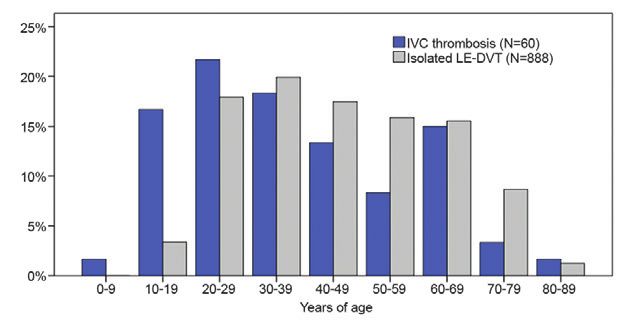
                                                                                                                                    Figure 1: Age distribution at the time of VTE manifestation among 60
                                                                                                                                                                                                                                        tially seen by an orthopaedic special-
                                                                                                                                    patients with IVC thrombosis and 888 patients with LE-DVT registered in
                                                                                                                                                                                                                                        ist. In these cases, a clinical assess-
                                                                                                                                    the MAISTHRO database.
                                                                                                                                                                                                                                        ment and an x-ray of the spine did
                                                                                                                                                                                                                                        not reveal any explanation for the
                                                                                                                                                                                                                                        pain. The median age of these patients
                                                                                                                                   inguinal ligament proximally. The         had a so-called tumour thrombosis                          was 26 years, and 6 patients were even
                                                                                                                                   patient ages at LE-DVT thrombosis         progressing into the IVC. In only one                      younger than 18 years. Another 12
                                                                                                                                   manifestation ranged from 17 to 87        case, IVC thrombosis was limited to                        patients (20 %) were initially seen
                                                                                                                                   years (median 44.0 years). The distri-    the IVC (1.6 %). In this case, symp-                       by general practitioners, surgeons
                                                                                                                                   butions of age at the time of throm-      tomatic hydronephrosis in a horse-                         or gynaecologists for lower back or
                                                                                                                                   bosis manifestation for both groups       shoe kidney led to the diagnosis of                        abdominal pain without finding the
                                                                                                                                   are presented in Fig. 1. Patients who     a circumscribed IVC thrombosis at                          source of the symptoms. In contrast,
                                                                                                                                   suffered IVC thrombosis as the first      the same time. In the majority of                          no patient with LE-DVT suffered
                                                                                                                                   thrombotic event were significantly       cases, IVC thrombosis extended to                          from lower back or abdominal pain as
                                                                                                                                   younger at the time of thrombosis         the iliac and lower-extremity veins.                       an initial symptom of DVT. Remark-
                                                                                                                                   manifestation than LE-DVT patients        Bilateral LE-DVT was detected in 17                        ably, only a minority of IVC throm-
                                                                                                                                   (31.5 vs. 44.0 years; p = 0.001). Twen-   cases (28 %), whereas involvement of                       bosis patients (30 %) reported classic
                                                                                                                                   ty-four out of 60 (40 %) IVC throm-       the left or the right lower extremity                      symptoms such as pain or swelling
                                                                                                                                   bosis patients were younger than 30       veins was observed in 26 (44 %) and                        of the lower extremities as their first
                                                                                                                                   years. No significant difference was      17 (28 %) cases, respectively. IVC                         symptom.
                                                                                                                                   observed between IVC thrombosis           thrombosis distally extended to the                        However, at the time when IVC
                                                                                                                                   and LE-DVT patients with regard to        iliac veins in 57 cases (95 %), to the                     thrombosis was finally diagnosed,
                                                                                                                                   sex (60 % and 55 % females, respec-       femoropopliteal veins in 48 cases                          the situation had changed. There were
                                                                                                                                   tively).                                  (80 %) and to the calf veins in 22                         53 IVC thrombosis patients (88 %)
                                                                                                                                                                             cases (37 %). In contrast, no patient                      who had developed leg symptoms
                                                                                                                                   Localisation and extension of             with an isolated LE-DVT had a bi-                          such as pain or swelling indicating
                                                                                                                                   thrombosis                                lateral thrombosis. Among patients                         thrombosis and resulting in specific
                                                                                                                                   IVC thrombosis was located in the         with isolated LE-DVT, there were 36                        diagnostic procedures. At the same
                                                                                                                                   suprarenal segment in 2 cases (3 %)       cases (63 %) with DVTs of the femo-                        time, 28 (52 %) and 16 patients (30 %)
                                                                                                                                   and the infrarenal segment in the         ropopliteal veins and 21 cases (37 %)                      also reported lower back and abdomi-
                                                                                                                                   remaining 58 cases (97 %). Suprare-       with isolated calf vein thromboses.                        nal pain, respectively. In contrast,
                                                                                                                                   nal IVC thrombosis occurred in one                                                                   leg pain and swelling were the most
                                                                                                                                   patient with Budd-Chiari syndrome         Clinical presentation                                      prevalent first symptoms in LE-DVT
                                                                                                                                   involving the hepatic veins and ex-       Table II presents the prevalence of                        patients (89 %).
                                                                                                                                   tending proximally into the IVC and       symptoms at the time of thrombosis                         Thrombosis manifestation oc-
                                                                                                                                   the right atrium. The other patient       manifestation. We differentiated be-                       curred with symptoms indicative
                                                                                                                                   with suprarenal IVC thrombosis suf-       tween the patients’ initial symptoms                       of PE (e.g., chest pain, dyspnoea)
                                                                                                                                   fered from renal cell carcinoma and       and the symptoms at the time when                          in 13 % and 9 % of patients with
Vasa 2013; 42: 275 – 283                            C. Kraft et al.: Clinical presentation of IVC thrombosis
                                                                                                                                © 2013 Hans Huber Publishers, Hogrefe AG, Bern

                                                                                                                                                                                                                                                         Original communication 279

                                                                                                                                Table II: Prevalence of the patients’ initial symptoms and the symptoms at the time of diagnosis

                                                                                                                                                                                             Initial symptom*                              Symptoms at the time of diagnosis*
http://econtent.hogrefe.com/doi/pdf/10.1024/0301-1526/a000288 - Sunday, October 04, 2015 3:01:52 PM - IP Address:144.76.86.22

                                                                                                                                                                                     IVC            Isolated                                 IVC            Isolated
                                                                                                                                                                                 Thrombosis          LE-DVT                              Thrombosis          LE-DVT
                                                                                                                                                                                   (N = 60)          (N = 57)                              (N = 60)          (N = 57)
                                                                                                                                                                                    N (%)             N (%)            p-value              N (%)             N (%)         p-value
                                                                                                                                  Lower back pain                                  18 (30)             0 (0)
C. Kraft et al.: Clinical presentation of IVC thrombosis                              Vasa 2013; 42: 275 – 283
                                                                                                                                                                                                                                               © 2013 Hans Huber Publishers, Hogrefe AG, Bern

                                                                                                                                280 Original communication

                                                                                                                                    Table IV: Prevalence of hereditary and acquired thrombophilia                                           Thus, lower back or abdominal pain
                                                                                                                                                                                                                                            was much more likely to be the ini-
                                                                                                                                                                         IVC                       Isolated              P value            tial symptom of IVC thrombosis than
http://econtent.hogrefe.com/doi/pdf/10.1024/0301-1526/a000288 - Sunday, October 04, 2015 3:01:52 PM - IP Address:144.76.86.22

                                                                                                                                                                     Thrombosis                     LE-DVT                                  leg pain or swelling. At the time of
                                                                                                                                                                       (N = 60)                     (N = 57)                                the definitive diagnosis of DVT,
                                                                                                                                                                                                                                            even more IVC thrombosis patients
                                                                                                                                                                     N          %              N              %                             (82 %), suffered from lower back pain
                                                                                                                                     FVL mutation                 17/58        29           21/56            38            0.428            and abdominal pain. Until the time
                                                                                                                                     PT mutation                   6/57        11            4/56             7            0.742            that DVT was diagnosed, i.e., one to
                                                                                                                                     Elevated FVIII activity       7/49        14            9/53            17            0.789            35 days after the start of symptoms,
                                                                                                                                     AT deficiency                 2/60         3            0/60             0            0.496            as many as 89 % of IVC thrombosis
                                                                                                                                     PC deficiency                 1/49         2            1/52             2            1.000            patients also developed leg symptoms
                                                                                                                                     PS deficiency                 3/47         6            3/49             6            1.000            which led to the suspicion of DVT
                                                                                                                                     Lupus anticoagulant           6/57        11            3/57             5            0.490            and finally to the correct diagnosis.
                                                                                                                                     ACL-IgG/-IgM                  2/59         3            1/57             2            1.000            In contrast, 91 % of patients with iso-
                                                                                                                                                                                                                                            lated LE-DVT initially became symp-
                                                                                                                                     FVL = Factor V Leiden; PT = prothrombin; FVIII = factor VIII; AT = antithrombin;                       tomatic with pain, swelling or cramp-
                                                                                                                                     PC = protein C; PS = protein S; ACL = anticardiolipin.
                                                                                                                                                                                                                                            ing of the leg. No LE-DVT patient
                                                                                                                                                                                                                                            experienced lower back or abdominal
                                                                                                                                    tients without IVC anomalies (35 vs.         ease was already known at the time                         pain as a first symptom.
                                                                                                                                    37 years; p = 0.695). In contrast, no        of thrombosis manifestation.                               In cases where thrombotic obstruc-
                                                                                                                                    IVC anomalies were detected among                                                                       tion of the IVC occurs, the ascending
                                                                                                                                    patients with isolated LE-DVT (Ta-                                                                      lumbar veins and lateral sacral veins
                                                                                                                                    ble III).                                    Discussion                                                 are important collaterals to achieve
                                                                                                                                                                                                                                            venous blood flow from the lower ex-
                                                                                                                                    Malignant disease                            In this retrospective case-controlled                      tremities into the azygous and hemia-
                                                                                                                                    Malignant disease was present in 16          study, we investigated the clinical                        zygos veins. The extensive blood flow
                                                                                                                                    IVC thrombosis patients (27 %) and           characteristics of IVC thrombosis                          via the ascending lumbar veins also
                                                                                                                                    was located in the kidneys (n = 2),          patients compared to patients with                         affects the vertebral venous system
                                                                                                                                    ovaries (n = 2), testes (n = 2), blad-       isolated LE-DVT. The main find-                            that consists of an internal and exter-
                                                                                                                                    der (n = 1), vulva (n = 1), colon            ings of our study are as follows: in                       nal venous network (Fig. 2). Radicu-
                                                                                                                                    (n = 2), lung (n = 1), mammary               the majority of patients, IVC throm-                       lar veins traverse the intervertebral
                                                                                                                                    (n = 1), and brain (n = 1). External         bosis occurs before the age of 40; an                      foramen and connect the internal and
                                                                                                                                    compression of the IVC by tumour             IVC thrombosis often presents with                         external vertebral veins and plexus.
                                                                                                                                    masses was observed in only 2 cases,         lower back or abdominal pain; IVC                          Thus, IVC thrombosis may lead to
                                                                                                                                    and in 2 patients with renal cell car-       thrombosis frequently involves both                        enlargement of these collateral veins
                                                                                                                                    cinoma, a tumour invasion into the           iliac or lower-extremity veins; in the                     and compression of the neighbour-
                                                                                                                                    renal vein caused a so-called tumour         acute situation, the risk of symptom-                      ing neural nerve roots. Consequently,
                                                                                                                                    thrombosis. Additionally, three IVC          atic pulmonary embolism is high;                           patients can present with radicular
                                                                                                                                    thrombosis patients suffered from            and regarding the prevalence of es-                        pain and sciatica. In most cases, lower
                                                                                                                                    haematologic malignancies. The               tablished VTE risk factors, malignant                      back pain associated with radicular
                                                                                                                                    prevalence of malignant disease was          disease is more frequent among IVC                         pain is caused by prolapse of an inter-
                                                                                                                                    higher when compared to LE-DVT               thrombosis patients when compared                          vertebral disc. However, compression
                                                                                                                                    patients (27 % vs. 9 %; p = 0.015). In       to those with isolated LE-DVT. Fur-                        of the peripheral nerve roots due to
                                                                                                                                    the LE-DVT group, cancer was lo-             thermore, a considerable number of                         vascular pathologies has also been
                                                                                                                                    cated in the mammary (n = 2), the            IVC thrombosis patients presented                          recognised as a possible cause of back
                                                                                                                                    ovaries (n = 2) and the lung (n = 1).        with a congenital anomaly of the                           and radicular pain. In particular, the
                                                                                                                                    In 4/16 IVC thrombosis patients and          IVC (i.e., segmental hypoplasia or                         enlargement of the epidural veins has
                                                                                                                                    2/5 LE-DVT patients, malignant dis-          aplasia).                                                  been associated with the occurrence
                                                                                                                                    ease was first diagnosed during the          Approximately every second patient                         of lower back pain, lumbar radicu-
                                                                                                                                    diagnostic work-up for DVT. In the           with IVC thrombosis suffered from                          lopathy, claudication, and even cauda
                                                                                                                                    remaining patients, malignant dis-           initial symptoms atypical for DVT.                         equina syndrome [5 – 8]. Paksoy et
Vasa 2013; 42: 275 – 283                         C. Kraft et al.: Clinical presentation of IVC thrombosis
                                                                                                                                © 2013 Hans Huber Publishers, Hogrefe AG, Bern

                                                                                                                                                                                                                                                 Original communication 281

                                                                                                                                                                                                                                            ment of DVT despite a compensatory
                                                                                                                                                                                                                                            enlargement of the lumbar, azygous,
                                                                                                                                                                                                                                            hemiazygos and other collateral veins
http://econtent.hogrefe.com/doi/pdf/10.1024/0301-1526/a000288 - Sunday, October 04, 2015 3:01:52 PM - IP Address:144.76.86.22

                                                                                                                                                                                                                                            that allow adequate venous outflow
                                                                                                                                                                                                                                            from the pelvis and lower extremities
                                                                                                                                                                                                                                            [10, 11, 14].
                                                                                                                                                                                                                                            Besides congenital anomalies, ma-
                                                                                                                                                                                                                                            lignant disease was observed more
                                                                                                                                                                                                                                            frequently in IVC thrombosis pa-
                                                                                                                                                                                                                                            tients when compared to patients
                                                                                                                                                                                                                                            with isolated LE-DVT (27 % vs. 9 %;
                                                                                                                                                                                                                                            p = 0.015). Among IVC thrombosis
                                                                                                                                                                                                                                            patients, 15 patients had solid tu-
                                                                                                                                                                                                                                            mours located in different organs
                                                                                                                                  Figure 2: Anatomy of the paravertebral venous system (from [5])                                           (i.e., genitourinary and gastrointes-
                                                                                                                                                                                                                                            tinal tract, lung, mamma, or brain). In
                                                                                                                                                                                                                                            addition, IVC thrombosis occurred
                                                                                                                                al. analysed MRIs from 9640 patients             of IVC anomalies in the general pop-                       in three patients with haematologic
                                                                                                                                experiencing back pain or sciatica               ulation. In the majority of cases, IVC                     malignancies. It may be possible that
                                                                                                                                and found that 13 (0.13 %) had IVC               anomalies usually are asymptomatic                         an obstruction of the IVC due to a
                                                                                                                                obstruction or occlusion [5]. In ten             due to a patent compensatory system                        neighbouring tumour process or a
                                                                                                                                of these patients, thrombosis of the             of venous collaterals and are detected                     tumour thrombosis due to invasive
                                                                                                                                infrarenal IVC was detected, and in              only when imaging studies are per-                         tumour growth could explain the
                                                                                                                                three patients, external compression             formed for other reasons. However,                         difference between groups. How-
                                                                                                                                of the IVC due to a neighbouring                 the anomaly most often observed in                         ever, only 2 cases of external IVC
                                                                                                                                pathologic process was diagnosed.                conjunction with IVC thrombosis                            compression by tumour masses and
                                                                                                                                MRI scans demonstrated enlarge-                  or proximal LE-DVT is a segmen-                            2 cases of tumour thrombosis from
                                                                                                                                ment of the epidural venous plexus in            tal aplasia or hypoplasia of the IVC                       renal cell carcinoma were identified
                                                                                                                                all of these cases. All of these patients        [10 – 22], which was also observed in                      in our cohort. Therefore, the majority
                                                                                                                                presented with acute onset of lower              our IVC thrombosis cohort.                                 of malignancy-associated VTE can
                                                                                                                                back pain followed shortly thereafter            Remarkably, a congenital anomaly                           be attributed to malignancy-induced
                                                                                                                                by acute radicular symptoms.                     of the IVC was present in 13 % of                          hypercoagulability.
                                                                                                                                The development of symptoms, i.e.,               the IVC thrombosis cases; however,                         Patients with IVC thrombosis did not
                                                                                                                                lower back pain as an initial symp-              these patients were not younger at                         differ from those with isolated LE-
                                                                                                                                tom, favours the hypothesis that in a            IVC thrombosis manifestation than                          DVT with regard to the other VTE
                                                                                                                                considerable number of patients, the             those without anomalies. In contrast,                      risk factors or the prevalence of he-
                                                                                                                                thrombotic process initially involves            no IVC anomalies were detected                             reditary or acquired thrombophilias.
                                                                                                                                the IVC and secondarily descends to              among patients with isolated LE-                           However, the patient numbers were
                                                                                                                                the pelvic and lower extremity veins.            DVTs. Unfortunately, the majority of                       too small to draw definitive conclu-
                                                                                                                                Bilateral thrombosis is a frequent               LE-DVT patients were diagnosed by                          sions.
                                                                                                                                finding among IVC thrombosis pa-                 ultrasonography or conventional ve-                        In our cohort, there was a trend to
                                                                                                                                tients. In a systematic review of 26             nography but did not receive a CT or                       a higher frequency of symptomatic
                                                                                                                                cases with IVC anomalies and DVT,                MRI scan, so we may have overlooked                        PE among patients with IVC throm-
                                                                                                                                Yun et al. observed a bilaterally lo-            some cases of IVC anomalies. In the                        bosis compared to LE-DVT patients.
                                                                                                                                cated DVT in 16/26 cases (54 %) [9].             general population, IVC anomalies                          Patients with IVC thrombosis have a
                                                                                                                                In the presence of an anomaly of the             are reported to occur in 0.3 % of oth-                     larger clot burden than patients with
                                                                                                                                IVC, bilateral iliofemoral thrombosis            erwise healthy subjects and generally                      isolated LE-DVT which may height-
                                                                                                                                has been reported in 35 % to 75 % of             result from aberrant development                           en the risk of symptomatic PE. It has
                                                                                                                                patients, respectively [10 – 13]. How-           during the sixth to eighth weeks of                        previously been shown that patients
                                                                                                                                ever, these numbers have to be inter-            embryogenesis [10]. IVC anomalies                          with proximal LE-DVT have higher
                                                                                                                                preted with caution because there is             are considered a condition that fa-                        PE rates than patients with isolated
                                                                                                                                still uncertainty about the prevalence           vours venous stasis and the develop-                       distal LE-DVT [23].
C. Kraft et al.: Clinical presentation of IVC thrombosis                                Vasa 2013; 42: 275 – 283
                                                                                                                                                                                                                                            © 2013 Hans Huber Publishers, Hogrefe AG, Bern

                                                                                                                                282 Original communication

                                                                                                                                   It is possible that massive thrombo-     Conclusions                                                4      Miyakis S, Lockshin MD, Atsumi
                                                                                                                                   sis may occur especially in cases of                                                                       T, BranchDW, Brey RL, Cervera R,
                                                                                                                                   recurrent DVT, but in 78 % of cases,     Inferior vena cava thrombosis should                              Derksen RH, De Groot PG, Koike
http://econtent.hogrefe.com/doi/pdf/10.1024/0301-1526/a000288 - Sunday, October 04, 2015 3:01:52 PM - IP Address:144.76.86.22

                                                                                                                                   the IVC thrombosis was the first         be considered a differential diagno-                              T, Meroni PL, Reber G, Shoenfeld
                                                                                                                                   VTE event, and these patients were       sis in cases of lower back pain or                                Y, Tincani A, Vlachoyiannopoulos
                                                                                                                                   significantly younger at the time of     sciatica, especially if the patient is                            PG, Krilis SA. International consen-
                                                                                                                                   thrombosis manifestation compared        young and no orthopaedic or other                                 sus statement on an update of the
                                                                                                                                   to all patients with isolated LE-DVT.    pathology can be identified. Lower                                classification criteria for definite an-
                                                                                                                                   A similar observation was made by        back or abdominal pain is the most                                tiphospholipid syndrome (APS). J
                                                                                                                                   Joffe et al. who compared patients       frequent initial clinical sign of IVC                             Thromb Haemost 2006; 4: 295 – 306.
                                                                                                                                   with iliac vein thrombosis to patients   thrombosis and often precedes the                          5      Paksoy Y, Gormus N. Epidural ve-
                                                                                                                                   with isolated LE-DVT and found that      development of typical symptoms                                   nous plexus enlargements present-
                                                                                                                                   patients with massive initial DVT        of LE-DVT. However, the definitive                                ing with radiculopathy and back
                                                                                                                                   were younger [24]. The reason that       diagnosis of DVT is often not made                                pain in patients with inferior vena
                                                                                                                                   younger patients tend to develop         until the development of typical leg                              cava obstruction or occlusion. Spine
                                                                                                                                   more massive thrombosis than older       symptoms. Bilateral LE-DVT extend-                                2004; 29: 2419 – 24.
                                                                                                                                   patients is unknown. In our cohort,      ing into the iliac or femoral veins is                     6      Dudeck O, Zeile M, Poellinger
                                                                                                                                   58 % of IVC thrombosis occurred          a common finding in patients with                                 A, Kluhs L, Ludwig WD, Hamm
                                                                                                                                   before the age of 40. However, the       IVC thrombosis.                                                   B. Epidural venous enlargements
                                                                                                                                   time interval between the start of                                                                         presenting with intractable lower
                                                                                                                                   symptoms and the diagnosis of DVT                                                                          back pain and scatica in a patient
                                                                                                                                   was similar between both groups. A       Conflicts of interest                                             with absence of the infrarenal in-
                                                                                                                                   genetic predisposition seems to be                                                                         ferior vena cava and bilateral deep
                                                                                                                                   unlikely as hereditary thrombophilia     There are no conflicts of interest                                venous thrombosis. Spine 2007; 32:
                                                                                                                                   and a family history positive for VTE    existing.                                                         E688 – E691.
                                                                                                                                   was even less frequent among IVC                                                                    7      Hammer A, Knight I, Agarwal A.
                                                                                                                                   thrombosis patients in our study,                                                                          Localized venous plexi in the spine
                                                                                                                                   although the difference between the      References                                                        simulating prolapse of an interver-
                                                                                                                                   groups was not significant. However,                                                                       tebral disc. A report of six cases.
                                                                                                                                   the patient numbers were too small to     1     Bates SM, Jaeschke R, Stevens SM,                          Spine 2003; 28: E5 – E12.
                                                                                                                                   draw definitive conclusions.                    Goodacre S, Wells PS, Stevenson                     8      Gormus N, Ustun ME, Paksoy Y,
                                                                                                                                                                                   MD, Kearon C, Schunemann HJ,                               Ogun TC, Solak H. Acute throm-
                                                                                                                                   Limitations                                     Crowther M, Pauker SG, Makdissi                            bosis of inferior vena cava in a
                                                                                                                                   The retrospective study design is a             R, Guyatt GH. Diagnosis of DVT.                            pregnant woman presenting with
                                                                                                                                   limitation in this study. However,              Antithrombotic Therapy and Pre-                            sciatica: a case report. Ann Vasc
                                                                                                                                   apart from six patients who died and            vention of Thrombosis, 9th ed:                             Surg 2005; 19: 120 – 2.
                                                                                                                                   one patient who could not be reached            American College of Chest Phy-                      9      Yun SS, Kim JI, Kim KH, Sung GY,
                                                                                                                                   for follow-up, all patients were con-           sicians Evidence-Based Clinical                            Lee DS, Kim JS, Moon IS, Lim KW,
                                                                                                                                   tacted again and were assessed using            Practice Guidelines. Chest 2012;                           Koh YB. Deep venous thrombosis
                                                                                                                                   a standardised questionnaire so that            141 (Suppl): e351S – e418S.                                caused by congenital absence of
                                                                                                                                   the interpretation of data was not        2     Lindhoff-Last E, Bauersachs R,                             inferior vena cava, combined with
                                                                                                                                   based on the analysis of medical re-            Jesgarz J Bergh B, Spannagl M,                             hyperhomocysteinemia. Ann Vasc
                                                                                                                                   cords alone.                                    Schramm W. The german thrombo-                             Surg 2004; 18: 124 – 9.
                                                                                                                                   However, this study is the first sys-           philia registry. Ann Hematol 2001;                  10     Gayer, G, Luboshitz J, Hertz M, Zis-
                                                                                                                                   tematic evaluation of a cohort of IVC           80(Suppl1): A43.                                           sin R, Thaler M, Lubetsky A, Bass A,
                                                                                                                                   thrombosis patients showing differ-       3     Linnemann B, Schindewolf M,                                Korat A, Apter S. Congenital anom-
                                                                                                                                   ences in clinical characteristics when          Zgouras D, Erbe M, Jarosch-Preu­                           alies of the inferior vena cava re-
                                                                                                                                   patients were compared to isolated              sche M, Lindhoff-Last E. Are pa-                           vealed on CT in patients with deep
                                                                                                                                   LE-DVT patients matched for sex                 tients with thrombophilia and pre-                         vein thrombosis. Am J Roentgenol
                                                                                                                                   and age.                                        vious venous thromboembolism at                            2003; 180: 729 – 32.
                                                                                                                                                                                   higher risk to arterial thrombosis?                 11     Chee YL, Culligan DJ, Watson HG.
                                                                                                                                                                                   Thromb Res 2008; 121: 743 – 50.                            Inferior vena cava malformation as
Vasa 2013; 42: 275 – 283                         C. Kraft et al.: Clinical presentation of IVC thrombosis
                                                                                                                                © 2013 Hans Huber Publishers, Hogrefe AG, Bern

                                                                                                                                                                                                                                                 Original communication 283

                                                                                                                                      a risk factor for deep venous throm-              cava and retroaortic left renal vein                22   Siragusa S, Anatasio R, Falaschi F,
                                                                                                                                      bosis in the young. Br J Haematol                 mimicking retroperitoneal neo-                           Bonalumi G, Bressan MA. Conge-
                                                                                                                                      2001; 114: 878 – 80.                              plasm. Abdom Imaging 2007; 32:                           nital absence of inferior vena cava.
http://econtent.hogrefe.com/doi/pdf/10.1024/0301-1526/a000288 - Sunday, October 04, 2015 3:01:52 PM - IP Address:144.76.86.22

                                                                                                                                12    Lambert M, Marboeuf P, Midulla M,                 403 – 6.                                                 Lancet 2001; 357: 1711.
                                                                                                                                      Trillot N, Beregi JP, Mounier-Vehi-        17     Lane DA. Congential hypoplasia of                   23   Kucher N, Tapson VF, Goldhaber
                                                                                                                                      er C, Hatron PY, Jude B. Inferior                 the inferior vena cava: an under-                        SZ. Risk factors associated with
                                                                                                                                      vena cava agenesis and deep vein                  appreciated cause of deep venous                         symptomatic pulmonary embo-
                                                                                                                                      thrombosis: 10 patients and review                thromboses among young adults.                           lism in a large cohort of deep vein
                                                                                                                                      of the literature. Vasc Med 2010; 15:             Mil Med 2005; 170: 739 – 42.                             thrombosis patients. Thromb Hae-
                                                                                                                                      451 – 9.                                   18     Tiesenhausen K, Amann W, Thal-                           most 2005; 93: 494 – 8.
                                                                                                                                13    Guanella R, Glauser F, Bounameaux                 hammer M, Aschauer M. Aplasia of                    24   Joffe HV, Kucher N, Tapson VF,
                                                                                                                                      H, Mazzolai L. Inferior vena cava                 the vena cava inferior as a cause for                    Goldhaber SZ. Few predictors of
                                                                                                                                      agenesis: association with bilateral              recurring thrombosis of the lower                        massive deep vein thrombosis.
                                                                                                                                      lower-limb deep vein thrombosis                   extremitites and pelvic veins. VASA                      Thromb Haemost 2005; 94: 986 – 90.
                                                                                                                                      in young males. Thromb Haemost                    1999; 28: 289 – 92.
                                                                                                                                      2009; 102: 795 – 8.                        19     Mirzaie M, Schorn B, Meyer T, Lotfi
                                                                                                                                14    Obernosterer A, Aschauer M,                       S, Dalichau H. Deep phlebothrom-                    Correspondence address
                                                                                                                                      Schnel W, Lipp RW. Anomalies of                   boses of iliac vein an aplasia of the
                                                                                                                                      the inferior vena cava in patients                inferior vena cava. VASA 1999; 28:                  PD Dr. Birgit Linnemann, MD
                                                                                                                                      with iliac venous thrombosis. Ann                 293 – 5.                                            Division of Vascular Medicine
                                                                                                                                      Intern Med 2002; 136: 37 – 41.             20     Mouton KT, Zehnder T, Wagner                        Department of Internal Medicine
                                                                                                                                15    Basile A, Certo A, Ascenti G, Lam-                HE, Mouton WG. Follow-up after                      Goethe University Hospital
                                                                                                                                      berto S, Cannella A, Medina JG.                   deep venous thrombosis in azy-                      Frankfurt / Main
                                                                                                                                      Embryologic and acquired anom-                    gos continuation. VASA 2005; 34:                    Theodor-Stern-Kai 7
                                                                                                                                      alies of the inferior vena cava with              266 – 8.                                            60590 Frankfurt / Main
                                                                                                                                      recurrent deep vein thrombosis.            21     Ruggeri M, Tosetto A, Castaman G,                   Germany
                                                                                                                                      Abdom Imagin 2003; 28: 400 – 3.                   Rodeghiero F. Congenital absence                    Birgit.Linnemann@kgu.de
                                                                                                                                16    Cizginer S, Tatli S, Girshman J,                  of the inferior vena cava: a rare
                                                                                                                                      Beckman JA, Silverman SG. Throm-                  risk factor for idiopathic deep vein                Submitted: 15.01.2013
                                                                                                                                      bosed interrupted inferior vena                   thrombosis. Lancet 2001; 357: 441.                  Accepted after revision: 03.03.2013
You can also read