Automated prediction of final infarct volume in patients with large-vessel occlusion acute ischemic stroke
←
→
Page content transcription
If your browser does not render page correctly, please read the page content below
NEUROSURGICAL
            FOCUS                                                                                                                   Neurosurg Focus 51 (1):E13, 2021

                           Automated prediction of final infarct volume in patients
                           with large-vessel occlusion acute ischemic stroke
                           *Rania Abdelkhaleq, BS,1 Youngran Kim, PhD,1 Swapnil Khose, MD,1 Peter Kan, MD,2
                           Sergio Salazar-Marioni, MD,1 Luca Giancardo, PhD,3 and Sunil A. Sheth, MD1
                           1
                             Department of Neurology and 3Center for Precision Health, UTHealth School of Biomedical Informatics, UTHealth McGovern
                           Medical School, Houston; and 2Department of Neurosurgery, University of Texas Medical Branch, Galveston, Texas

                           OBJECTIVE In patients with large-vessel occlusion (LVO) acute ischemic stroke (AIS), determinations of infarct size
                           play a key role in the identification of candidates for endovascular stroke therapy (EVT). An accurate, automated method
                           to quantify infarct at the time of presentation using widely available imaging modalities would improve screening for EVT.
                           Here, the authors aimed to compare the performance of three measures of infarct core at presentation, including an au-
                           tomated method using machine learning.
                           METHODS Patients with LVO AIS who underwent successful EVT at four comprehensive stroke centers were identi-
                           fied. Patients were included if they underwent concurrent noncontrast head CT (NCHCT), CT angiography (CTA), and
                           CT perfusion (CTP) with Rapid imaging at the time of presentation, and MRI 24 to 48 hours after reperfusion. NCHCT
                           scans were analyzed using the Alberta Stroke Program Early CT Score (ASPECTS) graded by neuroradiology or neurol-
                           ogy expert readers. CTA source images were analyzed using a previously described machine learning model named
                           DeepSymNet (DSN). Final infarct volume (FIV) was determined from diffusion-weighted MRI sequences using manual
                           segmentation. The primary outcome was the performance of the three infarct core measurements (NCHCT-ASPECTS,
                           CTA with DSN, and CTP-Rapid) to predict FIV, which was measured using area under the receiver operating character-
                           istic (ROC) curve (AUC) analysis.
                           RESULTS Among 76 patients with LVO AIS who underwent EVT and met inclusion criteria, the median age was 67
                           years (IQR 54–76 years), 45% were female, and 37% were White. The median National Institutes of Health Stroke Scale
                           score was 16 (IQR 12–22), and the median NCHCT-ASPECTS on presentation was 8 (IQR 7–8). The median time
                           between when the patient was last known to be well and arrival was 156 minutes (IQR 73–303 minutes), and between
                           NCHCT/CTA/CTP to groin puncture was 73 minutes (IQR 54–81 minutes). The AUC was obtained at three different
                           cutoff points: 10 ml, 30 ml, and 50 ml FIV. At the 50-ml FIV cutoff, the AUC of ASPECTS was 0.74; of CTP core volume,
                           0.72; and of DSN, 0.82. Differences in AUCs for the three predictors were not significant for the three FIV cutoffs.
                           CONCLUSIONS In a cohort of patients with LVO AIS in whom reperfusion was achieved, determinations of infarct core
                           at presentation by NCHCT-ASPECTS and a machine learning model analyzing CTA source images were equivalent to
                           CTP in predicting FIV. These findings have suggested that the information to accurately predict infarct core in patients
                           with LVO AIS was present in conventional imaging modalities (NCHCT and CTA) and accessible by machine learning
                           methods.
                           https://thejns.org/doi/abs/10.3171/2021.4.FOCUS21134
                           KEYWORDS computed tomography; machine learning; CT perfusion; ischemic stroke; cerebrovascular disease/stroke

ABBREVIATIONS AIS = acute ischemic stroke; ASPECTS = Alberta Stroke Program Early CT Score; AUC = area under the ROC curve; CTA = CT angiography; CTP =
CT perfusion; DSN = DeepSymNet; EVT = endovascular stroke therapy; FIV = final infarct volume; IV tPA = intravenous tissue plasminogen activator; LVO = large-vessel
occlusion; NCHCT = noncontrast head CT; NIHSS = National Institutes of Health Stroke Scale; ROC = receiver operating characteristic; TICI = Thrombolysis in Cerebral
Infarction.
ACCOMPANYING EDITORIAL DOI: 10.3171/2021.4.FOCUS21263.
SUBMITTED March 1, 2021. ACCEPTED April 6, 2021.
INCLUDE WHEN CITING DOI: 10.3171/2021.4.FOCUS21134.
* L.G. and S.A.S. contributed equally to this work.

©AANS 2021, except where prohibited by US copyright law                                                             Neurosurg Focus Volume 51 • July 2021              1

                                                                                                                    Unauthenticated | Downloaded 11/17/21 01:40 PM UTC
Abdelkhaleq et al.
E
ndovascular stroke therapy (EVT) is a highly ef- sis in Cerebral Infarction [TICI] grade of 2b or 3), and
fective intervention in patients with large-vessel to have had FIV imaging consisting of MRI at 24 to 48
occlusion (LVO) acute ischemic stroke (AIS), and hours after reperfusion. In all patients, NCHCT, CTA, and
its effectiveness is largely dependent on the extent of ir- CTP were acquired concurrently, which was at the time of
reversibly injured, or infarcted, tissue at the time of pa- presentation to the hospital.
tient presentation.1 At present, the optimal method to de- The study was reviewed and approved by the Commit-
termine infarct core at the time of presentation remains tee for the Protection of Human Subjects at The University
unknown. The Alberta Stroke Program Early CT Score of Texas Health Science Center at Houston, and waiver
(ASPECTS) has been developed to quantify the extent of of consent and Health Insurance Portability and Account-
ischemia using noncontrast head CT (NCHCT), which is ability Act (HIPAA) authorization was granted.
nearly universally available. On the other hand, ASPECTS
grading can be subject to high interrater variability and Measurements
can require a higher level of expertise for accurate reading All study data were collected and managed using Re-
than is available in many lower-volume centers at which search Electronic Data Capture (REDCap) tools.8,9 Quan-
AIS is evaluated.2 CT perfusion (CTP), on the other hand, tification of ischemic core at the time of presentation was
benefits from automated determinations of infarcted tis- made by three methods. First, ASPECTS was determined
sue and played a large role in recent clinical trials. CTP from NCHCT using a neuroradiology report, or if it was
is less widely available, however, and in addition has been not available, by an expert reviewer with experience in
shown to under- and overestimate the infarct core.3–5 A AIS imaging determinations. Second, CTP studies were
methodology that can accurately detect infarcted tissue, postprocessed using Rapid. Finally, CTA source images
in an automated fashion, using imaging techniques that were analyzed using DSN-v2 (see below). We defined FIV
are currently in widespread use at all hospitals that evalu- on diffusion-weighted MRI performed within 72 hours
ate AIS, including lower-volume centers, would improve after successful EVT, consistent with prior studies.3,10–12
screening for EVT. FIV was measured utilizing the manual region of interest
To this end, we previously developed a machine learn- measurements (Horos, https://horosproject.org).
ing model called DeepSymNet (DSN) that successfully
predicted infarct core, as compared with concurrently ac- DeepSymNet-v2
quired CTP, using a much more widely available modality,
CT angiography (CTA), in an automated fashion.6,7 In this DSN-v2 is a machine learning algorithm based on a
study, we hypothesized that NCHCT-ASPECTS and CTA 3D convolutional neural network that utilizes brain hemi-
with DSN perform adequately well in identifying the in- sphere symmetry information in order to learn imaging
farct core relative to CTP with Rapid (iSchemaView, Inc.) patterns that are predictive of outcomes related to stroke.
in patients with LVO AIS. We compared the performance In our previous works, we described the first iteration of
of these three modalities at predicting the final infarct vol- the algorithm, DSN, which was used to detect LVO, es-
ume (FIV) in patients who underwent successful EVT. timate infarct core, and detect hemorrhagic stroke.6,7,13
In this study, we utilized a new version of the machine
learning model, which has been improved by including a
Methods CTA-specific template, multiresolution pipeline, and skull
Study Population stripping. The model was further improved by using sym-
The data that support the findings of this study are metric and nonsymmetric paths, and by reducing the num-
available from the corresponding author on reasonable re- ber of parameters by using more efficient building blocks,
quest. Our study population consisted of a subset of the skip connections, and batch normalization.14,15
Practical Implementation of Mechanical Thrombectomy The algorithm for this machine learning model was
(PRIME) study. PRIME is a prospective observational trained and internally validated on an external CTA data
cohort study examining all patients diagnosed with AIS set acquired from patients outside this analysis cohort, al-
or transient ischemic attack at 11 Joint Commission–certi- lowing for a meaningful estimation of the algorithm gen-
fied stroke centers within the same health system across eralizability and the ability to fine-tune its architecture. At
the Greater Houston area. For this study, we identified a training time, the algorithm used a binary variable indi-
subset of this population that presented to four of these cating a large (> 50 ml) or small (< 50 ml) infarct core as
hospitals, which are comprehensive stroke centers, with a target, which was computed from the CTP image and
onsite CTP and EVT capabilities, between March 2016 the Rapid analysis software. At inference time (i.e., when
and April 2019. Patients were included if they were diag- the algorithm was evaluated in this analysis) the algorithm
nosed with anterior circulation LVO AIS and underwent generated a real number ranging from 0 to 1 in indicating
an imaging workup that included simultaneous NCHCT, the noncalibrated likelihood of having a small infarct core,
CTA, and CTP with Rapid postprocessing at the time of with values closer to 1 more indicative of a smaller core.
presentation. In all patients, CTA was used to assess the
presence of LVO. LVO was defined as an occlusion of the Primary Outcome
intracranial internal carotid artery, A1 or A2 segments of The primary outcome of this study was the performance
the anterior cerebral artery, or M1 or M2 segments of the of the three infarct core measures—NCHCT-ASPECTS,
middle cerebral artery. Patients were also required to have DSN-v2 model (using CTA), and CTP-Rapid—at predict-
undergone EVT with successful reperfusion (Thromboly- ing FIV in this cohort of patients with successful EVT.
2 Neurosurg Focus Volume 51 • July 2021
Unauthenticated | Downloaded 11/17/21 01:40 PM UTCAbdelkhaleq et al.
This outcome was measured using area under the receiver TABLE 1. Demographic and clinical characteristics
operating characteristic (ROC) curve (AUC) statistics at All Patients (n = 76)
three FIV cutoffs (10 ml, 30 ml, and 50 ml). These cutoff
values were chosen to show the performance of the algo- Median age, yrs (IQR) 67 (54–76)
rithm at different core sizes. They were based on clinically Female sex, % 44.7
relevant values while also ensuring we had sufficient data Race, %
with our distribution of core sizes to perform the analysis. White 36.8
The 10-ml cutoff was chosen as a lower value. The 30-ml Black 18.42
and 50-ml cutoffs were chosen to be consistent with the
Asian 5.26
core thresholds used in the DAWN trial.16
Other 39.47
Statistical Analysis Ethnicity, %
For the description of the characteristics of the study Hispanic 14.4
population, percentages are reported for categorical vari- PMHx, %
ables, and medians (IQR) for continuous variables. All Prior stroke 11.8
statistical tests were 2-sided, and conventional levels of Hyperlipidemia 21
significance (α = 0.05) were used for interpretation. The Hypertension 50
model performance was measured using AUC statistics.
Diabetes 22.3
The chi-square test was performed to test for significance
between AUCs for the predictors. All data analyses were Smoking 10.5
performed using Stata version 16 (StataCorp LLC) and Prestroke mRS score, %
Prism version 7 (GraphPad) software. 0–2 82.9
3–5 17.1
Results Median NIHSS score (IQR) 16 (12–22)
Among 76 patients with LVO AIS who underwent Median baseline ASPECTS (IQR) 8 (7–8)
EVT and met inclusion criteria, the median age was 67 Median Rapid ischemic core vol (IQR) 4.5 (0–21.5)
years (IQR 54–76 years), 45% were female, and 37% were Median FIV (IQR) 11.39 (2–37.5)
White. Fifty percent of the patients had a history of hy- Median time interval (IQR), mins
pertension, 21% had hyperlipidemia, and 12% had had LKW to arrival 156 (73–303)
a prior stroke. The median National Institutes of Health
CT to GP 73 (54–81)
Stroke Scale (NIHSS) score was 16 (IQR 12–22), and 65%
of patients had a prestroke modified Rankin Scale score CT to recanalization 111 (100–126.5)
of 0. The median ASPECTS on presentation was 8 (IQR Target occlusion site, %
7–8). The median time between when the patient was ICA 13.3
last known to be well and arrival was 156 minutes (IQR M1 66.6
73–303 minutes), and that between NCHCT/CTA/CTP to M2 16
groin puncture was 73 minutes (IQR 54–81 minutes). Fif- Other 4
ty-one percent of the patients received intravenous tissue
Occlusion laterality lt hemisphere, % 55.26
plasminogen activator (IV tPA), and 25% achieved a TICI
grade of 2b. More details on demographics and clinical IV tPA, % 51
characteristics can be found in Table 1. Final TICI grade, %
The distribution of infarct core predictions based on 2b 25.3
NCHCT-ASPECTS, CTP-Rapid, and DSN relative to FIV 3 74.6
can be seen in Fig. 1. The performance of these three mea-
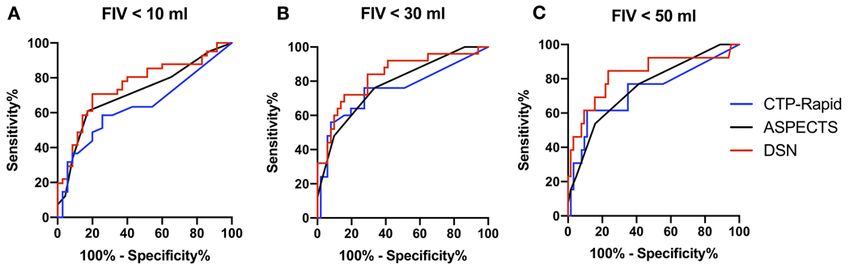
sures at predicting FIV can be seen in Fig. 2. The AUC GP = groin puncture; ICA = internal carotid artery; LKW = last known well;
mRS = modified Rankin Scale; PMHx = past medical history.
was obtained at three different cutoff points: 10-ml, 30-
ml, and 50-ml FIV. At the 10-ml FIV cutoff, the AUC of
ASPECTS was 0.67; of CTP core volume, 0.63; and of core at the time of presentation in patients with LVO
DSN, 0.75. Differences in AUCs for the three predictors AIS, all three measurements showed good performance
were not significant (p = 0.11). At the 30-ml FIV cutoff, at predicting FIV in patients with recanalization. Our au-
the AUC of ASPECTS was 0.76; of CTP core volume, tomated machine learning model analyzing CTA source
0.73; and of DSN, 0.82. Differences in AUCs for the three
images showed superior performance relative to NCHCT-
predictors were not significant (p = 0.36). At the 50-ml
ASPECTS and CTP-Rapid across the range of FIV sizes,
FIV cutoff, the AUC of ASPECTS was 0.74; of CTP core
volume, 0.72; and of DSN, 0.82. Differences in AUCs for although this difference was not statistically significant.
the three predictors were not significant (p = 0.44). The performances of all three models improved with iden-
tification of larger FIVs.
We chose to use FIV in patients with successful recan-
Discussion alization as the gold standard for infarct at presentation
In this study of different methods to measure infarct because of the known inaccuracies of other methods, par-
Neurosurg Focus Volume 51 • July 2021 3
Unauthenticated | Downloaded 11/17/21 01:40 PM UTCAbdelkhaleq et al.
FIG. 1. Distribution of FIVs against three measurements of infarct core at presentation. Scatterplots showing the distribution of FIV
versus ASPECTS (A), CTP-Rapid infarct core (B), and DSN (C) probability on imaging performed at the time of patient presentation.
ticularly in the earlier time windows.4 There are, however, Our study has several limitations. As mentioned above,
some limitations to this approach, as infarct volumes may it is possible that the infarct volume could grow even after
grow from the time of imaging to reperfusion, and pos- the < 72-hour MRI time point used here to define FIV. On
sibly even afterwards. On the other hand, this technique is the other hand, this time point has been used in many prior
consistent with prior studies evaluating the performance studies and has been shown to correlate closely with the
of machine learning methods for infarct core detection at 90-day clinical outcome.19 Furthermore, because we lim-
the time of presentation.10,17,18 Furthermore, testing imag- ited our cohort to patients who underwent EVT, the pro-
ing methods using FIV in reperfused patients may have portion of patients with very large infarcts was relatively
another advantage, as it measures the ability of the im- small. While our findings would certainly benefit from
aging methods to answer a clinically relevant question: if validation in another data set, we did observe improved
rapid reperfusion is achievable in this patient, what will be performance of ASPECTS and DSN with larger FIVs.
the expected outcome? Finally, our cohort contained primarily patients evalu-
In the current study, we found that NCHCT-ASPECTS ated within the first 6 hours after last known well, and it
and DSN using CTA images performed equivalently to is possible that these findings would not be replicated in
CTP-Rapid across a range of infarct sizes. These two im- patients who present in a later time window. Prior studies,
aging modalities, NCHCT and CTA, are far more widely on the other hand, have shown improved performance of
available than CTP. Of these three methods, DSN boasts NCHCT-ASPECTS with increasing time after last known
the advantage of being automated, thus requiring no ex- well.20
pert input. In addition, the algorithm can be run on CTA
source images in less than 1 minute. Given the known
wide interrater reliability of NCHCT-ASPECTS, an auto- Conclusions
mated approach to identify infarct is preferable, although In a cohort of patients with LVO AIS in whom reper-
recent advances in automated ASPECTS are improving in fusion was achieved, determinations of infarct core at
performance.10 In this cohort, the addition of CTP along presentation by NCHCT-ASPECTS and a machine learn-
with NCHCT and CTA did not add prognostic value to the ing model analyzing CTA source images were equivalent
imaging outcome. to CTP in predicting FIV. The machine learning model
FIG. 2. Performance of three infarct core measurement techniques at presentation to predict FIV. AUCs at three FIV cutoffs of 10
ml (A), 30 ml (B), and 50 ml (C) demonstrating the performance of three measures of infarct core at predicting FIV using imaging
performed at the time of presentation, prior to EVT.
4 Neurosurg Focus Volume 51 • July 2021
Unauthenticated | Downloaded 11/17/21 01:40 PM UTCAbdelkhaleq et al.
demonstrated superior performance, but this difference 13. Barman A, Lopez-Rivera V, Lee S, et al. Combining sym-
was not statistically significant. These findings suggest metric and standard deep convolutional representations for
that the information to accurately predict infarct core in detecting brain hemorrhage. In:Medical Imaging 2020:
Computer-Aided Diagnosis. Vol 11314. International Society
patients with LVO AIS is present in conventional imaging for Optics and Photonics;2020:113140D.
modalities (NCHCT and CTA) and accessible by machine 14. Huang G, Liu S, van der Maaten L, Weinberger KQ. Con-
learning methods. denseNet:an efficient DenseNet using learned group convo-
lutions. In: 2018 IEEE/CVF Conference on Computer Vision
and Pattern Recognition. IEEE;2018:2752–2761. Accessed
References May 10, 2021. https://ieeexplore.ieee.org/document/8578389
1. Goyal M, Menon BK, van Zwam WH, et al. Endovascular 15. Ioffe S, Szegedy C. Batch normalization:accelerating deep
thrombectomy after large-vessel ischaemic stroke:a meta- network training by reducing internal covariate shift. Proc
analysis of individual patient data from five randomised tri- Mach Learn Res. 2015;37:448–456.
als. Lancet. 2016;387(10029):1723–1731. 16. Nogueira RG, Jadhav AP, Haussen DC, et al. Thrombectomy
2. Gupta AC, Schaefer PW, Chaudhry ZA, et al. Interobserver 6 to 24 hours after stroke with a mismatch between deficit
reliability of baseline noncontrast CT Alberta Stroke Pro- and infarct. N Engl J Med. 2018;378(1):11–21.
gram Early CT Score for intra-arterial stroke treatment se- 17. Austein F, Riedel C, Kerby T, et al. Comparison of perfusion
lection. AJNR Am J Neuroradiol. 2012;33(6):1046–1049. CT software to predict the final infarct volume after throm-
3. Copen WA, Yoo AJ, Rost NS, et al. In patients with sus- bectomy. Stroke. 2016;47(9):2311–2317.
pected acute stroke, CT perfusion-based cerebral blood flow 18. Robben D, Boers AMM, Marquering HA, et al. Prediction of
maps cannot substitute for DWI in measuring the ischemic final infarct volume from native CT perfusion and treatment
core. PLoS One. 2017;12(11):e0188891. parameters using deep learning. Med Image Anal. 2020;59:
4. Boned S, Padroni M, Rubiera M, et al. Admission CT perfu- 101589.
sion may overestimate initial infarct core:the ghost infarct 19. Yoo AJ, Chaudhry ZA, Nogueira RG, et al. Infarct volume is
core concept. J Neurointerv Surg. 2017;9(1):66–69. a pivotal biomarker after intra-arterial stroke therapy. Stroke.
5. Geuskens RREG, Borst J, Lucas M, et al. Characteristics of 2012;43(5):1323–1330.
misclassified CT perfusion ischemic core in patients with 20. Nannoni S, Ricciardi F, Strambo D, et al. Correlation be-
acute ischemic stroke. PLoS One. 2015;10(11):e0141571. tween ASPECTS and core volume on CT perfusion:impact
6. Barman A, Inam ME, Lee S, et al. Determining ischemic of time since stroke onset and presence of large-vessel occlu-
stroke from CT-angiography imaging using symmetry- sion. AJNR Am J Neuroradiol. 2021;42(3):422–428.
sensitive convolutional networks. In:2019 IEEE 16th Inter-
national Symposium on Biomedical Imaging (ISBI 2019).
IEEE; 2019:1873–1877. Disclosures
7. Sheth SA, Lopez-Rivera V, Barman A, et al. Machine
Dr. Sheth reports grant funding from the National Institutes of
learning-enabled automated determination of acute ischemic
Health, American Academy of Neurology, and Society for Vascu-
core from computed tomography angiography. Stroke. 2019;
lar and Interventional Neurology.
50(11):3093–3100.
8. Harris PA, Taylor R, Thielke R, et al. Research electronic
data capture (REDCap)—a metadata-driven methodology Author Contributions
and workflow process for providing translational research Conception and design: Sheth, Abdelkhaleq, Kim, Giancardo.
informatics support. J Biomed Inform. 2009;42(2):377–381. Acquisition of data: Giancardo. Analysis and interpretation of
9. Harris PA, Taylor R, Minor BL, et al. The REDCap Consor- data: Sheth, Abdelkhaleq, Giancardo. Drafting the article: Sheth,
tium:building an international community of software plat- Abdelkhaleq, Kim, Khose, Salazar-Marioni, Giancardo. Critically
form partners. J Biomed Inform. 2019;95:103208. revising the article: all authors. Reviewed submitted version of
10. Bouslama M, Ravindran K, Harston G, et al. Noncontrast manuscript: all authors. Approved the final version of the manu-
computed tomography e-stroke infarct volume is similar script on behalf of all authors: Sheth. Statistical analysis: Sheth,
to RAPID computed tomography perfusion in estimating Kim. Administrative/technical/material support: Sheth. Study
postreperfusion infarct volumes. Stroke. 2021;52(2):634–641. supervision: Sheth, Giancardo.
11. Simonsen CZ, Yoo AJ, Sørensen LH, et al. Effect of general
anesthesia and conscious sedation during endovascular thera- Correspondence
py on infarct growth and clinical outcomes in acute ischemic Sunil A. Sheth: UTHealth McGovern Medical School, Houston,
stroke:a randomized clinical trial. JAMA Neurol. 2018;75(4): TX. ssheth@post.harvard.edu.
470–477.
12. Yoo J, Choi JW, Lee SJ, et al. Ischemic diffusion lesion
reversal after endovascular treatment. Stroke. 2019;50(6):
1504–1509.
Neurosurg Focus Volume 51 • July 2021 5
Unauthenticated | Downloaded 11/17/21 01:40 PM UTCYou can also read