Avian Bornavirus in Clinical Practice - MSPCA-Angell
←
→
Page content transcription
If your browser does not render page correctly, please read the page content below

Avian Bornavirus in Clinical Practice
                                    By Patrick Sullivan, DVM, DABVP (Avian Practice)
                                    MSPCA-Angell West
                                    avianexotic@angell.org
                                    617-989-1561
                                    January 2021

Avian Bornavirus (ABV) is an enveloped single-strand RNA virus with
worldwide distribution that is responsible for the condition we now know as
Proventricular Dilatation Disease (PDD). This condition has been identified
in psittacines since the 1970s, and it was originally described as “Macaw
wasting disease.” The causative agent, ABV, was only recently identified in
2008, although many other questions regarding this condition still remain.
While this disease has been identified in numerous types of birds, this article
will be focusing on psittacines, or parrot species, as they are the most
commonly seen birds in clinical practice.

Proventricular Dilatation Disease

The description given to the disease process that is caused by ABV is itself
somewhat controversial and contested. The pathology of the disease involves
nonpurulent inflammation of peripheral nerves. While the autonomic nerves
of the gastrointestinal (GI) tract are commonly affected, this inflammatory
process can also affect the central nervous system, as well as other organs
entirely, leading to symptoms that may not be related to the GI tract at all. The classic presentation of PDD is a bird
that is weak and lethargic, often with a normal appetite. If handled, the owner may notice a decrease in weight, but
often this is not noted until there is a significant change. The plumage may appear dull or tattered, due to chronic
malnutrition. Feces may contain undigested seeds or other food material, and vomiting or regurgitation is often
noted. Most of these symptoms are identified in the more advanced stages of the disease. Symptoms not related to
the GI tract may include ataxia, lameness, blindness, or even seizures. These may be seen concurrent with, or

 Angell Animal Medical Center • 350 S. Huntington Ave., Boston, MA 02130 • 617-522-7282 • fax: 617-989-1635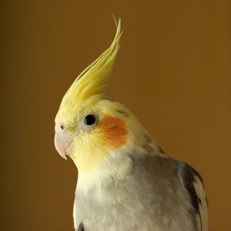
independent of GI changes. Sudden death has been reported, in the absence of clinical symptoms. This may be due
to changes or irregularities in cardiac conductivity, although this has not been proven.
Signalement for PDD is quite varied and can include any psittacine, although African grey parrots, Amazons,
cockatoos, macaws, and cockatiels appear overrepresented. No age predilection has been identified, although a
study in the early 1990s found the average age of affected birds to be 3.8 years old. Latent periods are suspected to
be possible, with lengths being quite variable. Single-housed birds have become symptomatic after years of no
known contact with, or exposure to, infected individuals.
Differential diagnoses for confirmed proventricular dilatation are numerous and should be ruled out prior to making
a diagnosis of PDD. A partial list should include bacterial or mycotic infections, particularly Macrorhabdus
ornithogaster, neoplasia, parasitism, heavy metal toxicity, or a gastrointestinal outflow obstruction. While
proventricular dilatation may be seen with these conditions, they do not typically cause thinning of the
proventricular wall as PDD does. If changes to the wall are noted, they usually involve a thickening secondary to
chronic inflammation.

Transmission

Viral RNA has been identified in several bodily fluids and materials, including
feces, urine, tears, and oral secretions. Due to these findings, transmission has
been assumed to be fecal-oral route. Several studies have found varying degrees
of success with this hypothesis. Infected birds introduced into “clean” flocks
resulted in spread of the disease to some, but not all, birds. Other studies have
shown that horizontal transmission through direct contact was not a sufficient
route in immunocompetent birds. Often these patients will test positive for the
virus on feathers, or skin biopsies, but will never seroconvert. Due to these
findings, Piepenbring et al concluded that exposure most likely did not achieve
persistent infection. This hypothesis has been strengthened by the fact that
multiple reports have identified infected and non-infected birds living together
for years, with no transmission.
At the time of this publication, transmission is not fully understood. Both horizontal and vertical transmission
appear possible, but several variables are thought to affect each of these, including individual health of the patient,
genotype of the virus, and any possible co-infections.

Diagnosis

Diagnosis of a clinical ABV infection is, in a word, challenging. Initial detection of the virus in the living patient is
typically done through reverse-transcription polymerase chain reaction (RT-PCR) testing. Samples submitted
include cloacal/choanal swabs, tissue, and whole blood. Submitting a swab from only one site, or blood without
accompanying swabs, dramatically reduced the likelihood of a positive sample. It is recommended to combine
swabs and blood into one tube, although individual labs may have different submission requirements. Less
commonly run tests to identify ABV include immunohistologic staining, and viral isolation. ABV-specific antibody
detection (ELISA, Western blot) may provide valuable information for diagnosis, although at the present time it is
unclear when antibodies are produced, or if they can potentially be correlated to clinical signs.
Another factor that adds even more confusion is the fact that some birds become infected with ABV, shed the virus,
but never seroconvert. Several theories for why this may happen have been introduced, although none have been
proven. Based on this information, the following protocols have been proposed to help identify positive and
negative birds. A bird that is found to be positive for ABV using both PCR and serology should be considered a
positive infection. A bird that is positive on PCR, but negative on serology, should be retested in 4-6 weeks to check

 Angell Animal Medical Center • 350 S. Huntington Ave., Boston, MA 02130 • 617-522-7282 • fax: 617-989-1635

for a false positive PCR and also to monitor for seroconversion. Repeated positive PCR tests, and/or seroconversion
should be considered a positive bird. Negative PCR paired with a positive serology are thought to be carriers,
although this has not been proven. Persistent elevated titers are indicative of infection in mammals infected with
bornavirus, and should be considered the same for birds. Some birds appear to be able to clear the infection, which
would explain the positive serology and negative PCR, although this too has yet to be proven.

Treatment

Several therapies have been proposed for treatment of both the symptoms of ABV, as well as for the virus itself. To
date, there are no successful antiviral therapies reported. Previous reports of nonsteroidal anti-inflammatory drugs
have failed to show significant, repeatable success. One study using meloxicam in infected cockatiels actually
showed more severe lesions, and increased mortality, when compared to the control group. Immunosuppressive
treatments, including the use of glucocorticosteroids, have been proposed at recent conferences. No published data
has been made available as of yet, and clinicians should be cautious of these currently unproven methods.
Symptomatic treatment, including diet modification, analgesia, and control of secondary infections should be the
basis of treatment for these cases.

Conclusion

Despite the recent advances in identifying and diagnosing this condition, there are still many questions left
unanswered. While diagnostics have improved considerably, interpretation of these tests can still be challenging. A
positive ABV diagnosis should not result in euthanasia, especially since many patients who test positive will never
show symptoms. That being said, once patients become symptomatic, the chances of long term survival are very
low, and quality of life should be assessed. Along with the discovery of additional symptoms, not related to the GI
tract, some have proposed a name change from proventricular dilatation disease to avian or neuropathic
ganglioneuritis. While this more accurately describes the condition, a name change has not been uniformly agreed
upon at this time.

References and Additional Resources:

    1. Wellehan J, Lierz M, Phalen D, et al. Infectious Disease. In: Speer B, Current Therapy in Avian Medicine
       and Surgery. 1st ed. St. Louis: Elsevier 2016; 28-47.
    2. Musser J, Heatley J. Parrot Bornavirus: Current Clinical Summary of Diagnosis, Treatment, and Control, in
       Proceedings. Exoticscon 2020
    3. Avian Bornavirus and Proventricular Dilatation Disease. Available at
       https://www.aav.org/global_engine/download.aspx?fileid=1F7E5DE5-43DD-44F6-B17B-2622787DCD09
       Accessed Jan 17, 2020.

 Angell Animal Medical Center • 350 S. Huntington Ave., Boston, MA 02130 • 617-522-7282 • fax: 617-989-1635
You can also read