Cytokeratins in Glandular Odontogenic Cyst - An Immunohistochemical Study of 4 Cases
←
→
Page content transcription
If your browser does not render page correctly, please read the page content below
Annals of R.S.C.B., ISSN:1583-6258, Vol. 25, Issue 6, 2021, Pages. 892-896
Received 25 April 2021; Accepted 08 May 2021.

                          Cytokeratins in Glandular Odontogenic Cyst – An
                               Immunohistochemical Study of 4 Cases
                                      Heena Sadiq1, Amani Mahajan2, Shahid Shaikh3

                 1. Assistant professor, Department of Dental Surgery, GMC Kathua;
                 2. Senior lecturer, Department of Oral Pathology, Swami Devi Dyal Hospital and Dental
                    College, Barwala, Panchkula, India;
                 3. Consultant pediatric dentist, Masina hospital, Byculla, Mumbai

             Corresponding author: Dr. Amani Mahajan, Senior lecturer, Department of Oral
             Pathology, Swami Devi Dyal Hospital and Dental College, Barwala, Panchkula, India, E
             mail: mahajan.amani18@gmail.com

             ABSTRACT:
             Introduction: Glandular odontogenic cyst (GOC) is a relatively rare cystic lesion of the
             jaws, which poses a diagnostic challenge as well as a challenge in treatment. This study was
             conducted to evaluate the expression of cytokeratins in GOC and also to evaluate if
             cytokeratins be used as a diagnostic tool for GOC?
             Materials and Methods: 4 cases were taken from archives of Department of Oral &
             Maxillofacial Pathology which were diagnosed as GOC histopathologically &
             Immunohistochemistry was done with 5 markers for each of 4 cases that are CK 7, CK 13,
             CK 14, CK 19 and CK8/18.
             Results: The microscopic features which aid in its differentiation included presence of
             variable thickness of the lining epithelium, epithelial plaques and whorls, hobnail cells,
             ciliated cells, clear cells and goblet cells. Immunohistochemistry showed that epithelium of
             GOCs stained for CK 7, CK 13, CK 14, CK 19 with slight changes in their patterns, and no
             reaction to and CK8/18
             Conclusion: Although GOC is relatively rare, correct diagnosis is of major clinical
             importance, since GOC has an aggressive potential, a high incidence of cortical perforation &
             a relatively high rate of recurrence, especially in cases treated with a conservative approach is
             observed.
             Key words: cytokeratin, Glandular odontogenic cyst, Immunohistochemistry,

             INTRODUCTION:
             Glandular odontogenic cyst is defined as “a cyst arising in the tooth bearing areas of the jaws
             and is characterized by an epithelial lining with cuboidal or columnar cells, both at the
             surface and lining, with crypts or cystlike spaces within the thickness of the epithelium.”1
             Glandular Odontogenic cyst is an uncommon cystic lesion arising in tooth bearing areas of
             jaws. In 1987 it was documented as “Sialo-odontogenic Cyst” by Padayachee A and Van
             Wyk CW.2 In 1988 it was termed as “glandular odontogenic cyst” by Gardener DG et al since
             its histological features are highly suggestive of an odontogenic origin.3,4
             GOC generally occurs in males over 40 years of age with a predilection for the anterior
             region of the mandible. Radiographically, it presents as either a unilocular or multilocular
             well-defined radiolucent lesion. The clinical and radiographic features are nonspecific, and it
             can mimic any other destructive lesion of the jaw.5
             A total of 20 different cytokeratins have so far been identified in human tissues. In epithelial
             cells cytokeratrin expression patterns differ according to cell type, developmental stage,
             differentiation status, anatomical site and degree of complexity, and have been regarded as a

             http://annalsofrscb.ro                                                                       892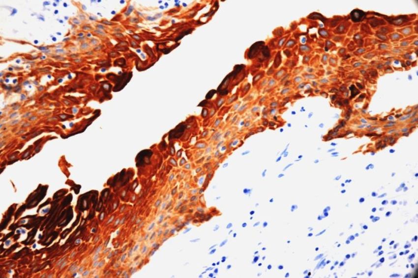
Annals of R.S.C.B., ISSN:1583-6258, Vol. 25, Issue 6, 2021, Pages. 892-896
Received 25 April 2021; Accepted 08 May 2021.

             useful tool in identifying different epithelial types and origins.6
             The present study was done to evaluate the expression of cytokeratins in GOC and also to
             evaluate if cytokeratins can be used as a diagnostic tool for GOC?

             MATERIALS & METHODS: 4 cases were taken from archives of Department of Oral &
             Maxillofacial Pathology which were diagnosed as GOC histopathologically &
             Immunohistochemistry done. The cases were retrieved with complete relevant clinical,
             radiographic and histopathological data. Clinical features such as age, sex, site of lesion and
             presenting features were analyzed. The GOC cases were diagnosed based on the classic
             criteria described by Gardner DG et al3 and WHO7 regarding histologic typing of
             odontogenic cysts and tumors. All tissue specimens were fixed in 10% neutral buffered
             formalin (18–48 h) and routinely processed and embedded in paraffin, including
             decalcification in 25% formic acid for 48–72 h if necessary. Histopathologic diagnosis was
             confirmed by two experiential pathologists on hematoxylin and eosin-stained section for each
             case, and periodic-acid Schiff (PAS) and Alcian Blue-stained slides were also used.
             Immunostaining for CK 7, CK 13, CK 14, CK 19 and CK8/18 was performed using a
             standard biotinstreptavidin immunoperoxidase technique on paraffin sections.
             Tissue sections of oral mucosal epithelium and salivary gland were stained as positive
             controls.
             Immunohistochemical reactivity for CKs was detected in the cytoplasm of the epithelial cells
             of GOC. Cytokeratin (CK) immunostaining was semi quantitatively analyzed in the epithelial
             lining of the cysts as: + = Weakly Positive, ++ =moderately Positive, +++ = strongly
             Positive and - = Negative

             RESULTS:

             Table 1: Clinical and radiographic details of the cases
                  CASES         AGE       SEX           LOCATION                                  R/F
                  Case 1        24 yrs    Male          Mandibular anterior region              Unilocular
                  Case 2        55 yrs    Female        Extending from 11 to18                  Unilocular
                  Case 3        19 yrs    Female        Extending from 12 to 16                 Unilocular
                  Case 4        32 yrs    Male          Extending from 31 to 36                 Unilocular

             Table 2: Cytokeratin (CK) immunostaining
                         CASES          CK 7    CK 13                  CK 14      CK 19     CK 8/18
                         Case 1          +++     +++                    +++         ++        _
                         Case 2           ++     +++                    +++        +++        _
                         Case 3          +++     +++                     ++        +++        _
                         Case 4           ++     +++                     ++        +++        _

             Table 3: Histopathologic features:
                      Squamous Intraluminal              Epithelial     Hobnail   Goblet    Micro     Ciliated   Clear
                      epithelial papillary               plaques        cells     cells     cysts     Cells      cells
                      lining      projections
                      with flat
                      interface
             Case 1 Present       Present                Present        Present   Present   Present Absent       Present
             Case 2 Present       Absent                 Present        Present   Present   Absent Present       Present
             Case 3 Present       Present                Present        Absent    Present   Absent Present       Present

             http://annalsofrscb.ro                                                                              893
Annals of R.S.C.B., ISSN:1583-6258, Vol. 25, Issue 6, 2021, Pages. 892-896
Received 25 April 2021; Accepted 08 May 2021.
Case 4 Present Present Present Present Present Present Absent Present
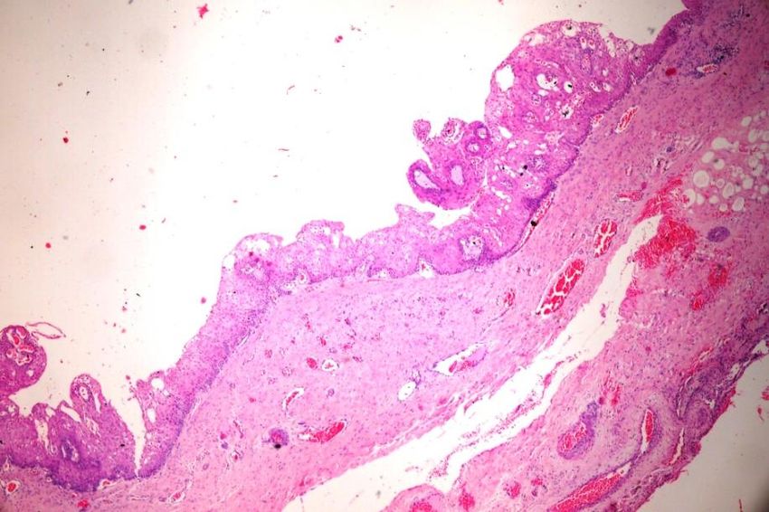
Figure 1 H&E staining showing GOC
Figure 2 Immunohistochemical staining in GOC
Discussion: GOC is an uncommon developmental cyst with a frequency of 0.012%–1.3% of
all the jaw cysts and its prevalence is 0.17%.8 The current case series is a step further to
correlate CK expression with origin of GOC and if cytokeratins can be used as a diagnostic
tool for GOC?
According to de Sousa SO et al9, GOC express CKs7, 13, 14 & 19 but not CKs 8 & 18.
Negative CK 8 & 18 differentiates GOC from Low grade Mucoepidermoid Carcinoma.
In present study, CK 7 is moderately to strongly positive all cases. This shows that CK 7 can
be used as a specific marker for knowing odontogenic origin. It is especially expressed in
cells of HERS & Stellate Reticulum and can be found in several simple epithelia or
occasional basal/ suprabasal cells of stratified epithelium.10
In present study, CK 13 is strongly positive in all cases indicating odontogenic origin of
GOC. CK 13 is predominantly present in Dental Lamina and is Restricted to suprabasal
layers of squamous stratified epithelium. It is also found positive in epithelial islands with
metaplastic squamous cells, some inner stellate cells & flattened cells lining cystic
http://annalsofrscb.ro 894Annals of R.S.C.B., ISSN:1583-6258, Vol. 25, Issue 6, 2021, Pages. 892-896
Received 25 April 2021; Accepted 08 May 2021.
structures.11
In this study, CK 14 is moderately to strongly positive indicating odontogenic epithelium. It
is the main intermediate filament of Odontogenic epithelium and observed in Dental Lamina,
in Reduced Enamel Epithelium & in almost all cells of Enamel Organ. It is expressed
intensely positive in basal cell layer & plaque like areas, less intensely in suprabasal layer of
cystic epithelium.12
In this study, CK 19 is showing positive results in all cases of GOCs. This supports
Odontogenic origin of GOC. Its Positive detection in preameloblasts & secretory ameloblasts
shows its association with secretory differentiation and can be used as a marker of
odontogenic differentiation.11
In present study, CK 8/18 shows negative results. This supports odontogenic origin &
differentiates GOCs from Mucoepidermoid Carcinoma. CK 8/18 is Not seen in epithelia of
Odontogenic Neoplasms. It is Positive in Salivary gland tumors and helps to differentiate
odontogenic epithelium from Salivary glands.13
According to Pires FB et al all GOCs expressed CK 5, CK 7, CK 8, CK 13, CK 14 & CK 19.
All Odontogenic cysts expressed CK 5, CK 13 & CK 14. 91% also expressed CK 19. Only
7% of Odontogenic lesions expressed CK 18, which was expressed by all Mucoepidermoid
Carcinomas of Central & Salivary gland types.
Histopathologically, it may prove to be a diagnostic dilemma due to its close resemblance to
lateral periodontal cyst, Botryoid odontogenic cyst, dentigerous cyst and most importantly
Central mucoepidermoid carcinoma. It is mandatory to differentiate GOC from the much
more aggressive lesions like Central mucoepidermoid carcinoma, and we recommend the use
of CK 7, CK 13, CK 14 and CK 19 antibody to establish odontogenic origin when in doubt.
Conclusion: Although GOC is relatively rare, correct diagnosis is of major clinical
importance, since GOC has an aggressive potential, a high incidence of cortical perforation &
a relatively high rate of recurrence, especially in cases treated with a conservative approach is
observed.
REFERENCES:
1. Nikalje TS, Shah K and Nerurkar S. Glandular Odontogenic Cyst of Mandible: A Rare
Case. Acta Scientific Medical Sciences. 2020;4(2):1-3.
2. Padayachee A, Van Wyk CW. Two cystic lesions with features of both the botryoid
odontogenic cyst and the central mucoepidermoid tumour: sialo-odontogenic cyst? J Oral
Pathol. 1987; 16: 499–504.
3. Gardner DG, Kessler HP, Morency R, Schaffner DL. The glandular odontogenic cyst: an
apparent entity. J Oral Pathol 1988;17: 359–66.
4. Koppang HS, Johannessen S, Haugen LK, Haanaes HR, Solheim T, Donath K. Glandular
odontogenic cyst (sialoodontogenic cyst): report of two cases and literature review of 45
previously reported cases. J Oral Pathol Med. 1998;27:455–62.
5. Urs AB, Kumar P, Augustine J, Malhotra R. Glandular odontogenic cyst: Series of five
cases. J Oral Maxillofac Pathol 2017;21:239-43.
6. Domingues MG, Jaeger MMM, Araujo VC, Araujo NS. Expression of cytokeratins in
human enamel organ. Eur J Oral Sci. 2000;108: 43–7.
7. Kramer IRH, Pindborg JJ, Shear M. Histological typing of odontogenic tumors. WHO
international histological classification of tumors, 2nd edn. Berlin, Germany: Springer-
Verlag, 1992; 38.
8. Krishnamurthy A, Sherlin HJ, Ramalingam K, Natesan A, Premkumar P, Ramani P, et
al. Glandular odontogenic cyst: Report of two cases and review of literature. Head Neck
Pathol 2009;3:153‑8.
9. de Sousa SO, Cabezas NT, de Oliveira PT, de Araujo VC. Glandular Odontogenic
http://annalsofrscb.ro 895Annals of R.S.C.B., ISSN:1583-6258, Vol. 25, Issue 6, 2021, Pages. 892-896
Received 25 April 2021; Accepted 08 May 2021.
Cyst: analysis of a case with cytokeratin expression. Oral Surg Oral Med Oral Pathol
Oral Radiol Endod 1997; 83(4):478-83.
10. Saluja P, Arora M, Dave A, et al. Role of Cytokeratin-7 in the pathogenesis of
odontogenic cysts - an immunohistochemical study. Med Pharm Rep. 2019;92(3):282-7.
11. Crivelini MM, de Araujo VC, de Sousa SOM , de Araujo NS. Cytokeratins in epithelia
of odontogenic neoplasms. Oral Diseases. 2003;9:1–6.
12. Shruthi DK, Shivakumar MC, Tegginamani AS, Karthik B, Chetan BI. Cytokeratin 14
and cytokeratin 18 expressions in reduced enamel epithelium and dentigerous cyst:
Possible role in oncofetal transformation and histogenesis- of follicular type of
adenomatoid odontogenic tumor. J Oral Maxillofac Pathol. 2014;18(3):365-71.
13. Pires FB, Chen SY, da Cruz Perez DE, de Almeida OP, Kowalski LP. Cytokeratin
expression in Central Mucoepidermoid Carcinoma and Glandular Odontogenic Cyst.
Oral Oncol 2004;40: 545-51.
http://annalsofrscb.ro 896You can also read