CLASSIFICATION OF FRACTURES AND DISLOCATIONS OF THE THORACIC AND LUMBAR SPINE - KEVIN J. DISILVESTRO MD AND ALAN H. DANIELS MD DEPARTMENT OF ...
←
→
Page content transcription
If your browser does not render page correctly, please read the page content below
Classification of Fractures and
Dislocations of the Thoracic and
          Lumbar Spine
        Kevin J. DiSilvestro MD and Alan H. Daniels MD
Department of Orthopaedics, The Warren Alpert Medical School of
               Brown University, Providence, RI.

                                                             Core Curriculum V5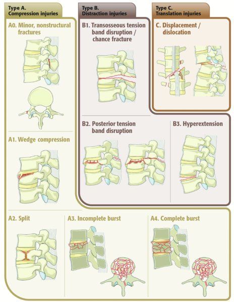
Objectives
• Understand the anatomy and its impact on pathology
• Learn the patterns of neurologic injury
• Understand the widely used classification systems
• Learn how to determine operative versus non operative management

                                                          Core Curriculum V5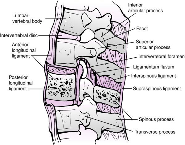
Introduction
• Males > females
   • 30% males in 30s
• High energy injury
• MVA >> Fall
• Spinal column injury, 10-25%
  neurologic deficit
• Thoracolumbar spine most
  common site of spinal injuries

                                   Core Curriculum V5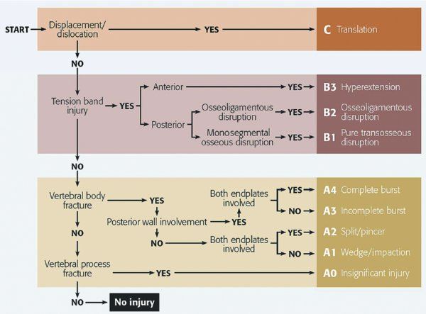
Introduction
• Frequency (Gertzbein 1994)
   • T11-L1(52%) > L1-5(32%) > T1-10(16%)
• Up to 50% have associated injuries
   • Intra-abdominal (liver, spleen)
   • Pulmonary injury- up to 20%
   • Up to 15% noncontiguous fractures

                                            Core Curriculum V5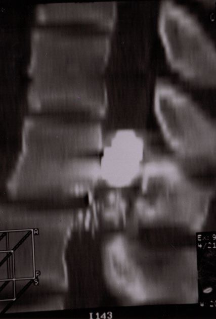
Clinical Anatomy
• Thoracic spine
   • Kyphotic
   • Facets in coronal plane
   • Stabilized by ribs
      • 2X Flexion stiffness
      • 3X lateral bending stiffness
      • Prevent rotation
   • ↓ canal:cord ratio (T2 to T10)
      • More susceptible to SCI

                                       Core Curriculum V5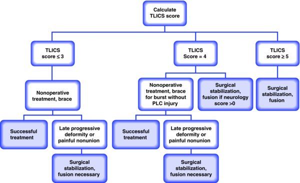
Clinical Anatomy
• Lumbar spine
  • Lordotic
  • Oblique/sagittal facet orientation
     • Prevent rotation
  • Wider canal
  • Cauda equina

                                         Core Curriculum V5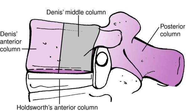
Clinical Anatomy
• Thoracolumbar
• Area of transition
• Decreased stiffness (below rib cage)
• Facet orientation (coronal sagittal)
• More vertical
• Coronal to 45º inward
• T11-12 “weak link”
• Predisposed to rotational injuries
• Absence of rib support with transitional facets
Core Curriculum V5Neuroanatomy
• Spinal Cord: C1-L1
• Conus medullaris: L1-2
• Cauda equina: L2-S5
Core Curriculum V5Patterns of Neurologic Injury
• Spinal cord injury
• Complete versus incomplete
• Conus medullaris syndrome
• Root injury
• Isolated root dysfunction
• Cauda equina syndrome
Core Curriculum V5ASIA Classification
• A = Complete
• No motor or sensory function is preserved in the sacral segments S4-S5
• B = Incomplete
• Sensory but no motor function is preserved below the neurological level and
includes the sacral segments S4-S5
• C = Incomplete
• Motor function is preserved below the neurological level, and more than half of
the key muscles below the neurological level have a muscle grade less than 3
• D = Incomplete
• Motor function is preserved below the neurological level, and at least half of the
key muscles below the neurological level have a muscle grade of 3 or more
• E = normal
• Motor and sensory function are normal
Core Curriculum V5Conus Medullaris Syndrome
• Thoracolumbar cord injury
• Presents with low back pain, lower extremity weakness, saddle
anesthesia, bowel/bladder dysfunction
• Injury to sacral myelomeres
• Can involve both upper and lower motor neurons (+/- root escape)
• Isolated: pure bowel, bladder & sexual dysfunction
• Combined root/cord injury: lumbar traversing roots
Core Curriculum V5Conus Medullaris Syndrome
• Exam
• Absent bulbocavernosus reflex
• Bowel/bladder/sexual dysfunction more frequent than lower extremity
weakness
• Treatment: urgent decompression
• Prognosis
• ? Improved with early decompression
• Better for root recovery (L1-4) than cord recovery (L5-S)
Core Curriculum V5Cauda Equina Syndrome
• Injury below L1/2 level
• Bowel and bladder dysfunction
• Urinary retention overflow incontinence
• Fecal incontinence
• Bilateral motor/sensory deficits
• Diminished perianal sensation and rectal tone
Core Curriculum V5Cauda Equina Syndrome
• Natural history: progressive weakness of lower extremities, loss of
bladder/bowel function
• Prognosis: presence of saddle anesthesia/bladder symptoms
associated with worse outcomes
• Treatment: Urgent decompression for best prognosis
• Treatment after 48 hours associated with worse outcomes
Core Curriculum V5Thoracolumbar Trauma
Classification
Core Curriculum V5Mechanisms of Injury
• Axial compression
• Flexion
• Lateral compression
• Flexion-Rotation
• Flexion-Distraction
• Shear
• Extension
Core Curriculum V5Thoracolumbar Trauma Classification
• Decision to operate often challenging
• >10 classification schemes described
• A useful classification system should:
• Be easy to remember, use, and communicate
• Predict patient outcome
• Drive treatment
• Have high inter- and intra-oberver reliability
Core Curriculum V5Denis Classification
• 1983: retrospective review of
412 thoracolumbar fractures
• 3 columns
• Anterior: anterior body, disc, ALL
• Middle: posterior body, disc, PLL
• Posterior: interspinous ligament,
supraspinous ligament, posterior
elements
• Middle column “crucial”
Core Curriculum V5Compression Fracture
• Failure of anterior column only
• Anterior (or lateral)
• Flexion (or lateral
bending/compression)
• Typically stable
• Brace when near thoracolumbar
junction
Core Curriculum V5Burst Fracture
• Involves middle column
• Axial load +/- rotation, etc
• Associated lamina fracture
70% dural tear
• Stable (low lumbar) vs. unstable
(thoracic, thoracolumbar, upper
lumbar)
Core Curriculum V5Flexion-Distraction
• Seat belt injuries
• Chance fracture (bony,
ligamentous, both)
• Distraction of posterior and
middle columns
• 0-10% neuro involvement
• Often requires surgery
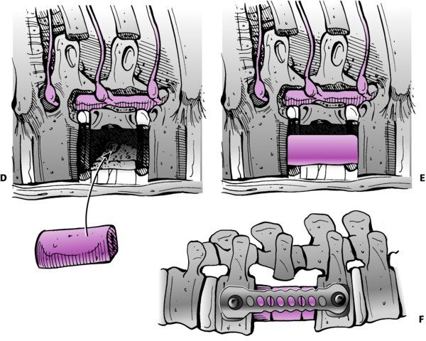
Core Curriculum V5Fracture Dislocation
• Injury to all three columns
• Combination of high energy
forces (shear)
• High likelihood of neuro deficit
• Always requires surgery
Core Curriculum V5Denis Classification: Fracture Dislocation
Core Curriculum V5AO Classification
Core Curriculum V5AO Classification
Core Curriculum V5Thoracolumbar Injury Classification and
Severity Score (TLICS)
• Developed to drive surgical versus nonoperative management of
thoracolumbar fractures (Vaccaro et al 2005)
• Score based on 3 factors
• Injury morphology/mechanism
• Posterior ligamentous integrity
• Neurologic injury
Core Curriculum V5TLICS: Injury Morphology
• Compression = 1 point
• Burst = +1 point
• Translation/rotational = 3 points
• Distraction = 4 points
Core Curriculum V5TLICS: PLC Integrity
• PLC disrupted in tension,
rotation, or translation
• Intact = 0 points
• Suspected/indeterminate = 2
points
• Injured = 3 points
Core Curriculum V5TLICS: Neurologic Status
• Involvement
• Intact = 0 points
• Nerve root = 2 points
• Cord, conus medullaris
• Complete = 2 points
• Incomplete = 3 points
• Cauda Equina = 3 points
Core Curriculum V5TLICS Score
• Relatively simple, shown to predict treatment, high IRR
Core Curriculum V5Stable vs. Unstable Injuries
• White and Panjabi (Spine 1978)
“the loss of the ability of the spine under physiologic conditions to maintain
relationships between vertebrae in such a way that there is neither damage
nor subsequent irritation to the spinal cord or nerve root and, in addition,
there is no development of incapacitating deformity or pain from structural
changes”
Core Curriculum V5Stable vs. Unstable Injuries
• Stable (Burst fracture)
• < 25-30º kyphosis
• < 50% loss of height
• < 30–50% canal compromise
• Neuro intact
• Unstable
• Neurologic deficit
• > 25-30º kyphosis
• > 50% loss of height
• >50-60% canal compromise
*No study to date has shown direct correlation between percentage of canal
compromise and severity of neurologic injury following burst fracture
Core Curriculum V5Surgical Decision Making
• Goals
• Maximize function
• Facilitate nursing care
• Prevent deformity/instability
• Potentially improve neurologic function
• Indications
• ? Instability
• Neurologic compression
• Posterior osteoligamentous disruption
Core Curriculum V5Surgical Approaches
• Anterior
• Transthoracic (T4- 9)
• Thoracoabdominal (T10- L1)
• Retroperitoneal (T12- L5)
• Posterior
• Indirect reduction possible: ligamentotaxis
• Posterolateral
• Transpedicular, costotraversectomy, etc
• Lateral
• XLIF, DLIF
Core Curriculum V5Surgery
• Indicated in Unstable injuries
• Anterior surgery for anterior compression at TL junction
• Posterior surgery in lower Lumbar spine
Core Curriculum V5Gunshot Wounds
• Non-operative treatment standard
• Steroids not useful (Heary, 1997)
• 10-14 days IV antibiotics for colonic perforations
ONLY
• Evidence for debridement and extraction of bullet
fragments is controversial, however patients with
incomplete injuries may improve (Scott 2019)
Core Curriculum V5Treatment
• Decompression rarely of benefit
except for intra-canal bullet from
T12-L5
• Better motor recovery than
nonoperative
• Fractures usually stable despite
“3-column” injury
Core Curriculum V5GSW to the spine Outcome and Complications
• Most dependent on SCI and associated injuries
• High incidence of CSF leaks with unnecessary decompression
• Lead toxicity rare, even with bullet in canal
• Bullet migration rare: late neurological sequelae
Core Curriculum V5Summary
• There are several patterns of neurologic injury for thoracolumbar
trauma
• There are multiple classification schemes
• Most thoracolumbar injuries, in the absence of neurologic deficit, are
stable and can be treated successfully nonoperatively
• Surgical intervention may be beneficial in improving patient
mobilization and early functional return for unstable spine fractures
• Indications for surgical intervention, timing of intervention, and
approach remain controversial
Core Curriculum V5Conclusions
• The goals of managing thoracolumbar injuries are to maximize
neurologic recovery and to stabilize the spine for early rehab
and return to a productive lifestyle
Core Curriculum V5Key References
• Denis F. The three column spine and its significance in the
classification of acute thoracolumbar spinal injuries. Spine (Phila Pa
1976). 1983;8(8):817-831. doi:10.1097/00007632-198311000-00003
• Vaccaro AR, Lehman RA Jr, Hurlbert RJ, et al. A new classification of
thoracolumbar injuries: the importance of injury morphology, the
integrity of the posterior ligamentous complex, and neurologic
status. Spine (Phila Pa 1976). 2005;30(20):2325-2333.
doi:10.1097/01.brs.0000182986.43345.cb
Core Curriculum V5Full References
Figures used with permission. Gendelberg D, Bransford RJ, Bellabarba C. Thoracolumbar Spine Fractures and Dislocations. In: Tornetta P, Ricci WM,
eds. Rockwood and Green's Fractures in Adults, 9e. Philadelphia, PA. Wolters Kluwer Health, Inc; 2019.
Magerl F, Aebi M, Gertzbein SD, Harms J, Nazarian S. A comprehensive classification of thoracic and lumbar injuries. Eur Spine J. 1994;3(4):184-201.
Brouwers E, van de Meent H, Curt A, Starremans B, Hosman A, Bartels R. Definitions of traumatic conus medullaris and cauda equina syndrome: a
systematic literature review. Spinal Cord. 2017;55(10):886-890.
Ahn UM, Ahn NU, Buchowski JM, Garrett ES, Sieber AN, Kostuik JP. Cauda equina syndrome secondary to lumbar disc herniation: a meta-analysis of
surgical outcomes. Spine (Phila Pa 1976). 2000;25(12):1515-1522.
Denis F. The three column spine and its significance in the classification of acute thoracolumbar spinal injuries. Spine (Phila Pa 1976). 1983;8(8):817-831.
Spine. Journal of Orthopaedic Trauma. 2018;32(1):S145-S160.
Vaccaro AR, Lehman RA, Hurlbert RJ, et al. A new classification of thoracolumbar injuries: the importance of injury morphology, the integrity of the
posterior ligamentous complex, and neurologic status. Spine (Phila Pa 1976). 2005;30(20):2325-2333.
White AA, Panjabi MM. The basic kinematics of the human spine. A review of past and current knowledge. Spine (Phila Pa 1976). 1978;3(1):12-20.
Heary RF, Vaccaro AR, Mesa JJ, et al. Steroids and gunshot wounds to the spine. Neurosurgery. 1997;41(3):576-583; discussion 583-584.
Scott KW, Trumbull DA, Clifton W, Rahmathulla G. Does surgical intervention help with neurological recovery in a lumbar spinal gun shot wound? A case
report and literature review. Cureus. 2019;11(6):e4978.
Core Curriculum V5You can also read