Clinical management of maggot wounds in Asiatic Black Bear (Ursus thibetanus)
←
→
Page content transcription
If your browser does not render page correctly, please read the page content below
Scientific Reports in Life Sciences 2021 2 (1): 7-12
                                DOI: http://dx.doi.org/10.22034/srls.2020.520564.1006
                                                                                                                           Short communication

Clinical management of maggot wounds in Asiatic Black Bear (Ursus
thibetanus)

Vipin Kumar
MD Instructor, HPFD , India
*
  Corresponding email: vipinvet@gmail.com

Received: 1 November 2020 / Revised: 28 January 2021 / Accepted: 30 January 2021 / Published online: 4 Februry 2021.
How to cite: Kumar V. (2021). Clinical management of maggot wounds in Asiatic Black Bear (Ursus thibetanus), Scientific Reports in Life Sciences 2(1), 7-
12. http://dx.doi.org/10.22034/srls.2020.520564.1006

Abstract
Like most other species, the Asiatic black bear is vulnerable to a wide range of diseases, including
infection, inflammation, parasitic infestation, and degenerative disease. The study reported the surgical
and conservative management of maggot wounds, including anesthetic protocol and postoperative care in
Himalayan Black Bear (Ursus thibetanus). Anesthesia was performed successfully by administering
Xylazine and Ketamineintramuscularly (IM) by remote injection using a tranquilizing gun with a 10CC
anesthetic dart. The animals' clinical evaluation showed an induction time of 4.5 minutes, duration of
anesthesia was 72.0 minutes, and recovery time of 38.0 minutes. All the physiological parameters were
within normal limits.

Keywords: Anesthesia, myiasis, Physiological parameters

Introduction
Zoos are an ex-situ form of conservation where animals are displayed in cages or enclosures for esthetic,
educational or research, and conservation purposes (Thawait et al. 2014) In captivity the health status of
the animals depends on many factors, like feeding, keeping conditions, animal management and
environmental conditions such as temperature and humidity.
The Asiatic black bear is distributed throughout the Himalayan ranges in the northwest (Jammu and
Kashmir; Himachal Pradesh), west (Himachal Pradesh and Uttaranchal), Central Sikkim, and Northern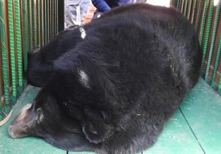
Kumar 2021 Scientific Reports in Life Sciences 2(1): 7-12
West Bengal) and east (Arunachal Pradesh) (Sathyakumar 1999). The Asiatic black bear, like most other
species, are vulnerable to a wide range of diseases, including infection, inflammation, parasitic
infestation, and degenerative disease. Myiasis is the parasitic infestation of tissue of a live mammal by
dipterans larvae (maggots) that grow inside the host while feeding on its tissue, causing more or less
every traumatizing injury (Hall and Farkas 2000). This infestation eventually turns into a maggot wound,
which annoys animals and disrupts regular feeding and resting habits. Left untreated, maggot wounds are
fatal as the animal may die due to the maggots tunneling into vital organs (depending on the wound's
site), blood loss, or secondary infections. The most affected hosts for myiasis are cattle (46.4%) followed
by dogs (15.3%), humans (14.7%), pigs (6.0%), horses (4.0%), and sheep (1.0%) (Sergio et al. 2007).
Myiasis causes a reduction in the production of meat, milk, and wool in livestock species, but in the case
of wildlife, it leads to wild animals' death in captivity and the free-ranging wildlife (Kumar and Raj
2012).
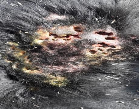
There is a shortage of information about the management of maggot wounds in wildlife. This case report
describes the medicinal management of maggot wound in Asiatic Black Bear (Fig. 1 and 2).
Figure 1: Maggot wound in Himalayan Black Bear
Figure 2: Maggots recovered from wounds
8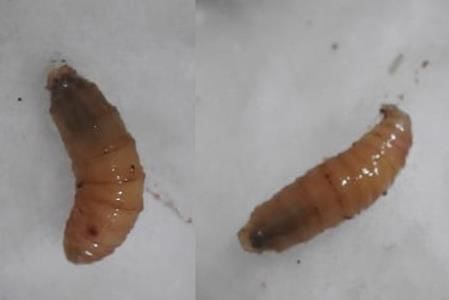
Kumar 2021                 Scientific Reports in Life Sciences 2(1): 7-12

Materials and Methods
Five years old male Asiatic black bear (weighing about 120 kg) was observed to be listless, tended to be
isolated, inappetence, restlessness, frequent head shaking, and used to vigorously rub his back regions
against wall or stones in the enclosure area. This all necessitated clinical investigation and treatment. The
animal had a history of infighting with other males in the enclosure.

Anesthesia, Surgical procedure, and Postoperative care
The sick bear was chemically immobilized for closer examination, sampling, and treatment. The bear was
moved to an indoor den with good lighting and ventilation to allow a quiet induction and recovery. The
feed and water were withheld for 24hrs and 8hrs, respectively, before immobilization. The animal was
anesthetized by combined injection of Xylazine hydrochloride (Ilium Xylazil®, Troy Laboratories Pvt.
Ltd., Australia) dosed at 2 mg/kg body weight (bwt), and Ketamine hydrochloride (Vetalar®, Boehringer
Ingelheim Vetmedica, Inc. St. Joseph, MO64506 USA) dosed at 5mg/kg bwt intramuscularly (IM) by
remote injection using a tranquilizing gun with 10CC anesthetic dart. The anesthesia was reversed with
Yohimbine dosed at 0.3mg/kg bwt intramuscularly (IM) (Reverzine® Bayer Australia Ltd (Animal
Health)) (Fig. 3). Physiological parameters like body temperature, respiration rate, heart rate, and mucous
membrane color were monitored throughout the anesthesia at frequent intervals.

                                 Figure 3: Anaesthetized Asiatic Black Bear

After immobilization, multiple maggot wounds with whitish/creamy maggots were observed streaming
out from the wounded parts, on his back and neck region area. The skin over and around the wound was
seen red, swelled, and inflamed indeed. The wounds were evaluated for the spreading and depth of tissue
involved. Some of the scars have formed pockets with the length, width, and depth varying between 2.5-
4.0 cm, 2.0-3.0 cm, and 1.2-1.8 cm. At the same time, other wounds did not form pocket instead passed
beneath the skin and formed a tract wound of 4.0-5.5 cm in length. Hairs around each wound were
clipped using curved scissors. The operative site was then painted with sterile gauze soaked in tincture
iodine for disinfection of the skin. Superficial maggots were removed from the wound pocket using
sterile tissue forceps. The wound pocket was also packed with a gauge dipped in medicinal turpentineoil
for 10 minutes to remove the deep-seated maggots. The live and dead maggots that came out to the
surface of the wound were removed using simple tissue forceps. After removal of all maggots present in

                                                     9
Kumar 2021 Scientific Reports in Life Sciences 2(1): 7-12
wound sterile gauze soaked in tincture, iodine was used to clean out the dead tissue debris and to induce
inflammation. Thereafter, maggoticidal and bacteriostatic wound dressing powder (Negasunt powder®)
was sprinkled into the wound pocket. Then to prevent flies from sitting and laying eggs on this wound
site,a layer of Himax( Ayurvet Ltd.) and Lorexane (Virbac India) mixed in the proportion of 1:5 was
applied together. Postoperatively a subcutaneous injection of 1% Ivermectin (HITEK®, VirbacIndia) at
200 micrograms /kgb.wt was given. A broad spectrum antibiotic viz., Single application Enrofloxacin
(Fortivir®, Virbac India) dosed at 3ml/40kg b.wt. was injected IM at the time of operation and then
repeated once at 72 h interval. Antihistaminic preparation (Chlorpheniramine maleate, Cadistin®, Zydus
animal health Ltd., India) at 1 mg/kg b.wt was administered IM once daily for three consecutive days.
The treated wounds were observed on the third day of operation, and no maggots were found. The
wounds wholly healed after two weeks following the operation.
Results and Discussion
Anesthesia was performed successfully by administering Xylazine (3mg/kg body weight) and Ketamine
(5mg/kg body weight). Bears are monogastric and may vomit during induction or recovery, or regurgitate
while anesthetized, therefore feed and water was withheld for 24hrs and 8 hrs, respectively, before
immobilization.
The induction of anesthesia was smooth with good muscle relaxation throughout the surgical procedure.
The induction time was observed to be 4.5 min, adequate depth of anesthesia was maintained for 72 min
without the need for supplementation. The recovery time was 38 min after Reverzine. In wildlife cases,
wound treatments require general sedation or anesthesia. This is particularly true if extensive cleaning
and debridement (surgical removal of dead and severely damaged tissue) are necessary.
During the immobilization period, the animal was monitored with great caution. The vital health signs
like heart rate, respiratory rate, temperature, and color of mucous membranes were monitored and
recorded at 15 min intervals (Morris 2001).
Bears are prone to hyperthermia because of their thick fat layer; close monitoring of body temperature
during anesthesia is essential. Physiological parameters on an average basis revealed {heart rate (48±12
/min), respiration rate (12±7/min), and rectal temperature (37±1.5oC)} normal values.
Infighting injuries during the breeding season and resultant abrasion/lacerations on the skin surface or
open wound is a predisposing factor for cutaneous myiasis. Myiasis is a parasitic infestation. Its
incidence rate is higher in tropics, South–East Asia, and subtropics of Africa, where the causative
predisposing factors are exposed to myiasis–causing flies and their increased aggressiveness (Bolognia et
al. 2008). The efficacy of oil of turpentine in the treatment of myiasis has been reported earlier as
turpentine oil creates an anoxic condition in the wound pocket, and as a result, the maggots crawl out of
the pocket within three to five minutes (Bowe etal. .1977).
In this case study, the application of medicinal turpentine oil to the maggots wound helped in removing
maggots from the wound. Moreover, the oil of turpentine enhances ceruloplasmin activities that inhibit
inflammatory injury by its antioxidant property (DiSilvestro 1989). Ivermectin injectable solution is a
highly active, broad-spectrum parasiticide for parenteral administration; however, and data is minimal
regarding the efficacy and safety of Ivermectin in wild animals. In this study, Ivermectin was
administered dosed at 200 μg/kg bwt like domestic animals. Single application Enrofloxacin was
10Kumar 2021 Scientific Reports in Life Sciences 2(1): 7-12
administered to avoid secondary bacterial infection. The antihistaminic drug was used to counteract the
histamine released by damaged tissue. Ascorbic acid (vitamin C) was given orally once daily for one
week to promote the healing of a wound by promoting keratinocyte differentiation (Savini et al. 2002,
Duarte et al. 2009)and stimulating the formation of an epidermal barrier (Boyce et al.. 2002).
In this case report, maggot wounds completely healed in 14 days (2 weeks) post-operation. Similarly,
(Rahman et al. 2009) reported that 90.3% wound areas were healed by day 17 of operation for maggot
wounds in cattle treated with Ivermectin and broad-spectrum antibiotic. Early wound healing, in this case
might be due to species variation and an additional supplement of ascorbic acid. All these factors might
have promoted the recovery of the wound.
Conclusion
It can be concluded that surgical management using oil of turpentine and tincture iodine and topical
dressings with maggoticidal and bacteriostatic along with parenteral administration of Ivermectin, Single
application of Enrofloxacin and chlorpheniramine maleate is sufficient for successful management of
maggot wounds in Himalayan black bear in captivity.
Acknowledgments
The author is thankful to all the supporting staff of Gopalpur Zoo for their help during surgical
procedures and further managing animals during the postoperative period.
References
Bolognia J.L., Jorizzo J.L., Rapini R. 2008. Cutaneous myiasis. In: Dermatology. Vol 1, 2nd ed.: Mosby
Elsevier
Bowe D.L., Amaro T.E., Sotolongo G.F., Alonso BP. 1977. Oticmyiasis in a newborn caused by
Caliphoridae larvae of genus Phaenicia. Revista Cubana de Medicina Tropical 29: 75-79.
Boyce S.T., Supp A.P., Swope V.B., Warden G.D. 2002. Vitamin C regulates keratinocyte viability,
epidermal barrier, and basement membrane in vitro and reduces wound contraction after grafting of
cultured skin substitutes. Journal of Investigative Dermatology 118: 565-572.
DiSilvestro R.A. 1989. Effects of inflammation on copper antioxidant enzyme levels: Advanced efficacy
of different insecticides in the treatment of cattle hypodermis in north-eastern Algeria. Veterinary
Research 29(1): 21-29.
Duarte T.L., Cooke M.S., Jones G.D. 2009. Gene expression profiling reveals new protective roles for
vitamin C in human skin cells. Free Radical Biology and Medicine 46: 78-79
Hall M.J., Farkas R. 2000. Traumatic myiasis of humans and animals. In: Contributions to a Manual of
Palae arctic Diptera. Budapest: Science Herald.
Kumar V., Raj A. 2012. Management of wound myiasis in a lion (Panthera leo). International Journal for
Agro Veterinary and Medical Sciences 6(1): 4-6.
Morris P.J. 2001. Chemical immobilization of felids, ursids, and small ungulates. Veterinary Clinics of
North America Exotic Animal Practice 4: 267-298.
11Kumar 2021 Scientific Reports in Life Sciences 2(1): 7-12
Rahman M.A., Hossain M.A., Alam M.R. 2009. Clinical evaluation of different treatment regimes for
management of myiasis in cattle. Bangladesh Journal of Veterinary Medicine 7(2): 348-352.
Sathyakumar S. 1999. Status and Management of Asiatic black bear in India. Bears: Status survey and
Conservation Action Plan. Servheen et al., 202-207.
Savini I., Catani M.V., Rossi A., Duranti G., Melino G., Avigliano L. 2002. Characterization of
keratinocyte differentiation induced by ascorbic acid: protein kinase C involvement and vitamin C
homeostasis. Journal of Investigative Dermatology, 118: 372-379.
Sergio E.B., Jose D.E., Angel B.C., Franklin C., Janina S., Sabina B., Enrique M. 2007. Incidence of
myiasis in Panama during the eradication of Cochliomyiahominivorax. seccion de EntomologiaMedica,
InstitutoConmemnorativo Gorgas de Estudios de laSalud, PO Box 0816-02593, Panama.
Thawait, V.K., Maiti, S.K. Dixit, A.A. 2014. Prevalence of gastro-intestinal parasites in captive wild
animals of Nandan Van Zoo, Raipur, Chhattisgarh. Vet. World, 7(7): 448-445.
12You can also read