Correlation of ER, PR, and HER2 at the protein and mRNA levels in Asian patients with operable breast cancer
←
→
Page content transcription
If your browser does not render page correctly, please read the page content below
Bioscience Reports (2022) 42 BSR20211706
https://doi.org/10.1042/BSR20211706
Research Article
Correlation of ER, PR, and HER2 at the protein and
mRNA levels in Asian patients with operable breast
cancer
Chih-Jung Chen1 , Ting-Hao Chen2,3 , Jason Lei4 , Ji-An Liang5 , Po-Sheng Yang6 , Chiun-Sheng Huang7 ,
Chia-Ming Hsieh8 , Ling-Ming Tseng9 , Liang-Chih Liu1,10 , Skye Hung-Chen Cheng11 and Kuan-Hui Shih2
Downloaded from http://portlandpress.com/bioscirep/article-pdf/42/1/BSR20211706/928027/bsr-2021-1706.pdf by guest on 09 February 2022
1 Department of General Surgery, China Medical University Hospital, Taichung, Taiwan; 2 Department of Medical Operation, Amwise Diagnostics Pte. Ltd., Singapore; 3 Institute of
Epidemiology and Preventive Medicine, National Taiwan University, Taipei, Taiwan; 4 Department of Product Development, Amwise Diagnostics Pte. Ltd., Singapore; 5 Department
of Radiation Oncology, China Medical University Hospital, Taichung, Taiwan; 6 Department of General Surgery, MacKay Memorial Hospital, Taipei, Taiwan; 7 Department of Surgery,
National Taiwan University Hospital, Taipei, Taiwan; 8 Department of General Surgery, Taiwan Adventist Hospital, Taipei, Taiwan; 9 College of Medicine, National Yang Ming Chiao
Tung University, Taipei, Taiwan; 10 College of Medicine, China Medical University, Taichung, Taiwan; 11 Department of Radiation Oncology, Koo Foundation Sun Yet-Sen Cancer
Center, Taipei, Taiwan
Correspondence: Kuan-Hui Shih (kasey.shih@amwisedx.com)
Breast cancer is the most common cancer and the leading cause of cancer-related deaths
in women. The estrogen receptor (ER), progesterone receptor (PR), and human epider-
mal growth factor receptor 2 (HER2) are the important biomarkers in the prognosis of
breast cancer, and their expression is used to categorize breast cancer into subtypes.
We aimed to analyze the concordance among ER, PR, and HER2 expression levels and
breast cancer subtyping results obtained by immunohistochemistry (IHC, for protein) and
reverse transcriptase-polymerase chain reaction (RT-PCR, for mRNA) and to assess the
recurrence-free survival (RFS) of the different subtypes as determined by the two methods.
We compared biomarker expression by IHC and RT-PCR in 397 operable breast cancer pa-
tients and categorized all patients into luminal, HER2, and triple-negative (TN) subtypes. The
concordance of biomarker expression between the two methods was 81.6% (κ = 0.4075)
for ER, 87.2% (κ = 0.5647) for PR, and 79.1% (κ = 0.2767) for HER2. The κ-statistic was
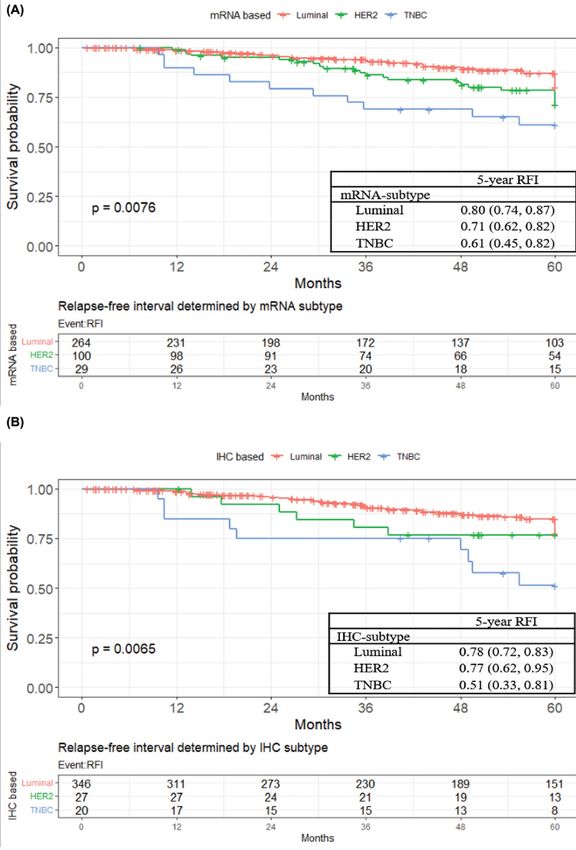
0.3624 for the resulting luminal, HER2, and TN subtypes. The probability of 5-year RFS was
0.78 for the luminal subtype versus 0.77 for HER2 and 0.51 for TN, when determined by IHC
(P=0.007); and 0.80, 0.71, and 0.61, respectively, when determined by the RT-PCR method
(P=0.008). Based on the current evidence, subtyping by RT-PCR performs similar to con-
ventional IHC with regard to the 5-year prognosis. The PCR method may thus provide a
complementary means of subtyping when IHC results are ambiguous.
Introduction
Breast cancer is the most common cancer and the leading cause of cancer-related deaths in women, with
approximately 1.7 million incident cases in 2016 [1]. The estrogen receptor (ER), progesterone receptor
(PR), and human epidermal growth factor receptor 2 (HER2) represent critical pathways for tumor growth
Received: 24 July 2021
and replication of breast cancer cells [2]. Molecular subtypes of breast cancer have been established based
Revised: 21 December 2021 on the biological expression of these three proteins [3]. They include the luminal (ER/PR-positive and
Accepted: 07 January 2022 HER2-negative), HER2 (HER2-positive regardless of ER/PR status), and triple-negative (TN; ER-, PR-,
and HER2-negative) breast cancer subtypes [3,4].
Accepted Manuscript online:
10 January 2022 Surgery to remove the tumor is usually the first line of treatment for breast cancer. To reduce the risk of
Version of Record published: recurrence, patients can consider adjuvant therapies, including hormonal and HER2-targeted therapies
18 January 2022 for luminal and HER2-enriched subtypes, respectively [3]. Correct identification of the different subtypes
© 2022 The Author(s). This is an open access article published by Portland Press Limited on behalf of the Biochemical Society and distributed under the Creative Commons Attribution 1
License 4.0 (CC BY).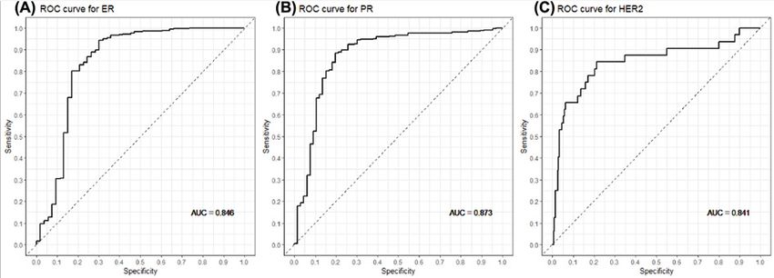
Bioscience Reports (2022) 42 BSR20211706
                                                                                                                                         https://doi.org/10.1042/BSR20211706

                                                                                                                                                                                           Downloaded from http://portlandpress.com/bioscirep/article-pdf/42/1/BSR20211706/928027/bsr-2021-1706.pdf by guest on 09 February 2022
           Figure 1. The expression levels of miR-24-3p and IL-1β in AMI patients

           is thus crucial for the management of breast cancer. Conventionally, oncologists identify this important clinical in-
           formation by estimating ER, PR, and HER2 protein levels using immunohistochemistry (IHC) for staining tumor
           cells [5,6]. Whereas it is relatively easy to perform IHC, reproducibility issues of the IHC method have been reported,
           probably due to the subjective nature of both the sampling process and the interpretation and scoring of the staining
           level [7–9]. Consequently, subtypes could be misidentified, especially in low-volume laboratories [10].
              Alternatively, genomic tests have been reported recently in determining the ER (ESR1), PR (PGR), and HER2
           (ERBB2) status in breast cancer [10–12]. In contrast with the protein levels determined by IHC, it is the gene expres-
           sion (mRNA) levels that are determined by the genomic approach, using the reverse transcriptase-polymerase chain
           reaction (RT-PCR) [13,14]. The reproducibility of the genomic method is very high due to its more homogeneous
           sampling process, and the automated quantification by instruments without human intervention ensures objectivity
           [15,16].
              In the present study, we compared the ER (ESR1), PR (PGR), and HER2 (ERBB2) status and the resulting subtypes
           of nearly 400 breast cancer specimens, as determined by the IHC and RT-PCR methods, and then evaluated their
           corresponding clinical performance in predicting differential breast cancer recurrence.

           Methods
           Study population
           The breast cancer patients treated with breast-conserving surgery (BCS) or mastectomy between 2005 and 2016 at
           multiple medical centers in Taiwan were included in the Amwise dataset (Amwise Diagnostics Pte. Ltd.). Figure 1
           shows the subject selection process. The institutional review board of each participating medical center approved the
           study protocol. The inclusion criteria were (1) with invasive breast cancer; (2) had received mastectomy or BCS as first
           treatment; (3) with ER, PR, and HER2 status confirmed by IHC and/or fluorescence in situ hybridization (FISH) at
           each participating center; and (4) with formalin-fixed paraffin embedded (FFPE) tissue sections for RT-PCR testing.
           Patients at a stage of N3 or M1 were excluded.

2   © 2022 The Author(s). This is an open access article published by Portland Press Limited on behalf of the Biochemical Society and distributed under the Creative Commons Attribution
    License 4.0 (CC BY).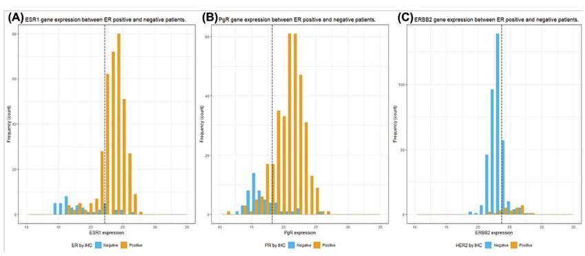
Bioscience Reports (2022) 42 BSR20211706
https://doi.org/10.1042/BSR20211706

Determination of the ER (ESR1), PR (PGR), and HER2 (ERBB2) status
The ER, PR, and HER2 results from the IHC/FISH testing were obtained from the medical charts of each participating
hospital and were considered the gold standard for positive/negative expression and subtype determination. For the
determination of positive/negative expression of the ESR1, PGR, and ERBB2 genes, receiver operating characteristic
(ROC) curve analysis was used. The cut-off value for positive expression of each of the three genes was determined
by the optimal value of both sensitivity and specificity with respect to the IHC results. We normalized the expression
of each of the three target genes (i.e., ESR1, PGR, and ERBB2) to the reference genes (ACTB, RPLP0, and TFRC)
by a proprietary algorithm, which the cycle threshold (C t ) is the output value from the ABI 7500Fast instrument:
                  25 − Ct (ESR1, PGR, ERBB2) + [Ct (ACTB) + Ct (RPLP0) + Ct (TFRC)]
                 Ct =
                                                         3
 Breast cancer subtypes were defined as follows: luminal = ER/PR-positive and HER2-negative; HER2 =

                                                                                                                                                                                           Downloaded from http://portlandpress.com/bioscirep/article-pdf/42/1/BSR20211706/928027/bsr-2021-1706.pdf by guest on 09 February 2022
HER2-positive regardless of ER/PR status; and TN = ER-, PR-, and HER2-negative.

Reverse transcription polymerase quantitative chain reaction
RNA was extracted from FFPE tissue sections (5–10 μm in thickness) with the RNeasy FFPE Kit (Qiagen, Valencia,
CA, U.S.A.). The extracted RNA was stored at −80◦ C until use after the concentration was determined by OD with a
Nanodrop spectrophotometer (Agilent RNA 6000 Nano Kit, Agilent Technologies, Santa Clara, CA, U.S.A.). A total of
2 μg RNA was used for RT-PCR using the RT2 First Strand and RT2 SYBR Green ROX qPCR MM Kits (Qiagen, Valen-
cia, CA, U.S.A.). Briefly, the RT reaction was performed at 42◦ C for 15 min before the reaction was terminated at 95◦ C
for 5 min. PCR was performed on the ABI 7500Fast instrument (Thermo Fisher, CA, U.S.A.) using the Standard mode
with 40 cycles at 95◦ C for 15 s and 60◦ C for 45 s. Primer sequences were as follows: 5 -cacagagaggtcattggttatagag-3 and
5 -tcacctgtgagagaacagaaac-3 for ESR1; 5 -gagtgggaaagacatttgagagta-3 and 5 -caggcatacacagatgaaagga-3 for PGR;
and 5 -agactgtccctgaaacctagta-3 and 5 - acaaagcctggatactgacac-3 for ERBB2. For data normalization, primers for
three housekeeping genes were also included in the assay: 5 -aatgcttctaggcggactatg-3 and 5 -ccaatctcatcttgttttctgcg-3
for ACTB; 5 -cttgtctgtggagacggattac-3 and 5 -ccacaaaggcagatggatca-3 for RPLP0; and 5 -gtacgtgctaacaggctcaata-3
and 5 - cgagaagacatctcaagaccag-3 for TFRC.

Concordance and clinical performance of subtyping by the IHC and
RT-PCR methods
To evaluate the classification, the concordance between subtype determination using the IHC and RT-PCR meth-
ods was analyzed by superimposing the frequency histograms. Concordance rates were analyzed by κ-statistics. κ
values of 0.4–0.6 were considered to represent moderate agreement. The clinical performance of the two methods
was evaluated by performing Kaplan–Meier survival analysis to measure the probability of recurrence over a 5-year
follow-up time for the three different subtypes as determined by the two methods. By using Cox proportional haz-
ards regression for the prognosis of recurrence-free survival (RFS), univariate and multivariate analyses of the two
methods were also performed with adjustment for various clinical factors, including age, tumor stage, tumor grade,
N stage, and lymphovascular invasion (LVI). All analyses were performed by using R-4.0.2 software, with P-values
Bioscience Reports (2022) 42 BSR20211706
                                                                                                                                                    https://doi.org/10.1042/BSR20211706

Table 1 Characteristics of included patients
    Characteristics                                                                                                                       n=3971

 Age
Bioscience Reports (2022) 42 BSR20211706
https://doi.org/10.1042/BSR20211706
Downloaded from http://portlandpress.com/bioscirep/article-pdf/42/1/BSR20211706/928027/bsr-2021-1706.pdf by guest on 09 February 2022
Figure 3. Gene expression of IHC-based positive and negative patients
(A) The distribution of ESR1 gene expression between ER-positive and negative patients. (B) The distribution of PgR gene ex-
pression between PR-positive and negative patients. (C) The distribution of ERBB2 gene expression between HER2-positive and
negative patients. Dash line: the cut-off value for each gene expression; x-axis: the gene expression after normalization with three
housekeeping genes.
Table 2 2 × 2 table for the concordance between IHC and mRNA expression
ER by mRNA Total κ P-value1
Positive Negative
ER by IHC 0.4075Bioscience Reports (2022) 42 BSR20211706
https://doi.org/10.1042/BSR20211706
Table 3 The cross-tabulation of subtype determined by mRNA and IHC
Characteristic mRNA-based Total κ P-value1
Luminal HER2 TNBC
IHC-based 0.3624Bioscience Reports (2022) 42 BSR20211706
https://doi.org/10.1042/BSR20211706
Downloaded from http://portlandpress.com/bioscirep/article-pdf/42/1/BSR20211706/928027/bsr-2021-1706.pdf by guest on 09 February 2022
Figure 4. Kaplan–Meier plot for probability of recurrence within 5 years
Kaplan–Meier plot for probability of recurrence within 5 years with subtype determined by (A) the genomic method or (B) the IHC
method. (A) The RFS with subtype determined by the genomic method. (B) The RFS with subtype determined by the IHC method.
© 2022 The Author(s). This is an open access article published by Portland Press Limited on behalf of the Biochemical Society and distributed under the Creative Commons Attribution 7
License 4.0 (CC BY).Bioscience Reports (2022) 42 BSR20211706
https://doi.org/10.1042/BSR20211706
Table 4 Cox proportional hazards regression model for RFS over 5 years
Characteristics Univariate Model 11 Model 22
HR 95% CI P-value HR 95% CI P-value HR 95% CI P-value
Age
0.9
T3 0.55 0.13, 2.32 0.418 0.30 0.07, 1.35 0.12 0.16 0.03, 0.82 0.028
N stage
N0 - - - - - -
N1 1.23 0.73, 2.08 0.443 1.15 0.63, 2.09 0.6 1.02 0.55, 1.91 >0.9
N2 3.83 1.85, 7.93Bioscience Reports (2022) 42 BSR20211706
https://doi.org/10.1042/BSR20211706
Conclusion
Subtyping of breast cancer by complementing IHC tests with genomic tests for determining the ER (ESR1), PR (PGR),
and HER2 (ERBB2) status could provide accurate classification results and further treatment of the patients can be
based on the subtyping.
Data Availability
The original contributions presented in the study are included in the article/supplementary material, further inquiries can be di-
rected to the corresponding author.
Competing Interests
The authors declare that there are no competing interests associated with the manuscript.
Downloaded from http://portlandpress.com/bioscirep/article-pdf/42/1/BSR20211706/928027/bsr-2021-1706.pdf by guest on 09 February 2022
Funding
This work was supported by the Amwise Diagnostics Pte. Ltd.
Ting-Hao Chen is the employee of Amwise Diagnostics Pte. Ltd; Jason Lei is Sr. Director of Product Development at Amwise
Diagnostics Pte. Ltd; and Kuan-Hui Shih is the Vice-President of Medical Operations at Amwise Diagnostics Pte. Ltd.
CRediT Author Contribution
Chih-Jung Chen: Conceptualization, Resources, Project administration. Ting-Hao Chen: Data curation, Formal analysis,
Writing—original draft. Jason Lei: Formal analysis, Methodology, Writing—original draft. Ji-An Liang: Resources. Po-sheng
Yang: Resources. Chiun-Sheng Huang: Resources. Chia-Ming Hsieh: Resources. Ling-Ming Tseng: Resources. Liang-Chih
Liu: Resources. Skye Hung-Chen Cheng: Conceptualization, Resources, Writing—review & editing. Kuan-Hui Shih: Funding
acquisition, Project administration, Writing—review & editing.
Ethics Approval
The study was approved by institutional review board of each participating medical center (IRB numbers: China Medical Univer-
sity Hospital-Radiation Oncology (CMUH106-REC1-151), China Medical University Hospital-Surgical (CMUH107-REC3-110),
MacKay Memorial Hospital (17CT040be), National Taiwan University Hospital (201610066RINA), Taiwan Adventist Hospital
(107-E-05), Taipei Veterans General Hospital (2020-09-004AC), Chia-Yi Christian Hospital (IRB2019060) and Cheng Hsin General
Hospital (108B-09)).
Abbreviations
BCS, breast-conserving surgery; CI, confidence interval; ER, estrogen receptor; FFPE, formalin-fixed paraffin embedded; FISH,
fluorescence in situ hybridization; HER2, human epidermal growth factor receptor 2; IHC, immunohistochemistry; LVI, lympho-
vascular invasion; PR, progesterone receptor; RFS, recurrence-free survival; ROC, receiver operating characteristic; RT-PCR,
reverse transcriptase-polymerase chain reaction; TN, triple-negative.
References
1 Fitzmaurice, C., Akinyemiju, T.F., Global Burden of Disease Cancer Collaboration et al. (2018) Global, Regional, and National Cancer Incidence, Mortality,
Years of Life Lost, Years Lived With Disability, and Disability-Adjusted Life-Years for 29 Cancer Groups, 1990 to 2016: a systematic analysis for the
Global Burden of Disease Study. JAMA Oncol. 4, 1553–1568, https://doi.org/10.1001/jamaoncol.2018.2706
2 Chung, Y.L., Sheu, M.L., Yang, S.C., Lin, C.H. and Yen, S.H. (2002) Resistance to tamoxifen- induced apoptosis is associated with direct interaction
between Her2/neu and cell membrane estrogen receptor in breast cancer. Int. J. Cancer 97, 306–312, https://doi.org/10.1002/ijc.1614
3 Goldhirsch, A., Wood, W.C., Coates, A.S., Gelber, R.D., Thurlimann, B. and Senn, H.J. (2011) Strategies for subtypes–dealing with the diversity of breast
cancer: highlights of the St. Gallen International Expert Consensus on the Primary Therapy of Early Breast Cancer 2011. Ann. Oncol. 22, 1736–1747,
https://doi.org/10.1093/annonc/mdr304
4 Arvold, N.D., Taghian, A.G., Niemierko, A. et al. (2011) Age, breast cancer subtype approximation, and local recurrence after breast-conserving therapy.
J. Clin. Oncol. 29, 3885–3891, https://doi.org/10.1200/JCO.2011.36.1105
5 Gandara-Cortes, M., Vazquez-Boquete, A., Fernandez-Rodriguez, B. et al. (2018) Breast cancer subtype discrimination using standardized 4-IHC and
digital image analysis. Virchows Arch. 472, 195–203, https://doi.org/10.1007/s00428-017-2194-z
6 Hagemann, I.S. (2016) Molecular testing in breast cancer: a guide to current practices. Arch. Pathol. Lab. Med. 140, 815–824,
https://doi.org/10.5858/arpa.2016-0051-RA
7 McCullough, A.E., Dell’orto, P., Reinholz, M.M. et al. (2014) Central pathology laboratory review of HER2 and ER in early breast cancer: an ALTTO trial
[BIG 2-06/NCCTG N063D (Alliance)] ring study. Breast Cancer Res. Treat. 143, 485–492, https://doi.org/10.1007/s10549-013-2827-0
8 Pinder, S.E., Campbell, A.F., Bartlett, J.M. et al. (2017) Discrepancies in central review re- testing of patients with ER-positive and HER2-negative breast
cancer in the OPTIMA prelim randomised clinical trial. Br. J. Cancer 116, 859–863, https://doi.org/10.1038/bjc.2017.28
© 2022 The Author(s). This is an open access article published by Portland Press Limited on behalf of the Biochemical Society and distributed under the Creative Commons 9
Attribution License 4.0 (CC BY).Bioscience Reports (2022) 42 BSR20211706
https://doi.org/10.1042/BSR20211706
9 de Gramont, A., Watson, S., Ellis, L.M. et al. (2015) Pragmatic issues in biomarker evaluation for targeted therapies in cancer. Nat. Rev. Clin. Oncol. 12,
197–212, https://doi.org/10.1038/nrclinonc.2014.202
10 Viale, G., Slaets, L., Bogaerts, J. et al. (2014) High concordance of protein (by IHC), gene (by FISH; HER2 only), and microarray readout (by TargetPrint)
of ER, PgR, and HER2: results from the EORTC 10041/BIG 03-04 MINDACT trial. Ann. Oncol. 25, 816–823, https://doi.org/10.1093/annonc/mdu026
11 Harbeck, N., Sotlar, K., Wuerstlein, R. and Doisneau-Sixou, S. (2014) Molecular and protein markers for clinical decision making in breast cancer: today
and tomorrow. Cancer Treat. Rev. 40, 434–444, https://doi.org/10.1016/j.ctrv.2013.09.014
12 Caan, B.J., Sweeney, C., Habel, L.A. et al. (2014) Intrinsic subtypes from the PAM50 gene expression assay in a population-based breast cancer
survivor cohort: prognostication of short- and long-term outcomes. Cancer. Epidemiol. Biomarkers Prev. 23, 725–734,
https://doi.org/10.1158/1055-9965.EPI-13-1017
13 Laible, M., Hartmann, K., Gurtler, C. et al. (2019) Impact of molecular subtypes on the prediction of distant recurrence in estrogen receptor (ER) positive,
human epidermal growth factor receptor 2 (HER2) negative breast cancer upon five years of endocrine therapy. BMC Cancer 19, 694,
https://doi.org/10.1186/s12885-019-5890-z
14 Wu, N.C., Wong, W., Ho, K.E. et al. (2018) Comparison of central laboratory assessments of ER, PR, HER2, and Ki67 by IHC/FISH and the corresponding
Downloaded from http://portlandpress.com/bioscirep/article-pdf/42/1/BSR20211706/928027/bsr-2021-1706.pdf by guest on 09 February 2022
mRNAs (ESR1, PGR, ERBB2, and MKi67) by RT-qPCR on an automated, broadly deployed diagnostic platform. Breast Cancer Res. Treat. 172, 327–338,
https://doi.org/10.1007/s10549-018-4889-5
15 Noske, A., Loibl, S., Darb-Esfahani, S. et al. (2011) Comparison of different approaches for assessment of HER2 expression on protein and mRNA level:
prediction of chemotherapy response in the neoadjuvant GeparTrio trial (NCT00544765). Breast Cancer Res. Treat. 126, 109–117,
https://doi.org/10.1007/s10549-010-1316-y
16 Sinn, H.P., Schneeweiss, A., Keller, M. et al. (2017) Comparison of immunohistochemistry with PCR for assessment of ER, PR, and Ki-67 and prediction
of pathological complete response in breast cancer. BMC Cancer 17, 124, https://doi.org/10.1186/s12885-017-3111-1
17 Kim, C., Tang, G., Pogue-Geile, K.L. et al. (2011) Estrogen receptor (ESR1) mRNA expression and benefit from tamoxifen in the treatment and
prevention of estrogen receptor-positive breast cancer. J. Clin. Oncol. 29, 4160–4167, https://doi.org/10.1200/JCO.2010.32.9615
18 Alfarsi, L., Johnston, S., Liu, D.X., Rakha, E. and Green, A.R. (2018) Current issues with luminal subtype classification in terms of prediction of benefit
from endocrine therapy in early breast cancer. Histopathology 73, 545–558, https://doi.org/10.1111/his.13523
19 Loi, S., Dafni, U., Karlis, D. et al. (2016) Effects of estrogen receptor and human epidermal growth factor receptor-2 levels on the efficacy of
trastuzumab: a secondary analysis of the HERA Trial. JAMA Oncol. 2, 1040–1047, https://doi.org/10.1001/jamaoncol.2016.0339
20 Pogue-Geile, K.L., Kim, C., Jeong, J.H. et al. (2013) Predicting degree of benefit from adjuvant trastuzumab in NSABP trial B-31. J. Natl. Cancer Inst.
105, 1782–1788, https://doi.org/10.1093/jnci/djt321
21 Mrozkowiak, A., Olszewski, W.P., Piaścik, A. and Olszewski, W.T. (2004) HER2 status in breast cancer determined by IHC and FISH: comparison of the
results. Polish J. Pathol. 55, 165–171
10 © 2022 The Author(s). This is an open access article published by Portland Press Limited on behalf of the Biochemical Society and distributed under the Creative Commons Attribution
License 4.0 (CC BY).You can also read