EHA-PTHIT HEMATOLOGY MINI TUTORIAL - SELF-ASSESSMENT CASE: DIFFUSE LARGE B-CELL LYMPHOMA - CASE 2
←
→
Page content transcription
If your browser does not render page correctly, please read the page content below
EHA-PTHiT Hematology Mini Tutorial

          Self-assessment Case: Diffuse Large B-cell Lymphoma – Case 2

                             Speaker: Jan Walewski

April 12-13, 2021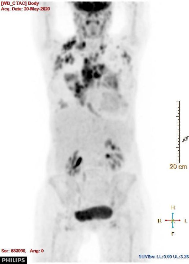
Jan Walewski, Disclosures
Roche (advisory board, research funding, lecture honoraria, travel expenses)
Abbvie (advisory board, lecture honoraria)
Takeda/Millenium (advisory board, lecture honoraria)
Gilead (advisory board, lecture honoraria)
Novartis (advisory board, lecture honoraria)
GSK/Novartis (research funding)
Servier (lecture honoraria)
Amgen (lecture honoraria)
DLBCL Case 2, learning objectives
1.   Understanding risk stratification by use of the international prognostic
     index (IPI) and NCCN-IPI
2.   Considering options of third-line therapy for relapsed/refractory DLBCL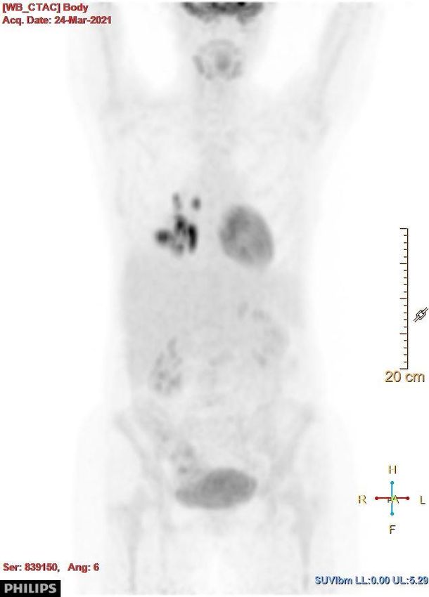
Patient case 2
‒ 44 year old female,
‒ April 2020: fever, cough, dyspnea, weight loss
   ➢ no improvement on oral amoxicillin
‒ Admitted to hospital with suspected COVID-19
   ➢ PCR test negative x2
‒ ECOG PS score 2/3
‒ Chest X ray: bilateral pleural effusion, pneumonia?
‒ Chest CT: bilateral pleural effusion, pericardial effusion, lymphadenopathy
‒ Abdominal CT: lymhadenopathy Ø 20 mm
Patient case 2
‒ transferred to ICU due to respiratory distress
    ➢ new supraclavicular lymph nodes noted (biopsy and aspiration
      for flow)
‒ History
    • Sarcoidosis (July 2019)
    • Hashimoto’s struma (Hashimoto’s thyroiditis)
    • Bilateral breast implants
Histopathologic examination
Lymphoma infiltration mainly with immunoblasts, partially centroblasts
Immunohistochemistry:
− CD20(+),
− Ki67(+++) in 85% of cells
− CD30(-/+) in 10% of cells
− CD3(-), -CD15(-/+), CD 23(-), CD 5(-)
− PAX5(+)
− CD10(-), BCL6(-), -BCL2(+/-) decreased expression of BCL2 on T cells (not seen
  in PMBL)
− MUM1(+/-), MYC(-) nuclear staining in 20% of cells,
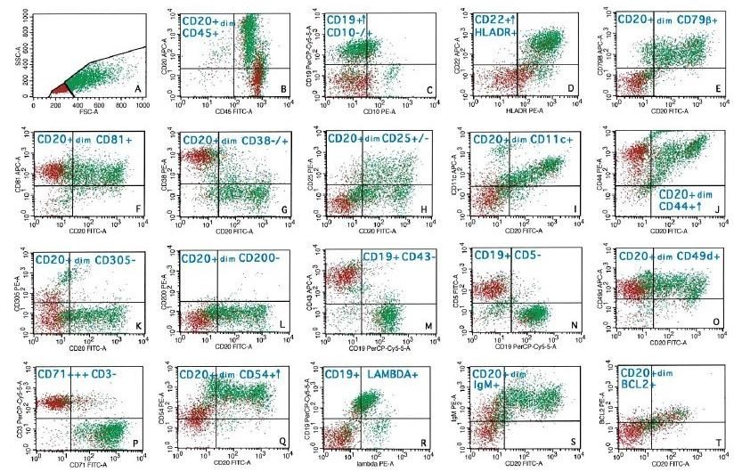
Cell suspension flow cytometry
Immunofenotype:
CD45+, CD20+ , CD19+ , CD22+,
CD79b+, HLADR+, CD54+, CD81+,
CD71+++, CD52+, BCL2+/-, lambda
+/-, IgM+, CD16/56-, BCL6-, CD5-,
CD43-, CD23-, CD10-, CD305-,
CD200-, CD30-/+

                        Fine needle aspiration biopsy of right supraclavicular nodes under USG guidance
Q1) Is this immunophenotype consistent with:
1. Primary mediastinal large B-cell lymphoma (PMBL)
2. T-lymphoblastic lymphoma (T-LBL)
3. Diffuse Large B-cell lymphoma (DLBCL), NOS, Germinal Centre B-cell (GCB)-
type
4. DLBCL, NOS, non-GCB-type
5. Marginal zone lymphoma (MZL) with mediastinal involvementCell suspension flow cytometry Immunofenotype: CD45+, CD20+ , CD19+ , CD22+, CD79b+, HLADR+, CD54+, CD81+, CD71+++, CD52+, BCL2+/-, lambda +/-, IgM+, CD16/56-, BCL6-, CD5-, CD43-, CD23-, CD10-, CD305-, CD200-, CD30-/+ Nodal DLBCL, NOS presenting as mediastinal large B cell lymphoma with peripheral lymph nodes involement. DLBCL, NOS with CD20+ dim, HLADR+, CD23-, CD200-, CD79b+ inconsistent with PMBL. Non-GCB sybtype Fine needle aspiration biopsy of right supraclavicular nodes under USG guidance
FISH and karyotype examination
FISH:
‒ no MYC, no BCL2, no BCL6 rearrangements
‒ duplication of BCL6 in 85% of cells
‒ Duplication of JAK2 in 92% of cells
‒ PAX5 rearrangement in 90% of cells
Karyotype:
47, XX,+del(3)(p12),t(9;14)(p13;q32)[10]
hyperdiploid karyotype with +3q (BCL6 duplication) and t(9;14) (PAX5 rearangement).
Q2) Plan for further work-up
1. CT scan
2. Bone marrow examination
3. MRI
4. PET-CT
5. Ultrasound
Staging and workup
‒ PET-CT (April 2020) involvement of
  multiple lymph nodes in the chest,
  abdomen, lungs, bones, bone marrow,
  pleural and cardiac effusion
‒ Bone marrow involvement (15%) on
  trephine biopsy
‒ CS IVB
‒ LDH 504 (N
Q3) IPI score is 4. What is the NCCN IPI score?

1. 1
2. 2
3. 3
4. 4
5. 5
Risk factor                                   score    The NCCN IPI
Age
   >40 do ≤60                                    1
   >60 do ≤75                                    2
   >75                                           3
LDH Ratio
   >1 do ≤3                                      1
   >3                                            2
CS III-IV                                        1
Extranodal sites:
   bone marrow, CNS, liver/G.I. tract, lung      1
ECOG PS≥2                                        1     Zhou Z et al.: Blood
                                                      2014, 123, 837-842.
Comparison of NCCN-IPI to IPI for risk stratification and outcomes
score 5-yr OS 5-yr PFS
Risk category NCCN-IPI IPI NCCN-IPI IPI NCCN-IPI IPI
Low 0-1 19% 0-1 38% 96% 90% 91% 85%
Low-interm. 2-3 42% 2 26% 82% 77% 74% 66%
High-interm. 4-5 31% 3 22% 64% 62% 51% 52%
High ≥6 8% 4-5 14% 33% 54% 30% 39%
Zhou Z et al.: Blood 2014, 123, 837-842.Q4) What is the optimal therapy 1. R-CHOP 2. R-CHOP + i.th. methotrexate 3. DA-EPOCH-R 4. CODOX-M/R-IVAC 5. R-CHOP + high-dose methotrexate
Treatment
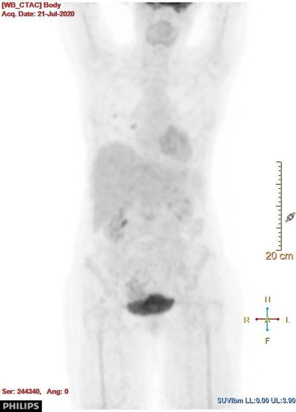
‒ May - July 2020: Interim PET post cycle 3:
➢ R-CHOP x 4 + high-dose methotrexate 3.5 g/m2 x 3 5PS - 2
‒ August 2020 – admitted for cycle 5
➢ Pleural bleeding after CVC insertion, respiratory
failure,
➢ admitted to ICU, recoveredQ5) How should this serious adverse event influence further treatment? 1. Change regimen to R-DHAP 2. Continue R-CHOP with HD MTX 3. Terminate chemotherapy 4. Continue R-CHOP alone 5. Proceed to high-dose therapy and autologous HCT
Treatment
‒ Continued R-CHOP x 2
PET scan post-cycle 6 (EOT)
‒ End of treatment PET-CT (07.10.2020): new lesion in
the right lung, 28x26mm maximum standard uptake
volume (SUV max) standardised to lean body mass
(lbm) 8.2
➢ misinterpreted as possible sarcoidosis → observation
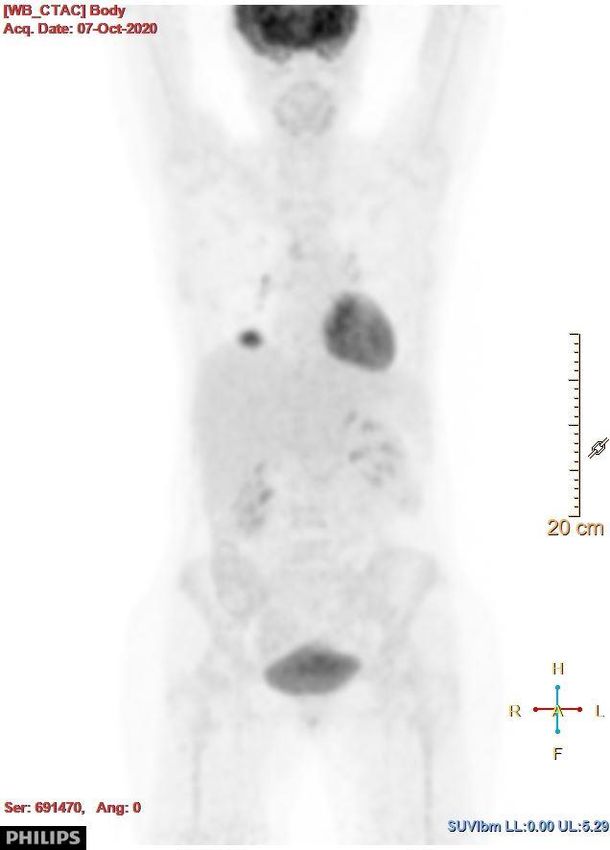
‒ Chest-CT (11.2020): tumor in the right lung 71x64mm
‒ Chest-CT (01.2021): tumor in right lung 83x78 mm,
new mediastinal tumor 32x30 mmTreatment
‒ ICE x 2 (02.2021 - 03.2021) PET-CT after ICE 2
‒ G-CSF
‒ apheresis
‒ PET-CT 24.03.2021: Progressive disease in
the right lungQ6) What options are available now 1. Consider clinical trial 2. Polatuzumab + bendamustine/rituximab (BR) 3. CAR-T cell therapy 4. Tafasitamab + lenalidomide 5. Pixantrone
Discussion Case 2

‒ Diagnostic difficulties: sarcoidosis, COVID-19 pneumonia, lymphoma
‒ Retrospectively, could induction therapy be better?
‒ Question of the third line treatment
References
‒   Rymkiewicz G et al.: Modern Pathol 2018; 31: 732-743
‒   Poppe B, i in. "PAX5/IGH rearrangement is a recurrent finding..", Genes, chromosomes, Cancer 2005;
    44: 218-223.
‒   Ohno H, i in. "Diffuse Large B-cell lymphoma carrying t(9;14)...". Hematol Oncol 2020; 38(2): 171-
    180.
‒   Zhou Z., Sehn L.H., Rademaker A.W., Gordon L.I., LaCasce A.S. et al. : An enhanced International
    Prognostic Index (NCCN-IPI) for patients with diffuse large B-cell lymphoma treated in the rituximab
    era. Blood 2014, 123, 837-842.
‒   Ruppert AS, Dixon JG, Salles G et al.: International prognostic indices in diffuse large B-cell
    lymphoma: a comparison of IPI, R-IPI, and NCCN-IPI. Blood 2020; 135 (23): 2041-2048.
‒   Sehn LH, Salles G: Diffuse Large B-Cell Lymphoma. N Engl J Med 2021; 384: 842-58.
Case 2 Acknowledgments Joanna Romejko-Jarosinska, MD, PhD – treating physician Grzegorz Rymkiewicz, MD, DSc - haematopathologist
You can also read