Infraocclusion: Prevalence, Characteristics, and Associated Dental Anomalies in Arabian Children
←
→
Page content transcription
If your browser does not render page correctly, please read the page content below
Hindawi
BioMed Research International
Volume 2022, Article ID 6624804, 7 pages
https://doi.org/10.1155/2022/6624804

Research Article
Infraocclusion: Prevalence, Characteristics, and Associated
Dental Anomalies in Arabian Children

          Saleh Ibrahim Alshaya,1 Abdulrahman Faleh Alanazi,1 Saleh Sulaiman Aldawish,1
          Mogren Mohmed Alsuhaim,1 Mohammad Saad Alomar,1 Yazeed Marzouq Almuaytiq,1
          Sami Abdulaziz Alfahad,1 Abdulrahman Abdulmohsen Suliman Almousa,1
          Abdullah Alassaf,2 and Sreekanth Kumar Mallineni 3,4
          1
            College of Dentistry, Majmaah University, Al-Majmaah 11952, Saudi Arabia
          2
            Department of Preventive Dental Science, College of Dentistry, Majmaah University, Al-Majmaah 11952, Saudi Arabia
          3
            Center for Transdisciplinary Research (CFTR), Saveetha Institute of Medical and Technical Sciences, Saveetha Dental College,
            Saveetha University, Chennai, 600077 Tamil Nadu, India
          4
            Division for Globalization Initiative, Liaison Center for Innovative Dentistry Graduate School of Dentistry, Tohoku University,
            Sendai, Japan

          Correspondence should be addressed to Sreekanth Kumar Mallineni; drmallineni@gmail.com

          Received 23 April 2022; Revised 27 June 2022; Accepted 4 July 2022; Published 23 July 2022

          Academic Editor: Andrea Abate

          Copyright © 2022 Saleh Ibrahim Alshaya et al. This is an open access article distributed under the Creative Commons Attribution
          License, which permits unrestricted use, distribution, and reproduction in any medium, provided the original work is
          properly cited.

          Aim. To analyze the distribution and characteristics of infraocclusion among Arabian children in primary dentition and its
          associated dental anomalies. Methods. A radiographic analysis was performed retrospectively using digital panoramic
          radiographs of children attending the pediatric dental clinic of College of Dentistry, Majmaah University, Saudi Arabia, from
          January 2019 to May 2021. The panoramic radiographs were analyzed to assess the distribution and characteristics of
          infraocclusion and its associated dental anomalies. Descriptive statistics were used for comparisons using SPSS version 21.0
          (IBM Corp., Armonk, N.Y., USA). The chi-square test was used to compare percentages. Results. Among the study population
          (542), only 40 children reported infraocclusion of 65 primary molars. Infraocclusion was common in males (90%) and very
          frequent in the mandibular arch (n = 48 teeth). In the primary dentition, unilateral infraocclusion (62.5%) was very frequent
          than bilateral presence (37.5%). Single molars were involved in 50% of the patients, while two, three, and four molars were
          involved in 42.5%, 2.5%, and 5% of cases. The mandibular second primary molar was frequently affected with infraocclusion,
          while the maxillary first primary molar was less commonly affected. In the mandibular arch, the second primary molar (28,
          58%) was more commonly affected with infraocclusion than the mandibular first primary molars and maxillary primary and
          secondary molars (p < 0:05). The majority of the infraoccluded molars were mild (75%), followed by moderate (23.5%) and
          severe (1.5%). Hypodontia (12.5%) is frequently associated with infraocclusion, followed by supernumerary teeth (5%) and
          radix entomolaris of the first permanent mandibular molars (5%). Infraocclusion was more in the second primary molar
          mandibular arch, while in the maxillary arch, the first primary molars were commonly affected (p > 0:05). Conclusion. In
          Arabian children, infraocclusion was commonly observed in mandibular second primary molars. Unilateral infraocclusion is a
          mild type of infraocclusion frequent in Arabian children. Numerical anomalies such as hypodontia and supernumerary teeth
          are associated with infraocclusion.

1. Introduction                                                         Numerous terms have been used, like half retention, arrested
                                                                        eruption, buried tooth, tooth depression, retained deciduous
Infraocclusion is a clinical finding in which teeth are found            tooth, shortened tooth, disillusion, impaction, incomplete/
below the occlusal surface compared to adjacent teeth.                  suppressed eruption, intrusion, secondary retention, and
2 BioMed Research International
reinclusion. Nonetheless, the most frequently used terms are graphs of children aged 4 to 12 years and those with eight
infraocclusion, ankylosed tooth, and submerged tooth [1], primary molars were considered for further analysis.
which refer to the chief visual feature of the abnormality.
They have become the term of preference for this positional 2.2. Exclusion Criteria. The children with blurred or poor-
anomaly of teeth [2, 3]. In most cases, infraocclusion can be quality radiographs, children other than the Arabian origin,
appreciated clinically, but radiographic examination is sel- and children with systemic problems or growth retardation,
dom required to diagnose this entity [1, 4]. This develop- cleft lip and palate, and other syndromes were excluded.
mental dental anomaly may occur if the tooth eruption Children more than 12 years of age and less than four years
mechanism fails and subsequently alters the preservation of age and with absence of any primary molars, incomplete
of its vertical position to the neighboring tooth [5–9]. Typi- records, and parental informed consent were not considered.
cally, the marginal ridges of the infraoccluded tooth are
below the adjacent teeth. Ankylosis is a common reason 2.3. Procedure. The 542 panoramic radiographs were
for infraocclusion. It happens because of the failure of the analyzed to assess the distribution and characteristics of
periodontal ligament to separate the root from the alveolar infraocclusion and its associated dental anomalies. The data
bone, which results in the fusion of the bone and root collection included is based on the gender of the child (male
[6–8]. The classification of infraocclusion has been discussed and female), age, the number of molars affected (1, 2, 3, 4, 5,
by various authors; however, the Brearley classification has 6, 7, and 8), arch (maxillary and mandibular), and type of
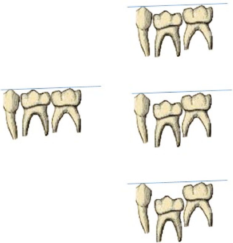
been used by various researchers in literature [10]. The clas- infraocclusion [10] (Figure 1) (mild, moderate, and severe).
sification involves three types that include (i) mild (occlusal Associated dental anomalies like tooth agenesis/hypodontia,
surface located approximately 1 mm below the occlusal a supernumerary tooth, odontomas, tooth transposition,
plane of the adjacent tooth), (ii) moderate (occlusal surface impacted teeth, and other dental anomalies were also
at the level of the contact point to the adjacent tooth), and evaluated.
severe (occlusal surface level below the interproximal gingi-
val tissue of adjacent tooth). The reported prevalence of 2.4. Statistical Analysis. All data tabulated and descriptive
infraocclusion varies broadly. An American study [11] statistics were used for comparisons using SPSS version
reported a prevalence of 1.3% in 2342 schoolchildren, a 21.0 (IBM Corp., Armonk, N.Y., USA). The chi-square test
study from Israel [10] reported 24.8% among 1530 children, was used to compare percentages, and a p value was consid-
and a Swedish study [1] reported 8.9% in 1059 children. ered less than 0.05. Kappa statistics were used to identify
Infraocclusion is very common in primary dentition rather intra- and interexaminer reliability.
than permanent dentition. It is primarily seen in the
mandibular arch as compared to the maxillary arch. Infraoc- 3. Results
clusion in primary molars has been linked with a few other
dental anomalies. These include an ectopic eruption of the Among the radiographs of 542 children included in the
first permanent molars, peg-shaped lateral incisors, palatal study, only 40 (7.38%) children reported infraocclusion of
displacement of maxillary canines, and enamel hypoplasia 65 primary molars. The infraocclusion was primarily seen
[12, 13]. Another aspect thought to play a role in infraocclu- in 36 (90%) males as compared to 4 (10%) females. Unilat-
sion is hypodontia. In a study, 65.7% of patients with miss- eral infraocclusion (62.5%) was very often compared to
ing permanent premolars reported having infraoccluded bilateral presence (37.5%) in the primary dentition
primary molars [14, 15]. The high occurrence of infraocclu- (Table 1). Single molars were involved in 50% (20) of the
sion in patients with hypodontia recommends a possible children, while two, three, and four molars were involved
common aetiological mechanism [16, 17]. The novelty of in 42.5% (17), 2.5% (1), and 5% (2) of the cases (Figure 2).
the present study is the lack of studies on the prevalence of In the mandibular arch, the second primary molar (28,
infraocclusion in primary dentition and its associated anom- 58%) was commonly affected with infraocclusion, followed
alies among Arabian children. Hence, the present study was by the first primary molar (20, 42%), while in the maxillary
aimed at analyzing the distribution and characteristics of arch, the first primary molars (9, 53%) were more frequently
infraocclusion among Arabian children in primary dentition affected than the maxillary second primary molar (8, 47%);
and its associated dental anomalies. the findings were statistically significant (p < 0:05). Among
infraoccluded primary molars, 55% were primary second
2. Materials and Methodology molars and 45% were first primary molars. Among the infra-
occluded molars, right second primary molars and maxillary
A retrospective cross-sectional radiographic analysis was right first primary molars were commonly affected. The
performed using digital panoramic radiographs of children majority (75%) of the infraoccluded molars were mild in
attending the pediatric dental clinic of College of Dentistry, nature followed by moderate (23.5%), and 1.5% (1) were
Majmaah University, Saudi Arabia, from January 2019 to the severe type of malocclusion (Figure 2); the comparison
May 2021. The institutional ethical committee of Majmaah was not statistically significant (p > 0:05). Hypodontia
University, Saudi Arabia, approved the study. (12.5%) was frequently associated with infraocclusion,
followed by supernumerary teeth (5%) and radix entomo-
2.1. Inclusion Criteria. Healthy children with clear pano- laris of the first permanent mandibular molars (5%)
ramic radiographs of Arabian origin were included. Radio- (Figure 3). The findings were not statistically significantBioMed Research International 3
Mild unusual pressure from the tongue, disturbance in the typ-
ical hard tissue resorption and deposition, and lack of
space [4, 18, 19].
Infraocclusion might be age-dependent, as it is closely
associated with root resorption due to premolar eruption
and the process of normal shedding. Peretz et al. [5]
Normal reported that there was a rise in the moderate form (8-10
Moderate
years old) and severe form (11-13 years) [5]. In a study by
Sidhu and Ali, the severe infraocclusion affected around
2.5–8.3% of the total infraoccluded primary molars [20]. In
line with previous findings, the present study included sub-
jects ranging from 2 years and inferred that infraocclusion
Severe
occurred at a mean age of 9:2 ± 3:8 years. The reported prev-
alence of infraocclusion among children varies from 2.8% to
38.5% according to various studies [1, 2, 16, 21–27] pub-
lished in the literature (Table 2).
Kurol [13] through clinical observation of 1059 children
between 3 and 12 years of age observed that females between
Figure 1: Reference used for diagnosis of infraocclusion. 3 and 6 years old showed more infraoccluded teeth, whereas
male children suffered from this condition more between 7
and 12 years of age [13]. The incidence of infraocclusion of
Table 1: Distribution of infraocclusion among Arabian children. deciduous second molars is more or less the same between
males and females, as described by Silvestrini Biavati et al.
Variables N % [21]. According to the findings reported by previous studies,
Mean age (years) Mean 9.2 SD 3.8 there was an insignificant difference in the prevalence of
Gender infraocclusion with regard to gender, according to the find-
Male 36 90 ings reported by previous studies [10, 16, 28]. On the con-
Female 4 10 trary, Steigman et al. described a higher incidence of
ankylosed mandibular second molars in males [22]. Ciftci
Occurrence
et al. [23] reported that there was no statistically significant
Unilateral 25 62.5 difference in the prevalence of infraocclusion between girls
Bilateral 15 37.5 (n = 51) and boys (n = 73). However, the present study
noticed more incidence in males compared to females. The
occurrence of infraocclusion of primary molars is reported
(p > 0:05). Kappa statistics confirmed substantial interrater to be in the 1.3-8.9% [1]; however, it can be as high as
reliability between the two examiners (kappa = 0:86). 38.5% [28]. Silvestrini Biavati et al. collected a group of
512 Italian subjects aged between 5 and 15 years and found
4. Discussion an incidence of ankylosis of 6.6% [21]. Furthermore, the
results of the present study revealed that the occurrence
Infraocclusion of the deciduous molar is a common finding of infraocclusion is more frequent in second primary
in which the tooth fails to reach the occlusal level compared molars; this anomaly has higher percentages in the man-
to the adjacent teeth. Though infraocclusion can be clinically dibular arch than in the maxillary arch. In children with
diagnosed, in children, assessing its severity is tough, so such developmental abnormalities, space issues might
dental radiographs are a boon in assessing such anomalies. pose and in such cases expansion may become essential
Panoramic or intraoral periapical radiographs and com- [18, 29, 30]. According to previous studies, the prevalence
puted tomography (CT) can determine the space between of mandibular infraocclusion was more than that of max-
the infraoccluded tooth surface and the normally occluded illary infraocclusion (189 : 36), and in the mandibular
adjacent teeth [18]. The present study was conducted to arch, the incidence is 2 to 10 times higher than that of
assess the prevalence of infraocclusion in Arabian children the maxillary arch [1, 2, 21–24]. Overall, infraocclusion
using panoramic radiographs attending a teaching hospital. affects predominantly mandibular molars up to 27 times
This retrospective analysis was done to determine the inci- more according to Odeh et al. [16]. In a study by Venza
dence and importance of infraocclusion of primary molars et al., subjects exhibited multiple infraoccluded teeth; the
and to report other associated dental anomalies in Arabian total number of infraoccluded teeth was 225, with a mean
children using panoramic radiographs, even though the eti- value of 1.7 infraocclusion per child [2]. Also, Zuñiga et al.
ology of infraoccluded teeth remains unclear. The following [24] and Salem and Mirzaee [25] reported 1.9 and 2.1 infra-
factors can be considered: disturbing local metabolism, peri- occlusions per child, respectively. However, Brearley and
odontal membrane disorders, trauma or infection, thermal McKibben [10] and Kurol [1] reported higher percentages
or chemical irritation, systemic diseases (like congenital of infraocclusion affecting only one molar per child (51%
syphilis), hereditary cause, local failure of bone growth, and 52%, respectively). This data is consistent with similar4 BioMed Research International
60 70
50 60
50
40
40
30
30
20
20
10 10
0 0
Male Female Maxillary arch Mandibular arch
First primary molar First primary molar
Second primary molar Second primary molar
(a) Based on gender (b) Based on arch
100 100
80 80
60 60
40 40
20 20
0 0
1 2 3 4 Mild Moderate Severe
First primary molar First primary molar
Second primary molar Second primary molar
(c) Based on number (d) Based on type
Figure 2: Distribution of 65 infraoccluded primary molars based on gender (a), arch (b), number of teeth (c), and type of infraocclusion (d).
are consistent with the report of Brearley and McKibben
12.50%
[10] and Cardoso Silva et al. [26] and Venza et al. [2]. They
used the same method, with infraoccluded molars mild in
nature (75%) followed by moderate (23.5%) and severe
(1.5%). Silvestrini Biavati et al. also described similar find-
ings using different classifications [21]. In the presence of
5% 5% infraoccluded primary molars, successor permanent teeth
may also get affected, and a delay in development can occur
in those teeth. In a study by Ciftci et al. [23], dental variation
was seen in 50.8% of children with infraocclusion. The
dental anomalies accompanying infraocclusion were mostly
agenesis, followed by dens invaginatus and supernumerary
Hypodontia Supernumerary teeth Radix entomolaris
tooth. Shalish et al. [3] reported an increased rate of dental
Figure 3: Dental anomalies associated with infraocclusion. anomalies associated with infraocclusion of primary molars,
palatally displaced canines, tooth agenesis, microdontia of
maxillary lateral incisors, and distal angulation of the man-
results of the present study, which showed that around 50% dibular second premolars [3]. Other associated anomalies
of subjects had multiple infraoccluded teeth. reported are aplasia of a successor, supernumerary teeth,
A Turkish study [23] examined 3.5% of the study popu- radix entomolaris of permanent teeth, and high prevalence
lation, and the authors reported that 45.2% involved one of agenesis [16, 25, 26]. Most of the authors observed that
tooth, 47.6% involved two teeth, 3.2% involved three teeth, the primary molars without successors have more chances
and 4% involved four or more teeth with infraocclusion. In of infraocclusion [22, 24]. The present study reported that
the present study, half of the children with infraocclusion hypoplasia is the most common associated anomaly,
involved a single tooth; 42.5% involved two infraoccluded followed by supernumerary teeth and radix entomolaris of
molars, while 3 and 4 infraoccluded molars were 2.5% and permanent teeth. Further, in a study by Venza et al., signifi-
5%, respectively. The present study included children with cant association was observed between the occurrence of
eight primary molars, and Turkish was mentioned regarding infraocclusion and impacted teeth (p < 0:001) [2]. However,
the presence of primary molars. According to Bjerklin and there was no significant relation evident in the association
Bennett’s method, the most observed category of infraocclu- between infraocclusion and dental anomalies in the present
sions was the mild one [31]. The results of the present study study (p > 0:05). Prior studies report an association withBioMed Research International 5
Table 2: Reported prevalence of infraocclusion.
Author Country Year Incidence/prevalence Most common tooth
Venza et al. [2] Italy 2018 2.8% Mandibular second primary molars
Ciftci et al. [23] Turkey 2021 3.25% Mandibular second primary molar
Brearley and McKibben [27] United States of America 1973 6.9% Mandibular first primary molar
Kurol et al. [1] Sweden 1984 8.9% Not mentioned
Silvestrini Biavati et al. Italy 2011 6.6% Mandibular second primary molars
Zúñiga-Tertre et al. Spain 2004 10.48% Mandibular first primary molar
Salem and Mirzaee [25] Iran 2009 15% Mandibular first primary molar
Cardoso Silva [26] Spain 2014 21.8% Mandibular first primary molar
Odeh et al. [16] Finland 2016 Maxilla:6 BioMed Research International
[8] J. Kurol, “Early treatment of tooth-eruption disturbances,” [25] K. Salem and B. Mirzaee, “Infraocclusion of primary molars
American Journal of Orthodontics and Dentofacial Orthope- and associated dental anomalies,” Journal of Biological Sci-
dics, vol. 121, no. 6, pp. 588–591, 2002. ences, vol. 4, no. 12, pp. 1217–1220, 2009.
[9] C. Dias, L. Q. Closs, V. Fontanella, and F. B. de Araujo, [26] C. Cardoso Silva, M. Maroto Edo, M. Soledad Alvaro Llorente,
“Vertical alveolar growth in subjects with infraoccluded and L. E. Barbería, “Primary molar infraocclusion: frequency,
mandibular deciduous molars,” American Journal of Ortho- magnitude, root resorption and premolar agenesis in a Spanish
dontics and Dentofacial Orthopedics, vol. 141, no. 1, sample,” European Journal of Paediatric Dentistry, vol. 15,
pp. 81–86, 2012. no. 3, pp. 258–264, 2014.
[10] L. J. Brearley and D. H. McKibben Jr., “Ankylosis of primary [27] L. Brearley and D. McKibben, “Ankylosis of primary molar
molar teeth. I. Prevalence and characteristics,” ASDC Journal teeth,” Journal of Dentistry for Children, vol. 40, no. 1,
of Dentistry for Children, vol. 40, no. 1, pp. 54–63, 1973. pp. 54–63, 1973.
[11] E. Koyoumdjisky-Kaye and S. Steigman, “Submerging primary [28] F. Krakowiak, “Ankylosed primary molars,” Journal of Den-
molars in Israeli rural children,” Community Dentistry and tistry for Children, vol. 45, no. 4, pp. 288–292, 1978.
Oral Epidemiology, vol. 10, no. 4, pp. 204–208, 1982. [29] V. Lanteri, A. Abate, D. Cavagnetto et al., “Cephalometric
[12] M. Hanisch, L. Hanisch, J. Kleinheinz, and S. Jung, “Primary changes following maxillary expansion with Ni-Ti leaf
failure of eruption (PFE): a systematic review,” Head & Face springs palatal expander and rapid maxillary expander: a
Medicine, vol. 14, no. 1, pp. 1–9, 2018. retrospective study,” Applied Sciences, vol. 11, no. 12,
[13] J. Kurol, “Infraocclusion of primary molars. An epidemio- p. 5748, 2021.
logical, familial, longitudinal clinical and histological study,” [30] V. Lanteri, M. Farronato, A. Ugolini et al., “Volumetric
Swedish Dental Journal. Supplement, vol. 21, pp. 1–67, changes in the upper airways after rapid and slow maxillary
1984. expansion in growing patients: a case-control study,” Mate-
[14] J. Kurol and B. C. Magnusson, “Infraocclusion of primary rials, vol. 13, no. 10, article 2239, 2020.
molars: a histologic study,” Scandinavian Journal of Dental [31] K. Bjerklin and J. Bennett, “The long-term survival of lower
Research, vol. 92, no. 6, pp. 564–576, 1984. second primary molars in subjects with agenesis of the premo-
[15] S. Peck, “Dental anomaly patterns (DAP),” The Angle Ortho- lars,” European Journal of Orthodontics, vol. 22, no. 3, pp. 245–
dontist, vol. 79, no. 5, pp. 1015-1016, 2009. 255, 2000.
[16] R. Odeh, S. Mihailidis, G. Townsend, R. Lahdesmaki, [32] S. K. Mallineni, C. K. Yung Yiu, and N. M. King, “Oral mani-
T. Hughes, and A. Brook, “Prevalence of infraocclusion of festations of Noonan syndrome: review of the literature and a
primary molars determined using a new 2D image analysis report of four cases,” Romanian Journal of Morphology and
methodology,” Australian Dental Journal, vol. 61, no. 2, Embryology, vol. 55, no. 4, pp. 1503–1509, 2014, PMID:
pp. 183–189, 2016. 25611289.
[17] R. Odeh, G. Townsend, S. Mihailidis, T. Hughes, and A. Brook, [33] S. K. Mallineni, G. K. Panampally, Y. Chen, and T. Tian,
“Infraocclusion: dental development and associated dental “Mandibular talon cusps: a systematic review and data analy-
variations in singletons and twins,” Archives of Oral Biology, sis,” Journal of Clinical and Experimental Dentistry, vol. 6,
vol. 60, no. 9, pp. 1394–1402, 2015. no. 4, pp. e408–e413, 2014.
[18] A. Arhakis and E. Boutiou, “Etiology, diagnosis, consequences [34] G. Shilpa, N. Gokhale, S. K. Mallineni, and S. Nuvvula,
and treatment of infraoccluded primary molars,” The Open “Prevalence of dental anomalies in deciduous dentition and
Dentistry Journal, vol. 10, no. 1, pp. 714–719, 2016. its association with succedaneous dentition: a cross-sectional
[19] H. S. Chen and J. D. Lieu, “An unusual primary first molar study of 4180 South Indian children,” Journal of Indian Society
impaction associated with a supernumerary tooth. Case of Pedodontics and Preventive Dentistry, vol. 35, no. 1, pp. 56–
report,” Case report. Australian Dental Journal, vol. 38, no. 4, 62, 2017.
pp. 277–279, 1993. [35] M. K. Sujon, M. K. Alam, and S. A. Rahman, “Prevalence of
[20] H. K. Sidhu and A. Ali, “Hypodontia, ankylosis and infraocclu- third molar agenesis: associated dental anomalies in non-
sion: report of a case restored with a fibre-reinforced ceromeric syndromic 5923 patients,” PLoS One, vol. 11, no. 8, article
bridge,” British Dental Journal, vol. 191, no. 11, pp. 613–616, e0162070, 2016.
2001. [36] M. K. Nanduri, T. P. Javangula, S. K. Mallineni, and
[21] B. A. Silvestrini Biavati, A. Signori, A. Castaldo, G. Matarese, S. Namineni, “Impacted primary mandibular second molar
and M. Migliorati, “Incidence and distribution of deciduous associated with late-formed second premolar: a rare entity of
molar ankylosis: a longitudinal study,” European Journal of reverse dentition,” Contemporary Clinical Dentistry, vol. 9,
Paediatric Dentistry, vol. 12, no. 3, pp. 175–178, 2011. Suppl 1, pp. S177–S179, 2018.
[22] S. Steigman, E. Koyoumdjisky-Kaye, and Y. Matrai, [37] S. Haque and M. K. Alam, “Common dental anomalies in cleft
“Submerged deciduous molars in preschool children; an epide- lip and palate patients,” The Malaysian Journal of Medical Sci-
miologic survey,” Journal of Dental Research, vol. 52, no. 2, ences: MJMS, vol. 22, no. 2, pp. 55–60, 2015.
pp. 322–326, 1973. [38] N. Venza, A. Borzabadi-Farahani, F. Fabi, C. Danesi, and
[23] Z. Z. Ciftci, Z. Kirzioglu, and A. Saritekin, “Prevalence of P. Cozza, “Dental anomalies: prevalence and associations
infraocclusion in primary molars and accompanying dental between them in a large sample of non-orthodontic subjects,
variations in a Turkish sample,” Journal of Oral Health and a cross-sectional study,” BMC Oral Health, vol. 17, no. 1,
Oral Epidemiology, vol. 10, no. 3, pp. 128–133, 2021. pp. 1–7, 2017.
[24] M. P. Zúñiga, T. Lucavechi, and E. Barbería, “Distribution and [39] M. Al-Abdallah, A. AlHadidi, M. Hammad, H. Al-Ahmad,
gravity of infraocclusion in temporary molars,” RCOE, vol. 9, and R. Saleh, “Prevalence and distribution of dental anomalies:
pp. 53–59, 2004. a comparison between maxillary and mandibular toothBioMed Research International 7
agenesis,” American Journal of Orthodontics and Dentofacial
Orthopedics, vol. 148, no. 5, pp. 793–798, 2015.
[40] D. G. Garib, S. Peck, and S. C. Gomes, “Increased occurrence
of dental anomalies associated with second-premolar agene-
sis,” The Angle Orthodontist, vol. 79, no. 3, pp. 436–441, 2009.
[41] S. Lochib, K. R. Indushekar, B. G. Saraf, N. Sheoran, and
D. Sardana, “Occlusal characteristics and prevalence of associ-
ated dental anomalies in the primary dentition,” Journal of
Epidemiology and Global Health, vol. 5, no. 2, pp. 151–157,
2015.
[42] C. Maspero, G. Begnoni, A. Magnani, M. Farronato,
N. Khomchyna, and C. Dellavia, “Rapid maxillary expander
and eruption guidance appliance therapy in skeletal class II:
cephalometric considerations,” European Journal of Paediatric
Dentistry, vol. 20, no. 4, pp. 280–284, 2019.
[43] M. McGeown and A. O'Connell, “Management of primary
molar infraocclusion in general practice,” Journal of the Irish
Dental Association, vol. 60, no. 4, pp. 192–198, 2014.
[44] C. de la Rosa Gay, E. Valmaseda-Castellón, X. Costa-Codina,
and C. Gay-Escoda, “Infraclusion of primary molars: reports
of cases,” ASDC Journal of Dentistry for Children, vol. 65,
no. 1, pp. 47–51, 1998.You can also read