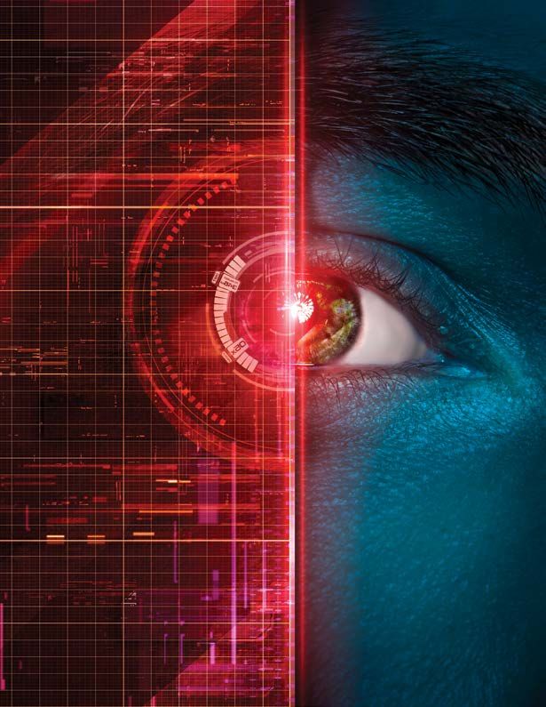
Innovative Vision Research Leadership in Action New Diagnostic Tools and Therapies - Bascom Palmer Eye Institute - University of Miami Health System
←
→
Page content transcription
If your browser does not render page correctly, please read the page content below
Bascom Palmer Eye Institute | University of Miami Health System

                                                                             U H E A LT H
                                                                      V O L U M E XXXI X
                                                                               IS S U E 1

                                                                      FEBRUARY 2021

                                                               Innovative
                                                          Vision Research

                                                                  Leadership
                                                                    in Action

                                                 New Diagnostic Tools
                                                       and Therapies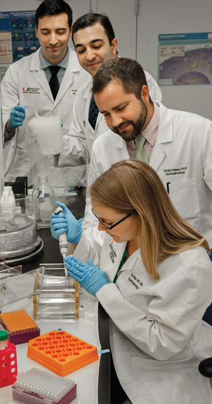
Bascom Palmer Eye Institute’s mission is to enhance the quality of life by
improving sight, preventing blindness, and advancing ophthalmic knowledge
through compassionate patient care and innovative vision research.

                                                   F E AT U R E

         2
                       Advancing the Frontiers
                       of Ophthalmology
                        Focus on Vision Research      2

                                                   Innovation and Invention                             12

                                     12
                                                   C L I N I CA L E XP E RT I S E

                                                   Saving Lives – Saving Vision                              14

                                                   On the Surface                   18

                                                   M E D I C A L E D U C AT I O N
                                     22
                                                   Passing the Baton                     22

                                                   B A S CO M PA L M E R E XCE L L E N C E

                                                   Awards and Honors                          27

                                                   Welcome New Faculty                             30

                                                   Profiles in Philanthropy                        32

                                                                                                        25

                31
Dear Friends and Colleagues:

                                                                          With the arrival of the new year, the dedicated
Eduardo C. Alfonso, M.D.
Kathleen and Stanley J. Glaser                                            professionals at Bascom Palmer Eye Institute
Chair in Ophthalmology                                                    are turning our vision to the future.
Director, Bascom Palmer Eye Institute                                        Our scientists and clinicians are working
Editor                                                                    closely together studying the diseases and
Marla Bercuson                                                            disorders of the eye, from the ocular surface
Executive Director of Business Operations                                 and cornea to the retina, macula, and optic
Bascom Palmer Eye Institute                                               nerve fibers leading back to the brain. This issue
Anne Bates Leach Eye Center                                               of Images highlights their remarkable work in
Miami
                                                                          our laboratories on gene therapy, stem cells,
900 NW 17 Street
Miami, Florida 33136                                                      and biomechanical devices to name just a few
305-326-6000                                of their leading-edge scientific programs.
Toll free in USA 800-329-7000                   Our far-sighted professionals are also developing advanced technology
Palm Beach Gardens                          applications like machine learning and artificial intelligence (AI) to improve
7101 Fairway Drive                          clinical care. These rapidly evolving tools will also allow our professionals
Palm Beach Gardens, Florida 33418           to screen and diagnose vision problems in clinics and remote locations,
561-515-1500                                extending our specialized ophthalmologic services to underserved
Naples                                      communities and help address disparities in access to care.
3880 Tamiami Trail North                        Support for our research comes from a variety of sources, including a
Naples, Florida 34103
                                            robust portfolio of grants from the National Institutes of Health (NIH), as
239-659-3937
                                            well as other federal and state programs. They help support our laboratory
Plantation
                                            infrastructure and contribute to our ability to keep our studies moving
8100 SW 10 Street
Plantation, Florida 33324                   forward, even through the ongoing COVID-19 pandemic.
954-465-2700                                    Private philanthropy is another vital source of support for our research,
Coral Gables                                as well as our clinical care and medical education programs. In this issue,
The Lennar Foundation Medical Center        you can read about the important contributions of these benefactors to our
5555 Ponce de Leon Boulevard                Institute, including our Ocular Surface Disease Service, which addresses
Coral Gables, Florida 33146                 problems like dry eye syndrome, as well as bacterial, fungal and viral
305-689-5555
                                            infections, tumors, and other diseases.
24-Hour Emergency                               As we look ahead to 2021, we are all hopeful that widespread adoption
305-326-6170                                of COVID-19 vaccines, coupled with the prudent safeguards recommended
Patient Appointments                        by the U.S. Centers for Disease Control and Prevention (CDC) will lead to
In-Office and Virtual Visits                a “new normal” for health care. We have learned many lessons from the
305-243-2020                                pandemic about safely delivering high-quality vision care in a variety of
Toll free in USA 888-845-0002               settings. That includes the excellent response to our telehealth program
                                            from patients and families who can access a Bascom Palmer professional
                                            from the comfort of their homes.
bascompalmer.org
                                                You can be assured that Bascom Palmer is committed to leadership in
                                            every field of ophthalmology. Thank you deeply for your continued support.
Images is produced by Bascom Palmer Eye
Institute with support from the George C.
Brosius Endowment Fund.

                                            Eduardo C. Alfonso, M.D.
                                            Kathleen and Stanley J. Glaser Chair in Ophthalmology
                                            Director, Bascom Palmer Eye Institute

                                                                                                  BASCOM PALMER EYE INSTITUTE   1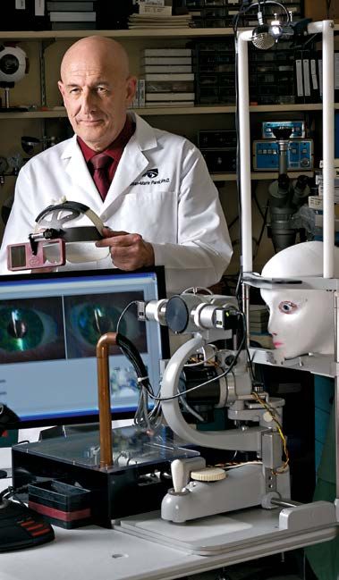
Dr. Vittorio Porciatti

                         A Conversation with Bascom Palmer’s Director of Scientific Research

        New Scientific Discoveries
        and Improved Clinical Care
                         For many decades, Bascom Palmer Eye Institute has been a global leader
                         in vision research. In a wide-ranging conversation, Vittorio Porciatti, D.Sc.,
                         the James L. Knight Professor in Ophthalmology, vice chair, and director of
                         scientific research, talks about what lies ahead for the dynamic research program.

    2     B A S C O M PA L M E R .O R G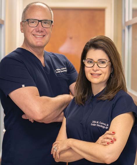
Q. What is your vision for laboratory research at         Q. How has COVID-19 affected Bascom Palmer’s
Bascom Palmer?                                            research?
Porciatti: Much of our research focuses on improving      Porciatti: COVID-19 has altered the balance between
clinical care through translational (bench to bedside)    bench and office time. Reduced bench time has
studies, as well as discovery research to explore new     slowed progress of ongoing experiments and
avenues with high clinical potential.                     development of new ideas. On the other hand,
                                                          increased office time has produced more scientific
Q. What challenges does vision research at                papers and grant submissions using available data. In
Bascom Palmer face?                                       the next period we may see a paradoxical increase of
Porciatti: We have three major challenges: funding,       scholarly productivity and grant awards that will be
talent recruitment, and space for our laboratories.       followed by a relatively greater reduction. Altogether,
                                                          there has been a substantial disruption.
Q. What are the highlights of your tenure as
scientific director in the past decade?                   Q. Tell us about your own research.
Porciatti: I am proud of having facilitated and           Porciatti: My own research focuses on prevention/
sustained the accomplishments made by Bascom              restoration of vision in glaucoma and optic
Palmer’s scientists with good administration, with        neuropathies such as Leber’s Hereditary Optic
an open door to listen to all their problems, and to      Neuropathy (LHON), a genetic condition which leads
search for viable solutions in a friendly environment.    to sudden loss of central vision. As my research
                                                          is relevant to other disciplines and pathological
Q. How has vision research changed in the past
decade?                                                   conditions, I have a number of interdisciplinary
                                                          collaborations.
Porciatti: We have seen dramatic changes in all
fields, leading to significant strategic choices.         Q. What about your work on diagnosing vision
Focusing on the translational aspects of research has     issues?
helped us orient our decisions.
                                                          Porciatti: To test vision, I use a non-invasive

Q. What is the size of Bascom Palmer’s research           electrophysiological tool called PERG (pattern

portfolio?                                                electroretinography) that I have pioneered and
                                                          continuously developed. It can identify problems with
Porciatti: Our currently active awards total $40
                                                          the optic nerve neurons before they die – a critical
million, including $18 million from the National
                                                          step in treating LHON to prevent loss of vision.
Institutes of Health (NIH) and $22 million from
other sponsors. Federal grants cover indirect costs       Q. What are some of the exciting things on the
to help sustain our research infrastructure. We           horizon?
greatly appreciate the vital support from industry,       Porciatti: We are looking at combining gene and stem
foundations and private philanthropy.                     therapies to sustain diseased eye tissues. Instead of
                                                          simply targeting defective genes or injecting stem
Q. What are your research priorities?
                                                          cells, we are studying how to perform gene editing
Porciatti: We are promoting high-impact
                                                          on the stem cells so they deliver the correct genes
research (gene therapy, stem cell therapy, gene
                                                          to the right place in the eye. We are also leaders in
editing therapy), developing new technology
                                                          developing new biomaterials and surgical devices,
(biomaterials, drug delivery, instruments for imaging,
                                                          as well as applying machine learning and artificial
electrophysiology, surgery, artificial vision, etc.),
                                                          intelligence (AI) to diagnosing and treating vision
generating interdisciplinary collaborations, and using
                                                          problems.
artificial intelligence to take advantage of large data
sets.                                                     Q. Looking ahead, what progress do you see in the
                                                          next five years?
Q. What is unique about Bascom Palmer’s
                                                          Porciatti: We have a very diverse research portfolio
research?
                                                          that combines our strengths in scientific discovery
Porciatti: Our key strengths include our excellent        and clinical care. Our professionals from many
reputation, a diverse patient population, and the         disciplines share ideas and collaborate on new
outstanding quality of faculty and administrative         strategies and tactics to improve patient care. I am
staff.                                                    confident that we will continue to recruit talented
                                                          young researchers and capitalize on our exceptional
                                                          history of leadership in vision research. •

                                                                                        B A S C O M PA L M E R E Y E I N S T I T U T E   3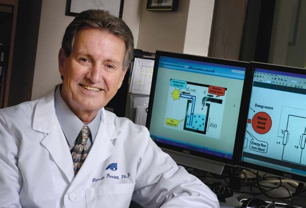
Bascom Palmer’s

    Laboratory
    Research
          Using the tools of 21st century medicine –
          including genetics, cellular biology, molecular
          diagnostics, advanced imaging and artificial
          intelligence, Bascom Palmer researchers are
          poised to understand why the eye may become
          susceptible to disease and how biotechnologies
          may help to prevent these conditions.

                                           William L. McKnight, the
                                           legendary leader of Minnesota
                                           Mining and Manufacturing
                                           Company (3M) valued research
                                           as “a key to tomorrow.” Bascom
                                           Palmer’s Evelyn F. and William
                                           L. McKnight Vision Research
                                           Center embodies his generosity
                                           and enthusiastic support of
                                           research on the nervous system
                                           and the eye as a “window to the
                                           brain and body.” Laboratories,
                                           primarily based in the McKnight
                                           Research Center, are home to
                                           Bascom Palmer’s scientists as
                                           they continue to advance the
                                           frontiers of ophthalmology.
                                           Meet some of Bascom Palmer’s
                                           researchers as they describe
                                           their work – in their own words.

        Dr. Daniel Pelaez and Dr. Galina Dvoriantchikova inspect a batch of freshly
       isolated stem cells they will use to create a new human retina in the lab as
                    part of the Pelaez lab program on regenerative ophthalmology.

4   BASCOMPALMER.ORG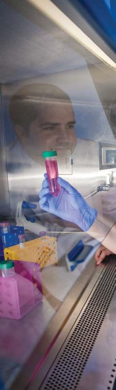
Mohamed Abou Shousha,
M.D., Ph.D.
Associate Professor of
Clinical Ophthalmology
With more than 580 patients in
clinical trials and 26 U.S. and
international patents, Bascom
Palmer’s Artificial Intelligence
and Computer Augmented Vision
Laboratory is leading the way to
transform vision diagnostics and
personalized vision correction.
The goal of the lab is to provide
physicians and patients access to
smarter, more accurate and portable
technology to diagnose and treat
visual field loss, double vision and
other vision disorders. Using our
AI-driven wearable technology
connected to the cloud, patients can
quickly and easily take vision tests
and the results will be transmitted
to their physicians in real time for
evaluation and diagnosis. In clinical
studies published in prestigious
ophthalmology scientific journals, our
wearable vision augmentation devices
enhanced vision and helped improve
mobility in 78% of enrolled patients.

         BASCOM PALMER EYE INSTITUTE   5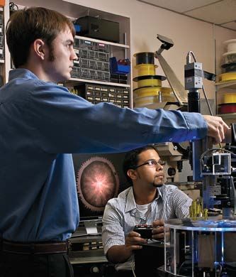
Delia Cabrera DeBuc, Ph.D.
                                                             Research Associate Professor of Ophthalmology
                                                             My research focuses on developing novel imaging
                                                             biomarkers of the onset and progression of
                                                             ophthalmic and neurological diseases. Our research
                                                             group has refined quantitative tools and introduced
                                                             innovative measures to analyze retinal images to
                                                             quantify retinal structure and function abnormalities
                                                             as well as treatment-induced changes in patients
                                                             with ocular and neurodegenerative diseases (e.g.,
                                                             diabetic retinopathy, multiple sclerosis, Parkinson’s
                                                             and Alzheimer’s disease). In our laboratory, we have
                                                             identified and evaluated low-cost, user-friendly,
                                                             and non-invasive technologies to screen the
Sanjoy K. Bhattacharya, Ph.D.                                eyes in the elderly at risk of
Professor of Ophthalmology                                   mild cognitive impairment
My research interests are restoration of visual function     using the eye-brain
in glaucoma and multiple sclerosis. Our research has two     connectome approach.
distinct arms: first, the long-distance axon regeneration,   We are also devoted to
reinnervation and functional restoration; and second,        artificial intelligence
achieving homeostasis of intraocular pressure. The           applications for disease
research in these areas is funded by grants from the         diagnosis, running a
U.S. Department of Defense and the National Institutes       multidisciplinary lab,
of Health, respectively. The axon regeneration involves      and collaborating
expansion of membranes that includes synthesis and           extensively with
transport of lipids in neurons. The membrane lipid           national and
composition of trabecular meshwork is consequential          international
for pressure homeostasis. Our group utilizes cell culture,   investigators.
experimental models and multiomics mass spectrometry,
imaging mass spectrometry, high-resolution imaging,
electrophysiology, and computer natural language
processing combined with usual bioinformatics tools for
these analyses.

                                                             Giovanni Gregori, Ph.D.
                                                             Research Associate Professor of Ophthalmology
                                                             My research focuses on the clinical applications of medical
                                                             image processing. My group has been a leader in the
                                                             development of quantitative tools for the analysis of retinal
                                                             images, in particular those acquired using Optical Coherence
                                                             Tomography (OCT) technology. Over the years we helped
                                                             introduce and study a number of new powerful mathematical
                                                             algorithms which have allowed us to characterize, for the first
                                                             time, the retinal anatomy and geometry in detail, advancing
                                                             our understanding of the pathophysiology associated with
                                                                  various ocular pathologies. Several of these algorithms
                                                                     have moved beyond clinical research and are now
                                                                     widely used in routine clinical settings to help diagnose
                                                                     and manage patients.

  6   BASCOMPALMER.ORG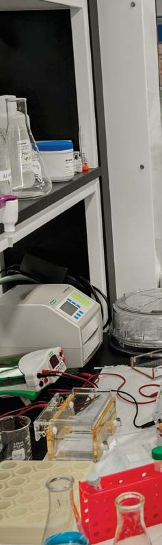
J. William Harbour, M.D.
                                                          Professor and the Mark J. Daily Chair
                                                          in Ophthalmology
                                                          My research focuses on improving
                                                          outcomes for patients with eye
                                                          cancers, such as uveal melanoma and
                                                          retinoblastoma. My team of scientists
                                                          from across the world uses a range of
                                                          scientific methods, including genetics,
                                                          genomics, cell biology, computational
                                                          biology, developmental biology, and
                                                          bioinformatics to make some of the most
                                                          important discoveries in the field. The
                                                          unique capabilities of Bascom Palmer allow       Dmitry V. Ivanov, Ph.D.
                                                          us to deliver the best patient care available,   Research Associate
 Abigail S. Hackam, Ph.D.                                 while continuing to make new discoveries         Professor of Ophthalmology
 Research Professor of Ophthalmology                      that further improve patient outcomes.           The ongoing studies in
 My laboratory investigates the response                  Among many contributions, we developed           my laboratory are aimed
 of retinal neurons and glia to damage                    the industry standard and most widely            at exploring the signaling
 from conditions such as inherited retinal                used prognostic test for uveal melanoma,         cascades and the epigenetic
 degenerations and optic nerve trauma. Our                and we recently discovered a new                 mechanisms involved in
 focus is on specifically modifying the retinal           immunotherapy target for uveal melanoma          ocular development and
 damage responses in order to halt disease                that has led to a groundbreaking new             regeneration, with a specific
 progression and promote tissue repair and                clinical trial.                                  focus on neurogenesis and
 regeneration. Current research topics include                                                             gliogenesis in the developing
 developing Wnt signaling pathway activators                                                               retina, and on strategies
 and innate immunity modulators as potential                                                               geared toward stimulating
 therapies. Additional research areas include                                                              neuronal regeneration in
 identifying molecules that contribute to                                                                  the adult retina. The second
 neuropathic pain in dry eye and characterizing                                                            important direction of my
 the effect of specific dietary supplements on                                                             laboratory research has
 retinal health. My lab’s research is supported                                                            been to understand the role
 by the National Institutes of Health and private                                                          and mechanism of sterile
 foundations.                                                                                              inflammation, or innate
                                                                                                           immune response in the
                                                                                                           absence of live pathogens, in
                                                                                                           the pathophysiology of retinal
                                                                                                           disorders, with a specific
                                                                                                           interest in the contribution
                                                    Xiang Run Huang, Ph.D.                                 of a signal for “danger” (the
                                                    Research Associate Professor of Ophthalmology          so called damage-associated
                                                    My research goal is to develop optical methods         molecular patterns or DAMPs)
                                                    for early diagnosis and sensitive monitoring of        and pattern recognition
                                                    glaucoma. My research interests include the            receptors.
                                                    optical properties of the retinal nerve fiber layer
                                                    (RNFL), their damage mechanisms in glaucoma
                                                    and imaging technologies for early detection of
                                                    the damage. My recent studies found that change
                                                    of RNFL optical properties was associated with
not only changes of axonal ultrastructure but also its functional activities. My current research
focuses on developing a new imaging method that measures the RNFL reflectance at different
wavelengths and proposing a new optical parameter, spectral contrast, for detection of glaucoma
at an early stage, before irreversible visual field loss has occurred.

                                                                                                            BASCOM PALMER EYE INSTITUTE   7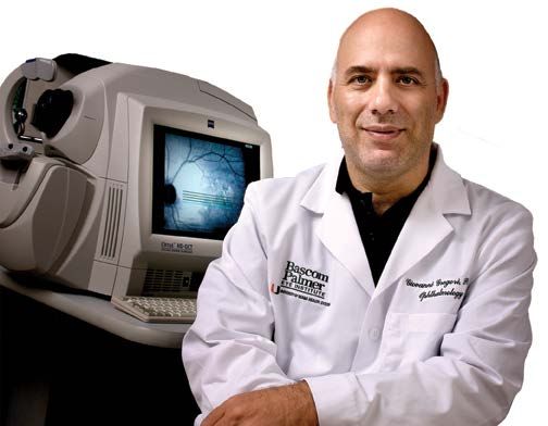
Stefan Kurtenbach, Ph.D.
                                 Research Assistant Professor of Ophthalmology
                                 My research focus lies on how genomic aberrations and resulting
                                 epigenetic changes foster tumor development. Using methylation
                                 profiling, RNA-seq, ChIP-seq, ATAC-seq, and single-cell sequencing,
                                 my projects aim to elucidate genetic and epigenetic changes that
                                 sub-characterize metastatic uveal melanoma (UM) and other
                                 cancers, to reverse these pharmacologically, for which in-vitro
                                 and experimental models are being used. My career goal is to
                                 make significant contributions to understanding the underlying
                                 mechanisms on how these genomic aberrations foster tumor
                                 development, using state-of-the-art genomics methods. Giving
                                 talks at patient retreats and with close contact with patients, I witness the suffering caused
                                 by notoriously hard to treat cancers. While we have gained comprehensive insight into the
Fabrice Manns, Ph.D.             chromosomal, mutational, and expression changes correlating with tumor sub-types and
Professor of Biomedical
                                 metastatic risk, we are still lacking knowledge about how these aberrations cause these cancers. I
Engineering and
                                 am also involved with projects related to myeloid malignancies and retinoblastoma.
Ophthalmology
I work closely with Dr.
Jean-Marie Parel, Dr. Marco
                                                              Richard K. Lee, M.D., Ph.D.
                                                              Associate Professor and the Walter G. Ross Distinguished Chair
Ruggeri, and clinicians to
                                                              in Ophthalmic Research
develop new devices to
image and measure the                                         As a clinician-scientist, my research focuses not only on the
eye. These devices provide                                    molecular, cellular, and proteomic mechanisms of retinal ganglion
data that will help improve                                   cell death (which results in vision loss through glaucoma), but also
the visual outcomes of                                        developing new surgical procedures and improving the outcomes of
procedures such as cataract                                   glaucoma surgeries, especially minimally invasive laser surgeries.
surgery or LASIK. In parallel,                                I have been involved in the major national and international genetic
our team studies the optical                                  studies for glaucoma risk. Our basic science understanding of how
system of the eye and its                                     nerve cells in the eye are damaged are resulting in translational
changes throughout our                                        research (such as the discovery of new molecular targets for
lifespan. The goal is to                                      improving optic nerve cell survival) which will lead to new
better understand how                                         paradigms for the treatment of glaucoma and optic neuropathies.
refractive errors, such as
myopia, develop during
childhood, and also how we
                                 Yiwen Li, M.D.
progressively lose our ability
                                 Research Assistant Professor of Ophthalmology
to focus on near objects         My research is focused on
later in life, a condition       neuroprotective treatment of retinal
known as presbyopia. This        neurons using neuroprotective
knowledge will help design       agents, including neurotrophic
new approaches for vision        factors. I am currently working
correction.                      on mesencephalic astrocyte drive
                                 neurotrophic factor (MANF), which
                                 has shown significant protection on
                                 photoreceptor and retinal ganglion
                                 cell survival in my experiments with
                                 disease models. I molecularly dissected
                                 MANF and showed that the first half of
                                 the MANF molecule (the N-terminal domain)
                                 possesses the full neurotrophic activity of MANF.
                                 My work now is focused on the development of MANF
                                 N-terminal domain as a novel neurotrophic factor for
                                 retinal degenerative disorders.

  8    BASCOMPALMER.ORG
Jean-Marie Parel, Ing.ETS-G, Ph.D.
Research Associate Professor and the Henri & Flore
Lesieur Chair in Ophthalmology
My research and that of the Ophthalmic Biophysics Center
team focuses on helping clinicians to improve patient care by
combining biophysics and engineering. Our center developed
a web-controlled robotized slit-lamp, an instrument to assess
the light sensitivity of patients undergoing gene therapy
and treatments for the effects of traumatic brain injuries.
The instrument is useful for remote patient examinations
from anywhere in the world and also in a pandemic. We also
developed an instrument using LEDs and a light-sensitive
drug to treat patients with corneal infections. Under
development are: an optical system to visualize aqueous
Darlene Miller,
humor scleral outflow, subminiature implants releasing
D.H.Sc., M.P.H.
medications to treat glaucoma, a biocompatible titanium
Research Professor
keratoprosthesis (artificial cornea) for patients not amenable
of Ophthalmology
to cornea transplant, and several diagnostic instruments.
The ocular microbiology
laboratory collaborates
with faculty, fellows, Daniel Pelaez, Ph.D.
residents, staff, and students Research Assistant Professor of Ophthalmology
to investigate new and The two main areas of research in my laboratory
innovative laboratory and are molecular biology of eye diseases and
clinical techniques for the regenerative ophthalmology. We harness the
detection and prevention of power of stem cells, advanced biomaterials, tissue
ocular infectious diseases. engineering, and molecular genetics to replace
My research interests cells and tissues and restore the functions that
include: investigation of are lost to disease or trauma. By applying the
microbial pathogenesis and latest and most innovative analytical techniques,
molecular epidemiology we explore the molecular events that trigger
of ocular infections, role the onset of disease processes, or promote their
of the ocular surface progression, in order to develop truly effective
microbiota and microbial therapies that go beyond halting the course of
microbiomes interactions a disease and actually help to regenerate what
and outcomes in contact has already been lost. We have ongoing projects
lens-associated microbial focusing on optic nerve, retinal neurons, and
keratitis, and use and impact lacrimal gland regeneration.
of pharmacodynamics and
pharmacokinetics of common
Marco Ruggeri, Ph.D.
ocular antimicrobials on the
Research Assistant Professor of Ophthalmology
management and emergence
of antibiotic resistance. A I work with physicians and other engineers to develop diagnostic
new and evolving focus of our and surgical devices that can have direct impact on patient eye
laboratory is the application care. The diagnostic devices produce images of the eye and data
of artificial intelligence and that will enable clinicians to better diagnose and treat diseases
other big data platforms such as pediatric retinopathies and keratoconus, a progressive
for the detection, diagnosis corneal disease. The surgical devices will improve the outcomes
and management of corneal of cataract surgery, corneal transplants, ocular tumors removal
ulcers. and other microsurgeries by guiding clinicians with images and
measurements of the eye during surgery, and with robots to
conduct delicate surgical maneuvers inside the eye. Our team also
engineers imaging technology that we use to better understand
the mechanisms of myopia and presbyopia.
BASCOM PALMER EYE INSTITUTE 9Valery I. Shestopalov, Ph.D.
Professor of Ophthalmology
My research projects in the retina
investigate cellular and molecular
mechanisms underlying injury, loss of
functionality, and death of retinal ganglion
cell, which is a fundamental clinical
problem and the cause of blindness in
many human diseases, such as glaucoma,
ischemic optic neuropathy, and other
optic nerve diseases. We have analyzed
transcriptomic and proteomic changes
in human glaucoma and used genetically
Alfonso L. Sabater, modified experimental models to
M.D., Ph.D. identify glial-specific and neuron-specific
Assistant Professor of pathways activated in response to ocular
Ophthalmology hypertension, the major risk factor of
My laboratory investigates glaucoma. These differentially activated pathways contribute to retinal stress and degeneration
new strategies to protect and often represent new drug targets for these blinding diseases. In particular, we have
and regenerate the cornea, identified new therapeutic targets in the glaucomatous, ischemic and post-traumatic retina,
which is the clear outer layer blockade, or therapeutic modulation of which suppresses or prevents degeneration of ganglion
at the front of the eye. Work cell, the underlying cause of blindness. Most of these targets are related to regulation and
by our lab and others has assembly of an innate immune complex, known as “inflammasome,” aberrant overactivation
shown that corneal damage of which has been demonstrated and linked in our studies to neuronal dysfunction and death.
due to multiple causes can be Inactivation of such activity completely protected retinal neurons, their functionality, and
slowed and even reversed by preserved vision in experimental glaucoma and traumatic optic nerve injury models.
a variety of approaches. These
approaches include activating Luis E. Vazquez, M.D., Ph.D.
cell survival and proliferation Assistant Professor of Clinical Ophthalmology
signaling, blocking strategic
Glaucoma is a leading cause of blindness worldwide that
cell death pathways, and
results from the degeneration of retinal ganglion cells
selectively replacing corneal
(RGCs) and the optic nerve. There is no cure for glaucoma,
layers through cell therapy
and current treatment is aimed at lowering the intraocular
and tissue engineering. In
pressure (IOP) to slow disease progression. The pathogenesis
doing so, we can protect
of RGC death remains unknown, but has been proposed to
the cornea and treat both
involve decreased vascular perfusion as a consequence of
rare and common diseases
elevated IOP. We hypothesize that the vascular tone held by
including dry eye syndrome,
vascular smooth muscle cells (VSMCs) controls blood flow
bullous keratopathy, corneal
and retinal perfusion and plays a major role in RGC health.
transplant failure, corneal
We aim to identify signal transduction pathways that control
infections and limbal stem
muscle tone in retinal VSMCs. Our goal is to find novel ways
cell deficiency.
to improve blood perfusion and open new avenues for
treatment of glaucoma and other eye diseases.
10 BASCOMPALMER.ORGJianhua (Jay) Wang, M.D., Ph.D., M.S.
Professor of Ophthalmology and Electrical and Computer Engineering
My research focuses on the development of advanced ophthalmic
imaging and its applications in eye research. A wide range of ophthalmic
imaging modalities has been developed at Bascom Palmer to study
structural and functional alterations in eyes during normal aging and
eyes with various disorders. Working with Dr. Hong Jiang, neurologist and
neuro-ophthalmologist, the team also focuses on microvasculature and
microstructure of the eye as a window of the central nerve system. The
research, funded by grants from the National Institutes of Health, National
Multiple Sclerosis Society, and Florida Department of Health, explores the
change in the eye in age-related dementia.
Rong Wen, M.D., Ph.D.
Professor of Ophthalmology
The goal of my research is to
understand the biology of the retina and
photoreceptors, the physiopathology
of retinal diseases, and to translate
the bench research to patient care.
I have been working on hereditary
retinal degenerations to understand
the mechanisms for photoreceptor
degeneration that are caused by
specific mutations, and to develop
neuroprotective therapy. In addition, I
am collaborating with a retinal imaging
colleague to develop novel non-invasive
imaging technologies that could provide
ophthalmologists with molecular
information in the retina.
Hong Yu, Ph.D.
Research Assistant Professor
Our long-term research objectives are to investigate mechanisms of neuronal death and test
potential therapeutics, with a focus on mitochondrial diseases. Our laboratory has three on-
going projects related to neurodegenerative diseases caused by mitochondrial gene mutation.
The first project is to determine the etiology and potential treatments for Leber Hereditary
Optic Neuropathy (LHON), the most common primary mitochondrial genetic disorder. Our
laboratory, which was directed by the late Dr. John Guy, has pioneered a novel technology
to efficiently introduce DNA directly into mitochondria, both in vitro and in vivo. This
technology allows us to restore the respiratory function in cells carrying LHON mutations,
create bona fide LHON experimental models, and develop and test potential therapeutic
strategies. The second project seeks to develop clinically relevant strategies for the treatment
of Maternally Inherited Leigh Syndrome and Neurogenic Ataxia and Retinitis Pigmentosa, a
pair of neurologic diseases renowned for causing death and blindness in children and young
adults. In the third project, preclinical studies for LHON intervention will be performed using
our mito-targeting technology to deliver wildtype ND4 directly into mitochondria. •
BASCOM PALMER EYE INSTITUTE 11Advancing patient care around the world with innovation and invention
Ophthalmic Biophysics Center
In 1969, Jean-Marie Parel,
Ph.D., Ing., ETS-G, a Swiss-born
biomedical engineer, joined
Bascom Palmer and helped change
the field of ophthalmology.
That year, he met retinal specialist Robert
Machemer, M.D., and Helmut Buettner,
M.D., a research fellow. Together they
conceived the idea for an instrument (later
called the Vitreous Infusion Suction Cutter
or VISC) that could remove the diseased
vitreous fluid while maintaining the shape
of the eye through an infusion of saline
solution. “This one tool could do three
different things,” said Parel. “It provided
the foundation for doing intraocular
microsurgery while preventing the collapse
of the eye.”
That remarkable success led to the 1971
founding of Bascom Palmer’s Ophthalmic
Biophysics Center for one single reason:
to help the doctors in the Institute deliver
better patient care. Through its five
decades, the OBC team has invented
or improved more than 350 surgical
instruments or devices with many more in
the works. The most notable include: the
In the early 1970s, Bascom Palmer’s
Ophthalmic Biophysics Center made this
rotating table for retinal detachment surgery.
Gas was injected in the eye and the patient Dr. Jean-Marie Parel in the
remained face down during the surgery. Ophthalmic Biophysics Center
12 BASCOMPALMER.ORGMarks 50 Anniversary th
VISC designed for the first vitrectomy, retinal tacks, a
glaucoma microshunt (half the size of an eyelash), and
developed an ocular drug transfer system known as
Ocular Coulomb-Controlled Iontophoresis. In addition
to these devices, the OBC creates novel imaging
technology for real-time imaging of the eye to study a
range of ocular structures and diseases.
“The doctors come to us with a problem and
then we go and dream up a solution,” said Parel.
“Sometimes it takes minutes and sometimes it takes
days. When we come up with a solution we ask the
clinicians to test it and give us their feedback. We then
go back to work to refine and improve the instrument
so that it satisfies the needs of the physicians and is
safe for the patients.” Research at the OBC addresses
all areas of ophthalmology ranging from the retina
and vitreous to cornea, glaucoma, cataracts, neuro-
ophthalmology and ocular oncology.
Located in the Evelyn F. and William L. McKnight
Vision Research Center adjacent to Bascom Palmer’s
flagship center in Miami, the Walter G. Ross
Ophthalmic Biophysics Center is a unique place. The
OBC team of scientists, engineers, and technicians
have their own machine shop, electronic lab, chemistry
lab, an optical lab to develop new lasers, and a fully
equipped “operating room” to test the safety and
efficacy of new instruments, implants, and surgical
techniques before they are used for actual clinical
or surgical care. “Not many places have all of these
capabilities in one location,” Parel said.
Biomedical engineers Derek Nankivil, M.S., Ph.D., (left) and Victor Hernandez, M.S.,
In the 1980s, Parel’s team designed the world’s Ph.D., are alumni of Bascom Palmer’s Ophthalmic Biophysics Center, who continue
smallest motorized scissors, which were used to cut to collaborate with the OBC: Nankivil on photosensitivity projects, and Hernandez
retinal membranes that obscure vision, as well as a on intraocular lenses.
fluid control system that improved surgical precision in
vitrectomies.
The OBC closely collaborates with Drs. Fabrice
Manns and Marco Ruggeri, as well as every other “Jean-Marie Parel’s innovative
scientist at Bascom Palmer who needs technology work at the OBC has been credited
support,” says Parel. In return, these gifted scientists
help the OBC by sharing their knowledge and
with opening up a wide range of
practical know-how. It is a synergistic relationship medical treatments for diseases,
that ultimately benefits patient care. Parel currently
holds more patents than any other member of the
and millions of patients around
University of Miami medical school community. the world have better vision as a
“Our founder, Dr. Edward Norton, recognized how result of his vision and expertise.”
biophysical engineering could make a huge difference
– Dr. Eduardo Alfonso
in ophthalmology,” said Parel. “He had a far-reaching
view of the future and we continue to look for new
ways to improve vision care.” •
BASCOM PALMER EYE INSTITUTE 13Saving Lives –
Saving Vision
Taking a Holistic Approach to Eye Cancer Care and Research
Dr. J. William Harbour and Dr. Zelia Correa
14 BASCOMPALMER.ORGA study analyzing new
evolutionary complexity in
uveal melanoma by
J. WILLIAM HARBOUR,
M.D., was recently
published in Nature
Communications, a highly
regarded, peer-reviewed
For J. William Harbour, M.D., and Zelia surgery, ophthalmic pathology, medical scientific journal. “This is
M. Correa, M.D., Ph.D., finding new education, and leadership development,” said the first-ever single cell
therapies for dangerous eye cancers is Eduardo C. Alfonso, M.D., director of Bascom analysis of uveal melanoma.
Palmer and the Kathleen and Stanley J. Glaser We discovered previously
truly a labor of love. The husband-and-
Chair in Ophthalmology. Correa’s current unrecognized genetic and
wife team take a coordinated approach research focuses on the use of artificial cellular complexity in this
to treating adult and pediatric intelligence to distinguish benign from deadly eye cancer,” said
patients in Bascom Palmer’s Ocular malignant ocular tumors based on imaging Harbour. Uveal melanoma is
Oncology Service, while pursuing new characteristics. the most common primary
discoveries in its leading-edge Ocular Along with their roles at Bascom Palmer, cancer of the eye, with
Harbour and Correa also serve on the faculty approximately 2,000 –
Oncology Laboratory.
of Sylvester Comprehensive Cancer Center 3,000 cases diagnosed each
“Bascom Palmer’s unique clinical and research of the University of Miami Miller School of year in the United States.
programs and our stellar faculty allow us Medicine, where Harbour is associate director It is highly metastatic and
to deliver the best possible patient care, for basic science. He is also a member of the unresponsive to checkpoint
while making new findings that improve Interdisciplinary Stem Cell Institute at the immunotherapy. This work
patient outcomes,” said Harbour, professor of University of Miami. “These close ties with was supported by National
ophthalmology, director of ocular oncology, Sylvester allow us to provide coordinated Cancer Institute grant R01
vice chair for translational research, and the holistic care for our ocular cancer patients and CA125970, Research to
Mark J. Daily Chair in Ophthalmology. Drawing to initiate highly innovative clinical trials to Prevent Blindness, Inc. Senior
on Bascom Palmer’s exceptional resources – benefit patients,” said Correa. Scientific Investigator Award,
including advanced imaging equipment and one In keeping with that comprehensive the University of Miami Miller
of the world’s top eye pathology laboratories approach, Harbour and Correa collaborate School of Medicine Medical
– the Florida Lions Ocular Pathology Laboratory with Sylvester specialists, such as Jose Lutzky, Scientist Training Program,
under the direction of Sander Dubovy, M.D., M.D., professor of medicine and director of the University of Miami Sheila
– Harbour and Correa are leading one of the the Cutaneous Oncology Services; and Lynn G. and David Fuente Graduate
world’s elite ocular oncology referral centers. Feun, M.D., professor of medicine and co-leader Program in Cancer Biology,
A world renown ocular cancer specialist with of the Melanoma Site Disease Group. the Center for Computational
extensive surgical experience, Correa joined “We are in a unique position to support our Science Fellowship, the
Bascom Palmer as professor of ophthalmology patients from the initial diagnosis through Melanoma Research
and co-director of ocular oncology in 2020. She surgery, medication or radiation treatment and Foundation Senior Investigator
previously held the prestigious Tom Clancy follow-up care if issues arise later in life,” said award, and a generous gift
Professorship at Wilmer Eye Institute of the Correa. “No other ocular oncology center in from Dr. Mark J. Daily. Bascom
Johns Hopkins University School of Medicine. the U.S. has the same resources and ability to Palmer received funding from
“Dr. Correa’s career has been characterized by provide coordinated support to patients whose NIH Core Grant P30EY014801
outstanding accomplishments, not only in the disease may have spread beyond the eye.” and a Research to Prevent
field of ocular oncology, but also in vitreoretinal Blindness Unrestricted Grant.
The Sylvester Comprehensive
“Bascom Palmer’s multidisciplinary team of experts is highly skilled Cancer Center also received
funding from the National
and experienced in treating every type of eye cancer. Since eye cancers
Cancer Institute Core Support
are uncommon and potentially life-threatening, they require treatment Grant P30CA240139.
at a highly skilled center providing the most advanced therapies.”
– Dr. J. William Harbour
BASCOM PALMER EYE INSTITUTE 15immune checkpoint inhibitor. “Our excellence in
patient care allows us to recruit participants from
around the world into our research program, helping
us turn laboratory discoveries into new treatments,”
he said.
Advanced ocular surgery
Today, patients with uveal melanomas may also
benefit from Correa’s innovative surgical skills. She is
an expert in pars plana vitrectomy procedures that
involve removing dying melanoma tissue from the
Dr. Stefan Kurtenbach eye after radiation therapy to preserve vision and
Improving clinical outcomes avoid enucleation following cancer treatment.
“We have made great progress in understanding
Since joining Bascom Palmer in 2012, Harbour has
and treating uveal melanomas,” she said. “Two
worked tirelessly to create a unique fusion of world-
decades ago, many patients became blind or lost their
class patient care and cutting-edge research to
eyes after life-saving radiation therapies. Now, we
revolutionize the care and treatment of patients with
have better drugs and more advanced surgery, such
eye cancers such as uveal melanoma in adults and
that we are able to salvage the majority of eyes we
retinoblastoma in children.
treat.”
“Our laboratory takes a wide approach to
To deliver good ocular outcomes after radiation,
research, looking at genetics, genomics, cell biology,
Correa takes a close look at the eye’s vascular
computational biology, developmental biology, and
structure and uses certain medications to control
bioinformatics to find new therapies for patients
fluid leaking and bleeding or reduce inflammation. In
with eye cancer,” said Harbour, who has recruited
selected cases, vitreoretinal surgery is an additional
and mentored a rising cadre of research stars
tool used to reattach the retina after tumor treatment.
at Bascom Palmer, including research assistant
“Our patients generally do very well with this
professors Stefan Kurtenbach, Ph.D., and Daniel
procedure,” she said. “In addition to salvaging more
Pelaez, Ph.D., who also serves as scientific director
eyes, we can safeguard peripheral vision, and even
of the Dr. Nasser Al-Rashid Orbital Vision Research
maintain a degree of central vision in some patients,
Center.
improving the quality of life of cancer survivors for
In the past decade, Bascom Palmer’s research has
many years.”
advanced the development of treatments for ocular
cancers. “Our team developed the most widely used Treating retinoblastomas
prognostic test for uveal melanoma and discovered Through Harbour’s leadership, Bascom Palmer
a new immunotherapy target for patients with this has become a national and international hub for
cancer,” said Harbour, who is now enrolling patients treatment and research for young children with
into a clinical trial to investigate a promising new retinoblastoma, a rare but highly malignant cancer
16 BASCOMPALMER.ORG“We have made great progress in understanding
and treating uveal melanomas. Two decades
ago, many patients became blind or lost their
eyes after life-saving radiation therapies.
Now, we have better drugs and more advanced POSSIBLE SYMPTOMS
OF OCULAR CANCER.
surgery, such that we are able to salvage the IF YOU HAVE ANY OF
majority of eyes we treat.” THESE SYMPTOMS,
CONTACT YOUR EYE
– Dr. Zelia Correa DOCTOR.
• Floaters (spots or wavy
lines in your vison) or
flashes of light
that develops inside the eye early in life. In state-of-the-art patient • Blurry vision
the provision of world-class patient care for care. We provide research
• A spot on the colored
retinoblastoma patients, Harbour and Correa work opportunities for an in- part of eye that is getting
closely with other experts at Sylvester, including depth understanding of bigger
Fernando Corrales-Medina, M.D., associate the science behind eye
professor of clinical medicine and a pediatric cancer, as well as clinical
hematology-oncology specialist. “We also offer the and surgical training to
benefit of an in-house pharmacy team to infuse provide the best possible
medications so these procedures can be done Dr. Eric Hansen care for ocular cancer
safely on even young infants,” Harbour said. patients.” • Bulging of your eye
Harbour and Correa work closely with other The program’s first fellow, Eric D. Hansen,
• A lump or sore on your
Bascom Palmer ocular cancer specialists, including M.D., will soon be returning to a faculty eyelid or on your eye
Carol L. Karp, M.D., a specialist in corneal and position at the University that’s increasing in size
conjunctival tumors, as well as the Institute’s of Utah, where he will be
ophthalmic, plastic and reconstructive surgeons the first fully trained ocular
who treat eyelid and orbital cancers: Chris R. oncologist in that part of
Alabiad, M.D.; Thomas E. Johnson, M.D.; Bradford the country. The second
W. Lee, M.D., M.Sc.; Wendy W. Lee, M.D., M.S.; Brian fellow will be Bascom
C. Tse, M.D.; David T. Tse, M.D.; and Sara T. Wester, Palmer’s chief resident,
• Pain in or around
M.D. Nathan L. Scott, M.D., your eye
M.P.P., who will begin the Dr. Nathan Scott
• In a child, the pupil
A unique new fellowship fellowship later this year.
(the dark central part
To continue advancing care for patients with Reflecting on their personal and professional of the eye) looks white
ocular cancers and education of the next collaboration, Harbour and Correa said they rather than black, or in a
generation of leaders in ocular oncology, often discuss cases and bounce ideas off each photograph the red reflex
Harbour and Correa last year launched a new other. “There is a great deal of professional is seen in one eye but not
the other
ocular oncology fellowship that emphasizes the synergy between us,” Harbour said. “Together,
importance of vitreoretinal skills in the complete we have expanded our clinical reach to
management of patients with eye cancer. extend throughout Florida and beyond, while
“We have a growing volume of patients and advancing our life-saving research initiatives.” •
can offer an excellent training opportunity,”
said Harbour, who was a vitreoretinal fellow photo courtesy of AAO
at Bascom Palmer in 1994-95. “We offer an
innovative fellowship that emphasizes the critical In-office and virtual visits available.
role of vitreoretinal skills in the optimal care of Appointments 305-243-2020
ocular oncology patients and the increasingly Toll free in USA 800-329-7000
vital scientific knowledge required to provide
BASCOM PALMER EYE INSTITUTE 17New Diagnostic Tools and Therapies for
Ocular Surface Diseases
Like Sherlock Holmes, the vision detectives at Bascom issues involving the
Palmer Eye Institute search carefully for clues when mucin, aqueous or
diagnosing and treating ocular surface diseases. They lipid layers of the
know there can be many causes of discomfort, pain, corneal surface.
blurry vision and other symptoms associated with the However, the
transparent multi-layer surface of the cornea. most common is a
“We are leading the way mixed dry eye with
in clinical care, with a wide deficiencies in tears
spectrum of personalized or lipid production.”
treatments to preserve and At Bascom Palmer,
restore our patients’ vision,” specialists in ocular
said Guillermo Amescua, M.D., surface disorders
associate professor of clinical have developed
ophthalmology and medical an algorithm to
director of the ocular surface rapidly identify Dr. Alfonso Sabater
Dr. Guillermo Amescua program. “Our researchers and address the
have developed special medications, biological causes of dry eye. Once that has been determined,
therapies, and photodynamic treatments, while treatments may include artificial tears, medications to
gaining a better understanding of this important, but stimulate tear production, plugs to slow the drainage
often neglected, aspect of vision care.” of tears, or stimulating the meibomian glands to
Blurry vision, eye discomfort or pain, redness, or remove blockages. For dry eye patients who do not
itching may be symptoms of ocular surface disorders respond to these treatments, Sabater and his team
of the surface of the cornea—the transparent layer have developed a biological therapy program using
that forms the front of the eye. Unfortunately, cases the patient’s blood plasma or serum to regenerate tear
often go undiagnosed and undertreated due to a cells and protect the cornea.
lack of understanding of symptoms and inaccurate
evaluation. Effective diagnosis, care, and management
of ocular surface disorders demand highly specialized
expertise.
Dry Eye
Dry eye is a chronic progressive disease associated
with aging that affects about 34 million adults in the
U.S. Symptoms can vary from mild discomfort to
severe pain and blurry vision, and the causes include
lack of tear production, blockages in the meibomian
glands, eyelid abnormalities that can abrade the
surface, allergies, scarring, and immunological
conditions.
“Dry eye is a complex disease that must be
carefully evaluated in order to apply the appropriate
treatment,” said Alfonso L. Sabater, M.D., Ph.D.,
assistant professor of ophthalmology. “There can be
Dr. Terrence O’Brien
18 BASCOMPALMER.ORGIn some cases, surgical treatments may be discomfort and affect performance and quality of
necessary, including corneal transplants to replace life.” In collaboration with ophthalmologists at the
damaged tissue. “We are also developing therapies Asociacion Para Evitar la Ceguera in Mexico, Martinez
to take cells from the patient, grow them in the validated questionnaires for Spanish-speaking
laboratory and bring them back to the cornea, without patients that will benefit patients not only in South
waiting for a donor graft,” said Sabater. Florida, but worldwide as well.
To reduce the risk of dry eye, Sabater suggests gently The relationship between symptoms and signs
washing the eyelids with warm water and a compress of dry eye is not linear and varies according to
each day, and removing makeup or other debris on the individuals and dry eye types. This stresses the
lashes as well as the lids. Both techniques can help importance of accurately quantifying ocular surface
keep the meibomian glands healthy. symptoms as a screening tool that can assist in
“Through the years, my work has largely been establishing the medical necessity for additional dry
supported from philanthropic donations,” said Sabater. eye evaluation, monitoring the progression of the
“That has helped us launch several projects and condition and response to treatment. It is crucial to
generate preliminary results or a proof of concept that validate translated dry eye questionnaires, given the
is vital for applying for federal research grants.” increased use of telemedicine. This requires those
“Dry eye is one of the most common ocular surface involved to speak the same language to achieve a
problems, affecting millions of people worldwide,” said correct diagnosis and excellent clinical follow-up
Terrence P. O’Brien, M.D., professor of ophthalmology
Scleral Lens
and the Charlotte Breyer Rodgers Chair in
Patients suffering from dry eye or a variety of other
Ophthalmology. O’Brien leads the ocular surface team
conditions may also benefit from scleral contact
at Palm Beach Gardens.
lenses. A scleral lens is a large contact lens that rests
“Thanks to the generosity of Carl Shapiro and his
on the sclera, (the “white” portion of the eye), and
family, we have recently established a Center for
creates a tear-filled vault over the cornea so that
Ocular Surface Diseases on our Palm Beach Gardens
the eye remains in a liquid environment. The ocular
campus,” said O’Brien, who has treated Shapiro for
surface disorders team includes Stephanie Frankel,
more than a decade. “This wonderful gift brings
O.D., and Priscilla Sotomayor, O.D. “Dr. Frankel and
a greater focus on these pervasive sight-stealing
Dr. Sotomayor take great care of our ocular surface
problems. With the Shapiro family donation, patients
patients,” said Amescua. “Their expertise with the
have access to advanced automated equipment to
scleral lens provides many patients with vision and
provide thermal pulsation of the meibomian gland
ocular surface protection.”
as well as automated equipment used for infrared
imaging. Ocular Pain
“These are dynamic imaging and treatment systems Problems with tears and lipids are far from the only
that allow us to assess the function of the meibomian cause of dry eye pain. “Many patients report their
glands that spread a thin oily layer over the eye’s eyes feel dry or dirty, but still have a normal flow
surface,” said O’Brien. “If the glands are congested, we of fluids,” said Anat Galor, M.D., associate professor
can provide a safe and effective thermal treatment of ophthalmology. “In some
to improve flow and provide long-lasting relief of cases, the cause might not
symptoms. This technology is an important addition even be in the eye.”
to our comprehensive approach for treating dry eye In her role as a pain
syndromes and ocular surface diseases.” detective, Galor focuses on
the peripheral nerves serving
Dry Eye Questionnaire
the eye, as well as the brain
“Dry eye questionnaires are
itself. That’s because some
essential tools to diagnose dry
eye medications can increase
eye disease in our patients,”
Dr. Anat Galor the sensitivity of nerves
said Jaime D. Martinez, M.D.,
serving the ocular surface. Lasik or cataract surgery
assistant professor of clinical
can also affect peripheral nerves, and a traumatic
ophthalmology. “They are
brain injury may lead to problems with the central
essential for diagnosis,
nervous system.
as dry eye may cause
Dr. Jaime Martinez
BASCOM PALMER EYE INSTITUTE 19When asked if dry eye can cause migraine pain,
Galor said there may be a connection through
the nerves, especially for migraine sufferers who
are very sensitive to light. “One of the biggest
challenges we face is when a patient comes with an
ocular surface that looks perfect but still feels pain
or dryness in the eye,” said Galor. “We may need
to run tests of the nerves during the evaluation to
try to identify the cause of the pain. We also need
support for further research to find new treatment
modalities to combat ocular pain.” Dr. Carol Karp
Ocular Infections Conjunctival Tumors
With year-round heat and humidity, South Florida Several types of cancerous tumors can occur on the
offers an ideal environment for bacteria, fungi, conjunctiva, the membrane that covers the front
viruses, and parasites that can infect the surface of of the eye and the eyelids. As with other forms of
the eye. If improperly diagnosed or left untreated, cancer, early diagnosis and treatment is vital for good
these ocular infections can lead to permanent outcomes.
damage or loss of vision. “It often takes serious “It is my job to determine if a patient has cancer,
detective work to determine the cause of an determine the type of cancer, and get rid of the
infection and find the right medication,” said malignancy,” said Carol L. Karp, M.D. Karp holds the
Amescua. “Because we see so many patients with Richard K. Forster Chair in Ophthalmology and the
infections – including referrals from the Caribbean Dr. Ronald and Alicia Lepke Endowed Professorship
and Latin America – our ocular pathology and in Corneal and Ocular Surface Diseases. “Thanks to
microbiology laboratories have extensive experience the donors who have supported our research, we have
in making a diagnosis.” made great strides in diagnosing and treating these
To treat these tropical infectious organisms, dangerous conjunctival tumors.”
the Institute’s pharmacy has developed special When Karp suspects squamous cell or melanoma
compound medications, along with more standard cancer, she can do optical biopsies using ultra-high-
therapies. “Unfortunately, many organisms are resolution optical coherence tomography (OCT)
becoming increasingly resistant to our drugs, so we techniques developed by Jianhua (Jay) Wang, M.D.,
are researching new ways to treat infections,” said Ph.D., M.S., professor of ophthalmology and electrical
Amescua. and computer engineering. “We can look at the
“With mentorship from Dr. Jean-Marie Parel and cell patterns to determine the type of cancer, and
Bascom Palmer’s Ophthalmic Biophysics Center, our track the progress of treatment until the cancer is
team has developed a photodynamic therapy for eradicated,” she said.
treating aggressive infections. We have found that Since the 1990s, Bascom Palmer has been a leader
Rose bengal activated by green fluorescent light can in curing squamous cell cancers without surgery. “We
make the cornea stronger and more resistant to the use medications compounded by our pharmacy that
enzymes produced by microorganisms,” he added. are very effective in curing many of these cancers,”
Amescua is collaborating with ophthalmologists she said. “We also offer surgical options for treating
at the LV Prasad Eye Institute in India and the melanomas and other tumors that don’t respond to
University of California San Francisco Proctor medications.
Foundation for Research in Ophthalmology on To reduce the risk of ocular cancers, Karp
studies to validate the findings in advance of recommends wearing a hat and protective sunglasses
potential clinical trials in the United States. “We are when spending time outdoors. “If you see anything
grateful for the support we received to begin this unusual on the surface of your eye, get it checked
project, which will benefit many patients around right away. Cancers are almost always easier to treat
the world.” when they are small.”•
20 BASCOMPALMER.ORGYou can also read