"Think Tank" Meeting April 5, 2014 San Francisco, California - Built On Leadership, Vision, And Commitment To Find A Cure
←
→
Page content transcription
If your browser does not render page correctly, please read the page content below
Built On Leadership, Vision,
And Commitment To Find A Cure

 “Think Tank” Meeting
     April 5, 2014
San Francisco, California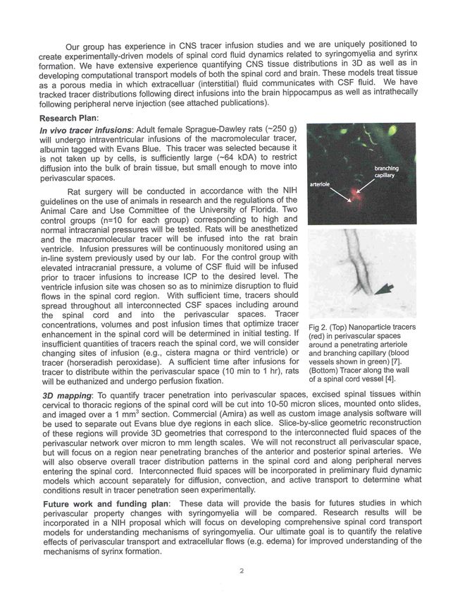
Meeting AM Agenda
Grand Hyatt San Francisco - Meeting Room – Union Square
Meeting Moderator: Dr. Allison Ashley-Koch
8:30 Breakfast
9:00 Welcome - Dorothy Poppe, Executive Director
Page
9:05 7 CSF Financial Update - Raymond Cornelius
9:10 10 unite@night Walks/CSF Educational Lecture Series - Cathy Poznik
9:20 12 2014 Consider Chiari Lecture Series - Lory Watson
9:30 13 2014 Bobby Jones Classic - Rob Noble
9:35 14 2013 – 2014 Night of Light Gala - Scott Gregerson
9:40 15 Overview of CSF's Research Strategic Plan - Dr. Rick Batzdorf
9:45 18 2013-2015 CSF Hydrodynamic Symposium/IHIWG - Dr. Harold Rekate
10:00 22 Discussion of 2013 – 2014 Colloquium - Dr. Fraser Henderson
• Review of Consensus - Dr. Ulrich Batzdorf, Dr. Edward C. Benzel
• Discussion on publication of book
10:15 Break
10:30 24 CSF Grant Recipient Presentations & Other Research Presentations -
Dr. Allison Ashley-Koch
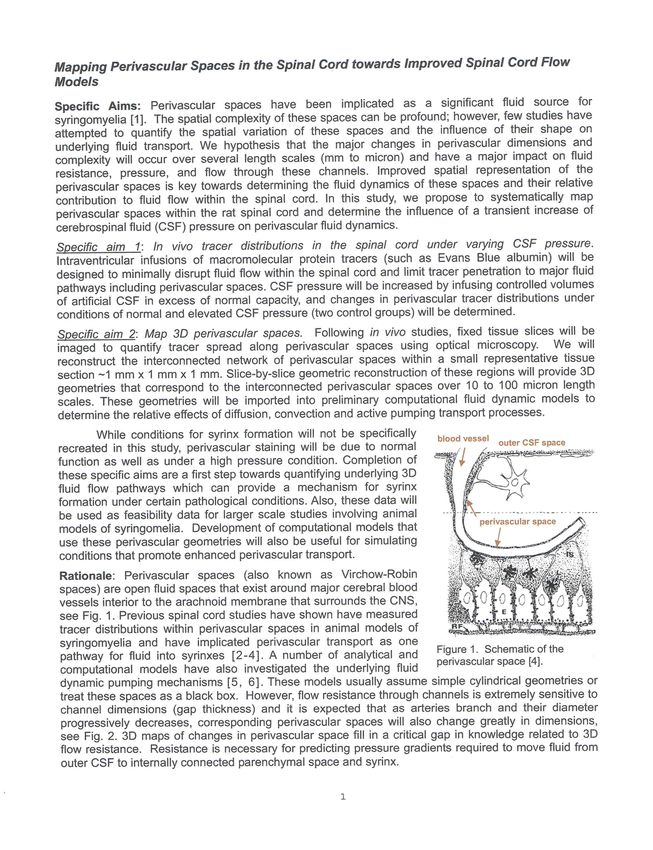
10:35 25 Mapping Perivascular Spaces in the Spinal Cord Toward Improved
Spinal Cord Flow Models - Dr. Malisa Sarntinoranont
11:00 28 NIH/NINDS Perspective / CDE Elements - Dr. Joanne Odenkirchen
(Skype)
11:25 35 Automated MRI-Based Parcellation of the Posterior Cranial Fossa –
(Dr. Ahmet Murat Bagci) Presented by Dr. Noam Alperin
41 Development of a Numerical Model of Spinal CSF Dynamics –
Dr. Christopher David Bertram - Please see information in PDF/booklet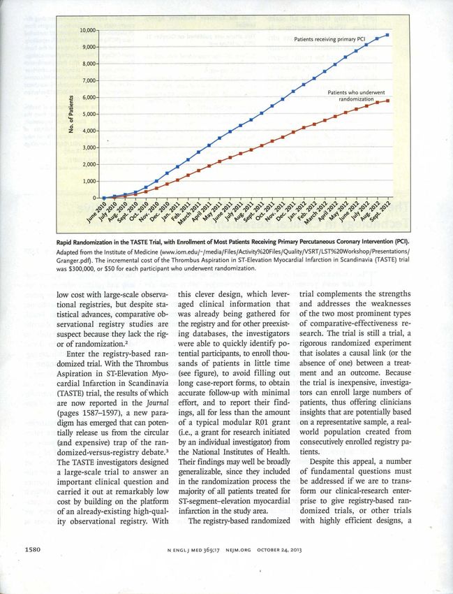
Meeting PM Agenda
Afternoon Session Moderator: Dr. Rick Batzdorf
12:00 Lunch (Room: Sunset A/B)
Page
1:00 42 CSF International Patient Registry Project
Steering Committee: Dr. Allison Ashley-Koch, Dr. Ulrich Batzdorf,
Dr. David Limbrick, Dr. Mark Luciano, Dr. Brandon Rocque,
Dr. Cormac Maher, Dr. Roger Kula, Dorothy Poppe
1:00 Overview of the Project - Dr. Mark Luciano
1:15 43 The Randomized Registry Trial – The Next Disruptive Technology in
Clinical Research? (Michael S. Lauer, MD, & Ralph B. D’Agostino, Sr, PhD)-
Discussed by Dr. Ulrich Batzdorf
1:30 46 Sub–committee Appointments - Dr. Mark Luciano
1:45 Seven Breakout Groups/Skype
2:45 Groups back in main meeting room for Committee Chair summaries
and final questions
3:15 54 Evaluation of gene expression in blood and dura mater from patients
with Chiari malformations - (Dr. Christina Markunas)
Presented by Dr. Allison Ashley-Koch
3:45 Break
4:00 79 SEA Board Business – Dr. Allison Ashley Koch / Dr. Ulrich Batzdorf
80 Review By-Laws
84 Rotate members of SEA Board, per CSF By-Laws
Rotate Chairman to Senior Advisory Panel, rotate Vice-Chairman to
Chairman’s position, and elect new Vice-Chairman (2-year term)
Elect/rotate Executive Committee members, as needed
85 Sign Conflict of Interest Statements
4:30 Closing of Meeting - Dr. Ulrich Batzdorf
7:00 Dinner: Jardinière, 300 Grove Street, San Francisco, California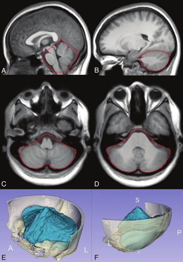
Our Mission, Vision, and Core Values
Mission:
To advance knowledge through research and to educate the medical,
allied sciences, and lay community about Chiari malformation,
syringomyelia and related disorders.
Vision:
Within a generation, we will be the premiere world-wide resource for
professional and lay people seeking accurate and current information
about treatments for and best practices surrounding the management of
CM and SM. With our unique resources, both financial and intellectual,
we will be the premiere, world-wide resource for accurate and current
information surrounding CM, SM and related disorders and the driving
force promoting ongoing programs and research focused on earlier
diagnosis and better outcomes for people suffering with these disorders.
Core Values:
Honesty and integrity is foremost in everything we do
Commitment to quality is central to all activities
Dedication to exceed the expectations of patients and physicians
Recognition of the critical role played by the Board of Trustees and
the Medical Research Board in terms of providing support and
guidance to the Board of Directors
Our volunteers and members are a respected source of knowledge and
experience
Social responsibility
Fiscal responsibility
1
Scientific Education & Advisory Board
(SEA Board)
Allison Ashley-Koch, PhD                  Bermans Iskandar, MD
Executive Committee Chair                 University of Wisconsin Hospitals
Duke University Medical Center            Madison, Wisconsin
Durham, North Carolina
                                          Roger W. Kula, MD
Ulrich Batzdorf, MD                       The Chiari Institute
Executive Committee Vice-Chair            Great Neck, New York
David Geffen School of Medicine at UCLA
Los Angeles, California                   David Limbrick, MD, PhD
                                          St. Louis Children’s Hospital
Paolo A. Bolognese, MD                    St. Louis, Missouri
The Chiari Institute
Great Neck, New York                      Mark Luciano, MD
                                          Cleveland Clinic
Richard G. Ellenbogen, MD                 Cleveland, Ohio
Executive Committee
University of Washington                  Dominic J. Marino, DVM, DACVS
Seattle, Washington                       Executive Committee
                                          Long Island Veterinary Specialists
Clair Francomano, MD                      Plainview, New York
Executive Committee
Greater Baltimore Medical Center          John Oró, MD
Baltimore, Maryland                       Neurosurgery Center of Colorado
                                          Aurora, Colorado
David Frim, MD
University of Chicago Medical Center      Harold Rekate, MD
Chicago, Illinois                         Executive Committee
                                          The Chiari Institute
John D. Heiss, MD                         Great Neck, New York
National Institutes of Health
Bethesda, Maryland                        Marcus Stoodley, MD
                                          Macquarie University Hospital
Fraser Henderson, MD                      New South Wales, Australia
Executive Committee
Doctors Community Hospital                Shane Tubbs, MD
Lanham, Maryland                          University of Alabama at Birmingham
                                          Birmingham, Alabama

                                                                                2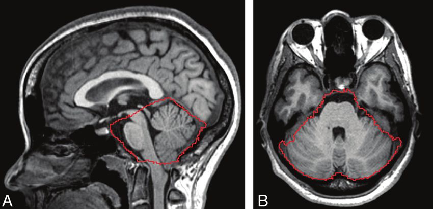
SEA Board Senior Advisory Panel

                Edward C. Benzel, MD
                  Cleveland Clinic
                  Cleveland, Ohio

                Timothy M. George, MD
    Dell Children’s Medical Center of Central Texas
                     Austin, Texas

                 Barth A. Green, MD
        The Miami Project to Cure Paralysis
       University of Miami School of Medicine
                    Miami, Florida

                Victor Haughton, MD
               University of Wisconsin
                 Madison, Wisconsin

                 Arnold Menezes, MD
        University of Iowa College of Medicine
                    Iowa City, Iowa

             Misao Nishikawa, MD, Japan
                 The Chiari Institute
                Great Neck, New York

                                                      3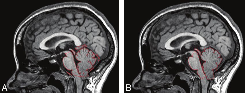
Board of Directors and Staff
               Board of Directors
                  Paul Farrell
                   Chairman

               Joseph Fitzpatrick
                 Vice Chairman

               Raymond Cornelius
                   Treasurer

                Scott Gregerson
                   Director

                  Robert Noble
                    Director

                  Lory Watson
                Director at Large

                      Staff
                Dorothy Poppe
               Executive Director

                Kaitlyn Esposito
               Program Assistant

                  Andrea Grosz
        Marketing & Development Director

                 Cathy Poznik
              Chapter Coordinator
                                           4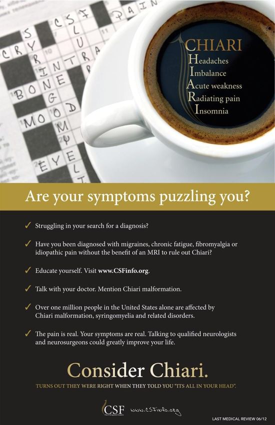
Board of Trustees

           Simon J. Archibald, PhD

          Robert Tyre Jones IV, PsyD

               Edwin B. Jordan

                 Adam Korn

            Richard E. Kuntz, MD

         Lady Trish Malloch-Brown

        J. Thomas Megerian, MD, PhD

             Michael N. Mikula

             Robert E. Rumphrey

         Board of Trustees (Emeritus)

             Douglas E. Kindlon

             Sean Lilienfeld, MD

                                        5
CSF Old and New Business

                           6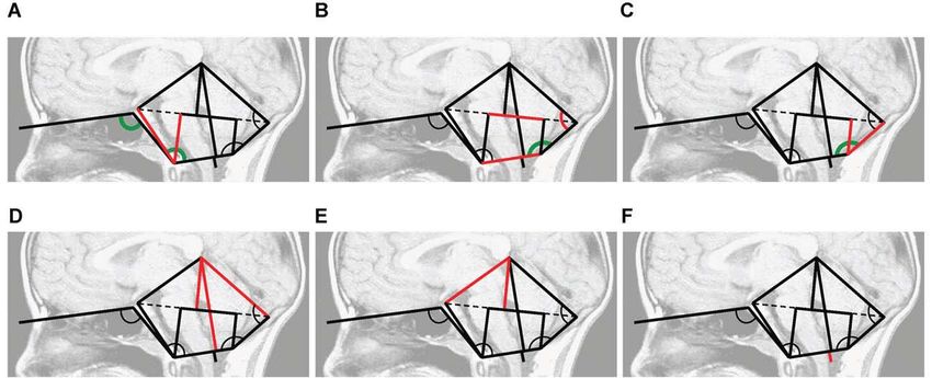
CSF Financial Update

  2014 Revenue Goals                2014 Revenue Goals       2013 Actual          2012 Actual

 Night of Light Children’s Gala                  $392,500           $390,637             $331,300

 unite@night Walks                               $225,000           $149,815              $77,465

 Bobby Jones Classic                             $163,250           $144,540              $83,050

 Charity Ball                                   $ 63,500*               $46,120           $80,725

 Dinner Dance for a Cure                          $40,000               $36,865           $27,592

 Chicago Event                                   $125,000           -                    $102,465

 Direct Mail                                      $41,500               $31,093           $24,585

 Other                                            $76,900               $38,140           $79,217

 Total Revenue                                  $1,127,650          $837,210             $806,399

*$73,000 2014 YTD actual

                                  2008 Gross Revenue
                                       $471,217

                                                                                                7
CSF 2014 Public Budget
Revenue: Amount
Direct Public Grants $ 30,000.00
Direct Public Support $ 5,000.00
Government Grants $ 5,000.00
Indirect Public Support $ 20,000.00
Program Income $ 52,100.00
Special Events Income $ 1,009,250.00
Restricted Donations $ 5,000.00
Investments $ 1,300.00
Total Revenue: $ 1,127,650.00
Expenses:
Research Grants $ 100,000.00
Educational Programs $ 100,000.00
Research Programs $ 207,788.00
Special Event Expense $ 329,603.00
Business Expense $ 11,350.00
Contract Services $ 34,000.00
Employee Benefit $ 18,000.00
Employee Salary $ 240,000.00
Operations $ 63,000.00
Insurance $ 11,660.00
Facilities & Equipment $ 9,000.00
Total Expense: $ 1,124,401.00
89
unite@night Walks

unite@night is a one-mile casual evening walk, in various locations around
 the country and in Canada during the month of June that brings together
       people who are suffering with the devastating effects of Chiari
            malformation, syringomyelia and related disorders.

unite@night supports CSF Chapters to provide education to physicians and
   the lay community and increase awareness, while funding important
              educational programs and research projects.

                National unite@night Sponsor

                                                                         10
Chapters/Educational Lecture Series
Chapters are set up across the United States to offer support for patients
and their families. Chapters are also responsible to set up free,
educational lectures for patients, their families,
and medical professionals, alike.
These lectures are always taped and posted on the CSF website so
patients and physicians from around the world have immediate
access to this material.
What exactly does a CSF Chapter do?
A Chapter will increase awareness, provide support, institute educational
programs, and raise funds in your area.
There have been over 40 educational lectures to date
These lectures are all available online for
free access on the CSF website
11Consider Chiari Campaign
Some doctors are failing to
Consider Chiari when patients
present textbook symptoms.
Our goal is to educate medical
professionals on the symptoms
and prevalence of Chiari so that
when symptoms present, MRI
technology will be utilized and
a proper diagnosis can be given.
Objective: To spark awareness,
putting Chiari on the radar of
medical professionals. We want
people to Consider Chiari.
Goal: We want 100% of the
participants to leave with a
clear picture of the symptoms
of Chiari and that proper
diagnosis requires an MRI.
12Bobby Jones Classic
www.bobbyjonesclassic.com
The third annual Bobby Jones Classic for CSF will strive to honor, celebrate
and exemplify the life and legacy of Bobby Jones. The event's activities will
include a private tour of Bobby Jones landmarks, an Alexa Stirling putting
competition and a cocktail reception on the evening prior to the
tournament. Monday's activities will include a full day of golf at East Lake
Golf Club followed by an awards dinner featuring Bob Jones IV,
Sid Matthew and Charles Harrison.
13Night of Light Gala
Guests gathered on November 9, 2013 at the James Burden Mansion in
New York City for this exclusive, white-tie event.
Gala Honorees:
Mr. and Mrs. Michael and Denise Mikula, Professor and Mrs. Rodney and
Barbara Grahame, Lord and Lady Mark and Trish Malloch-Brown.
Keynote Speaker:
Mr. Duncan Niederauer, Chief Executive Officer and Director,
NYSE Euronext.
2014 Night of Light Gala
November 22, 2014
Anderson House, Washington DC
14Overview of Research Strategic Plan Conquer Chiari Research Conference: New Developments & Controversies (2010) CSF sponsored this two-day professional research conference on November 11 and 12, 2010 in Chicago, Illinois, which focused on new developments in Chiari research and controversies in diagnosis and care. The event brought together the top physicians and researchers involved with Chiari malformation to share recent developments, discuss and debate controversial topics, and foster collaboration for future work. The proceedings are available through a video web archive. CSF Ehlers-Danlos Syndrome Colloquium (2011) CSF sponsored the Ehlers-Danlos Syndrome Colloquium which was held on October 1-2, 2011 at the Cosmos Club in Washington DC. Video of the Colloquium can be seen by clicking the link above. Project to Analyze the Prevalence of Chiari Malformation Within the ASD Population There appears to be an underserved population of children and adults with Chiari malformation and autism – the size of which is yet to be determined. Recent data suggests that a greater than expected overlap of findings in children diagnosed with Chiari Malformation and those diagnosed with Autism Spectrum of Disease (ASD). 1st CSF Hydrodynamics Symposium (2011) On July 8 and 9, 2011, CSF proudly sponsored the first CSF Hydrodynamics Symposium in Zurich, Switzerland, thanks to a very generous grant from the Monkton Institute and Ms. Candida Lancaster. CSF Think Tank Meeting (2013) - New Orleans, Louisiana Members of the CSF SEA Board, Board of Directors, Board of Trustees, and staff attended the annual CSF "Think Tank" meeting, in conjunction with the AANS annual meeting. 15
Overview of Research Strategic Plan
2nd CSF Hydrodynamics Symposium (2013)
The number of investigators conducting numerical and experimental
simulations to better understand the dynamics of cerebrospinal fluid (CSF)
has continued to increase since the 1st International CSF Dynamics
Symposium held in Zurich, Switzerland. Building on this momentum, we
held the 2nd International CSF Dynamics Symposium to continue exchange
of ideas toward modeling of CSF.

CSF Research Colloquium (2013)
Colloquium/consensus conference to facilitate a retrospective analysis of
patient images to determine whether Spinal Cord Stress Analysis Assessment
is helpful in determining which patients may need reductions, fusion,
stabilization

CSF Think Tank Meeting (2014) – San Francisco, California
Members of the CSF SEA Board, Board of Directors, Board of Trustees, and
staff attending the annual CSF "Think Tank" meeting, in conjunction with
the AANS annual meeting.

CSF Research Colloquium (2014) - Boston, Massachusetts

CSF International Patient Registry Meeting (2014) - Boston, Massachusetts

3rd CSF Hydrodynamics Symposium (2015) - Amiens, France

CSF International Patient Registry Project (Ongoing)
Chiari malformation (CM), syringomyelia (SM) and related disorders (RD)
affect millions of people worldwide. Starting on the path to improved care
can be a daunting task. An important early step is to establish a national
patient registry, an invaluable tool for improving the lives of people with
CM/SM/RD.

                                                                              16
CSF Funded Research Grants
CSF Funded Grants (2012)
Upon scientific review by experts from the CSF Scientific Education
& Advisory Board, CSF is pleased to fund the following grants
which were submitted for review in response to our 2012-2013
Request for Proposals.
• Mapping Perivascular Spaces in the Spinal Cord Toward Improved
Spinal Cord Flow Models (PDF) - Dr. Malisa Sarntinoranont
• Automated MRI-Based Parcellation of the Posterior Cranial Fossa -
Dr. Ahmet Murat Bagci (Published paper)
• Development of a Numerical Model of Spinal CSF Dynamics -
Dr. Christopher David Bertram
• Genetic Dissection of Chiari Type I Malformation - Christina
Markunas, PhD (Published paper)
CSF has also funded a Chiari Fellowship at the Duke Center for
Human Genetics.
172 N D C S F DY N A M I C S SY M P O S I U M
Feinstein Institute For Medical Research, Manhasset, New York
June 24 & 25, 2013
T HE M ONKTON
I NSTITUTE
!
!
ORGANIZED BY BRYN MARTIN, LYNNE BILSTON, & SHAOKOON CHENG 18SC H EDU L E OF E VE NT S
D AY 1 – M ONDAY , J UNE 24, 2013
7:00 B R E A K FA S T
Opening Remarks
8:30
Bryn Martin, Lynne Bilston, Shaokoon Cheng
PLENARY TALK
Chair: Lynne Bilston
Fine Structure Of CSF And Interstitial Fluid Spaces And Their Drainage Pathways From The Human
8:45
Central Nervous System
Roy Weller
SESSION A: EFFECT OF MICROANATOMY ON CSF
Chair: Lynne Bilston
9:45 Spinal Cord Nerve Roots And Denticulate Ligaments Alter CSF Dynamics In The Upper Cervical Spine
Bryn Martin
Effect Of Spinal Micro-anatomy On CSF Flow Patterns
10:15
Andreas Linninger
10:45 MORNING COFFEE BREAK
SESSION B: MODELING
Chair: Shaokoon Cheng
CSF Dynamics Society
11:15
Vartan Kurtcuoglu
Quantitative Assessment Of The Differences In Spinal CSF Dynamics In Chiari Malformation
11:30
Frank Loth
The Spinal Cord And Meninges As A Fluid-filled Elastic Waveguide In Syringomyelia
12:00
A. (Tony) Lucey
12:30 LUNCH
SESSION C: IMAGING
Chair: Mark Wagshul
4D MR Flow Imaging: Experiences In Hemodynamics And Potentials In CSF Hydrodynamics
13:30
Oliver Wieben
Blood And CSF Flow: What We Can See And What We Would Like To See Soon!
14:00
Olivier Balédent
2nd International CSF Dynamics Symposium 5 June 24 & 25, 2013, Manhasset, NY, USA
19Novel MRI-based Measurements Of CSF Flow Dynamics In Pediatric Patients With Chiari Malformation
14:30
John Oshinki
15:00 AFTERNOON COFFEE BREAK
SESSION D: CLINICAL
Chair: Frank Loth
What Role Does CSF Play In Vision Impairment In Astronauts
15:30
Michael Keith Sharp
Mathematical Models Of CSF Dynamics: Uses And Challenges
16:00
Harold Rekate
Dynamics And Solute Transport In CSF In Non-human Primates As Seen By Positron Emission
16:30 Tomography
Mikhail Papisov
19:00 S Y M P O S I U M D I N N E R AT 7 P M AT L I M A N I
D AY 2 – T UESDAY , J UNE 25, 2013
7:30 B R E A K FA S T
2ND PLENARY TALK
Chair: Vartan Kurtcuoglu
Pathogenesis And Pathology Of Hydrocephalus
8:30
Marc Del Bigio
SESSION E: IMAGING 2
What Can Animal Models Teach Us About CSF Flow Dynamics?
9:30
Mark Wagshul
Biomechanics Of Demyelination Processes: How Shear Wave Propagation Can Reveal
10:00 Microarchitectural Changes
Ralph Sinkus
10:30 MORNING COFFEE BREAK
SESSION F: MODELING 2
Chair: Shaokoon Cheng
How To Use Experimental Data Effectively In Modeling
11:00
Lynne Bilston
C ONTINUED ON NEXT PAG E
2nd International CSF Dynamics Symposium 6 June 24 & 25, 2013, Manhasset, NY, USA
20SC H EDU L E C O N T I N UE D . . .
D AY 2 – T UESDAY , J UNE 25, 2013
Near-Wall Ventricular Cerebrospinal Fluid Dynamics
11:30
Vartan Kurtcuoglu
On The Assumption Of Laminar CSF Flow In The Spinal Canal
12:00
Kent-Andre Mardal
12:30 LUNCH
SESSION G: SPINAL CORD
Chair: Bryn Martin
Potential Cerebrospinal Fluid Flow Pathways In The Development Of Syringomyelia
13:30
Shaokoon Cheng
Cerebrospinal Fluid And Spinal Cord Morphology Changes In The Hours After Spinal Cord Injury: Results
14:00 From Novel Porcine Model
Peter Cripton
Dynamic Cerebrospinal Fluid Pressure During Experimental Contusion Spinal Cord Injury: Results From
14:30 Novel Porcine And Synthetic SCI Models
Claire Jones
15:00 AFTERNOON COFFEE BREAK
SESSION H: MODELING 3
Chair: Lynne Bilston
A Fractional Pressure-Volume Model Of Cerebrospinal Fluid Dynamics: Marmarou’s Model Revisited
15:30
Corina S Drapaka
A Pilot, Multi-scale Numerical Framework For Brain Mechanics
16:00
Diane Dezelicourt
Closing Remarks
16:30
Bryn Martin & Lynne Bilston
17:00 DISCRETIONARY PLENARY DISCUSSION AND CLOSING COFFEE
2nd International CSF Dynamics Symposium 7 June 24 & 25, 2013, Manhasset, NY, USA
21CONSENSUS STATEMENT
1. Ventral brainstem compression, medullary kinking and deformation of the upper spinal cord and/or
brainstem over the odontoid process are potentially deleterious to the brainstem and upper spinal cord.
2. Deformation of the brainstem may manifest clinically as the Cervical Medullary syndrome.
3. The clinical findings of Cervical Medullary Syndrome may include, but are not limited to, the following:
i) headaches, suboccipital pain and neck pain,
ii) Bulbar and related symptoms: altered vision, diplopia, nystagmus, decreased hearing, tinnitus,
imbalance, vertigo, dizziness, choking, dysarthria, dysphagia dysautonomia, postural orthostatic
tachycardia, pre-syncopal or syncopal episodes disordered sleep architecture, sleep apnea,
iii) Symptoms of myelopathy: weakness, clumsiness, spasticity, altered sensation, paresthesias,
dysesthesia, change in gait, constipation, urinary urgency and frequency
4. In assessing the potential for cranio-cervical instability, it is reasonable to measure the angle between
the clivus and the spine. This angle has been termed the clivus canal angle, the clivus vertebral angle,
the clivus spinal angle, the clivus cervical angle and the clivus-axial angle.
In keeping with the greater part of the literature, we recommend the uniform adoption of the term
clivo-axial angle .This angle may be abbreviated CXA.
5. The clivo-axial angle is the angle between the clivus line and the posterior axial line. The clivus line is
drawn along the lower third of the clivus -from the spheno-occipital synchondrosis to the basion, or in
the case of basilar invagination, the superior most aspect of the odontoid .
When assessing the CXA with sagittal CT scan or X-ray, the posterior axial line may be drawn along the
posterior edge of the odontoid .
When assessing the CXA with MRI, the posterior axial line should be drawn from the posterior edge of
the tectorial membrane to the inferior posterior edge of the posterior ligament of the C2 vertebra.
The CT and MRI measurements may differ in the same patient: the CXA determined by CT reflects the
more traditional means of measurement ; the CXA determined by MRI will necessarily include thickening
of the ligament due to pannus.
6. The literature suggests that a clivo-axial angle of 135 degrees or less is potentially pathological. That is
a CXA of135 degree, may in some circumstances, result in harmful deformative stress upon the
brainstem and upper spinal cord and, therefore, warrants consideration for further evaluation and
possible treatment.
227.The CXA can be measured on sagittal CT or MRI, with the patient assuming moderate flexion of the
cranio-cervical junction. If a flexion view is not available, a neutral position will suffice in most
circumstances. An upright dynamic MRI may be desirable in some circumstances – but, such is often not
available.
8. In assessing the potential for cranio-cervical instability, it is reasonable and appropriate to measure
the BpC2 line, also known as the Grabb-Oakes measurement or line, or the Grabb Mapstone Oakes
Measurement, as one method to approximate the potential presence and magnitude of ventral
brainstem compression. We use the term Grabb-Oakes measurement herein. The Grabb-Oakes
measurement is the distance in millimeters from the dura to the line drawn from the basion to the
posterior inferior edge of the C2 vertebra. A Grabb Oakes measurement of 9mm represents the
diagnostic threshold for ventral brainstem deformity. Some clinicians may choose 8 mm as the
diagnostic threshold at which there may is potential ventral brainstem deformity.
9. The Harris measurement, also known as the Basion Axial Interval (BAI = distance from tip of basion to
posterior axial line), when drawn horizontally, should be less than 12 mm. The basion to dens interval
(BDI = distance from basion to tip of odontoid) drawn vertically, should be less than 12 mm. The
posterior axial line should be drawn along the posterior ligamentous surface of the C2 vertebra. In
keeping with the literature, a Harris measurement exceeding 12 mm is considered potentially
pathological, and reflects cranio-cervical instability.
10. In the presence of known ligamentous instability, such as a hereditary hypermobility connective
tissue disorder, the BAI (the Harris measurement) may be measured with the cervical spine in the flexion
and extension positions. This will assess and quantify translation of the basion with respect to the dens
(odontoid process). In keeping with the literature, any translation noted on dynamic imaging that
exceeds 2mm (the delta BAI > 2 mm), will be considered abnormal and potentially pathological.
11. Cranio-cervical hypermobility is common, and defined by the presence of hyper-extensibility of the
connective tissue, and in particular, hyper extensibility of the joints. While hypermobile joints occur
frequently in healthy children, such can also be severely disabling in others. Ehlers Danlos syndrome,
cleidocranial dysostosis, Down syndrome, Marfan syndrome, Morquio syndrome and several other less
well known connective tissue disorders are associated with ligamentous laxity. A pathological Lax
Ligament Syndrome may result in cranio-vertebral instability, kyphosis of the clivo-axial angle and
ventral brainstem compression.
The growing body of knowledge regarding the prevalence of hypermobility connective tissue disorders
should lead to more widespread recognition of the impact of ligamentous laxity on the health of
sufferers of hypermobility syndromes.
23CSF Grant Recipient
Presentations
2425
26
27
Guidelines
                          Developing Common Data Elements for NINDS

Goals and Timeline
The goal is to establish neurological disorder specific Common Data Element (CDE) Working Groups
(WGs) in order to recommend disorder/disease-specific elements commonly used in clinical research
studies. We anticipate that the disorder/disease specific CDEs will be drafted by WGs and either ready
to share with the other WGs or ready to share with the broader research community in one year after
initial meetings. The Working Groups will have an opportunity to review feedback from the research
community prior to the posting of version 1.0 of the specific disorder/disease CDEs. We also consider
in-person meeting(s) at national or international to facilitate this process.

End Products
Each CDE Working Groups should ultimately provide the NINDS CDE Team with the following products:
   1. Dictionary of CDEs that includes these specifications for each CDE (see attachment):
           a. Name
           b. Definition
           c. Code list/Permissible values
           d. Definitions for Code list/ Permissible values (PVs)
                   i. Additional specifications for the PV (e.g., min and max values, datatype, units)
           e. Comments/Special instructions
           f. Reference(s)
           g. Classification (1=Core, 2=Supplemental, 3=Exploratory)*
   2. Case report forms (CRFs) for the CDEs
   3. List of recommended standardized instruments**, including these details for each instrument:
           a. Name of Instrument
           b. Short description
           c. Scoring
           d. Comments/ Special Instructions
           e. Copyright Information
           f. Reference(s)
           g. Classification (1=Core, 2=Supplemental, 3=Exploratory)*
   4. Manual of Procedures (where applicable)
   5. Summary of Recommendations

* Classifications
          Core CDE: Used by the majority of the disorder/disease specific studies, strongly encouraged for
          use in a study
         Supplemental CDE: Used by a significant amount of disorder/disease specific studies, not as
         crucial to have in a study compared to Core CDEs as their relevance depends upon the study
         design (i.e., clinical trial, cohort study, etc.) or type of research involved
                  Within Supplement CDEs are annotated in recommendations for the following:
                  Highly Recommended: For outcome measures these should have excellent psychometric
                  properties and clinical utility
                                                                                               1|Page

                                                                                                             28
Guidelines
Developing Common Data Elements for NINDS
Recommended: For outcome measure, it has good psychometric properties and good
clinical utility.
Exploratory CDE: Will likely be used by the disorder/disease specific studies but the data
elements require further validation before they are ready for prime-time use. Reasonable to use,
but limited study in target group; the outcome measure has good or excellent psychometric
properties and clinical utility in a related population, but insufficient study in target population to
support higher recommendation.
** Standardized instruments may not be applicable to all subgroups.
Copyrighted Instruments
The working groups may recommend copyrighted instruments; however, at the current time the
instruments may not be posted on the public Web site. We are currently reaching out to the publishers
to find out if we may post the instruments with specific guidelines; however, this process will take some
time.
Development Process
The NINDS CDE Team will do their best to provide suggestions and assistance throughout the
development process and will assist in the development of the end products, in conjunction with the
critically important scientific expertise of the WGs.
It is recommended that you address the use of different instruments for different visit types (e.g., which
instruments would be used for screening versus longitudinal follow-up versus a single assessment).
In addition, standardized instruments should be classified for their use in different datasets: the basic
minimum dataset (e.g., conducted at each visit, similar to the Core CDEs), a comprehensive dataset
(similar to the supplemental CDEs), or a dataset for a defined area of interest (e.g., electrodiagnostics).
Each WG should consider any efforts that have already been undertaken to standardize how research
and/or clinical data are collected about the disorder/disease as the CDE Project does not want to
“reinvent the wheel” but rather strives to reuse relevant work that has already been done in this area.
2|Page
29Guidelines
Developing Common Data Elements for Neurological Disorders and Stroke
Template: CDE Data Dictionary for Elements
Permissible
Code List / Definitions for Additional Comments /
Definition / Values
CDE Name Permissible Codes / Special Instructions References
Description Additional Classification
Name of the data Values Permissible Values Other important References that contain
Definition Fields (e.g., 1 = Core
element being Describes the Further defines the information about the additional information
and/or Datatype; Min. 2 = Supplemental
recommended as standard way code list / CDE that will help ensure about the CDE and/or were
description of and Max. 3 = Exploratory
a CDE the CDE should permissible values it is collected used to define the CDE
the CDE Values; Units;
be coded consistently
and Format}
1= Core; should be used in all studies of this
disease
2= Supplemental; extended set that are
“common”, but supplemental, i.e. not
required - choose from a “menu”
3= Exploratory; not yet validated, or under
development
Template: CDE Data Dictionary for Instruments
Copyright Information
References
Instrument / Scale Name Explains whether the
Description References that contain Classification
Name and acronym of the Scoring Information instrument / measure has
Brief description of Comments / Special additional information 1 = Core
instrument/measure that Total range of scores and range copyright protection and if
the instrument / Instructions about the CDE and/or 2 = Supplemental
is recommended for of subscales if appropriate so, provides information on
measure were used to define the 3 = Exploratory
inclusion in the CDEs how to obtain it from the
CDE
publisher
3|Page
30Guidelines
Developing Common Data Elements for Neurological Disorders and Stroke
Example: Data Dictionary for Elements (Stroke CDEs)
Additional Comments / References
CDE (Listed here as
Definitions for Codes / Special Instructions References that contain Classification
Suggested Question Definition / Description Code List / Permissible Values
Permissible Values Other important additional information 1 = Core
Text) for data element Definition and/or Describes the standard way the
Further defines the code list / information about the CDE about the CDE and/or 2 = Supplemental
being recommended as a description of the CDE CDE should be coded
permissible values that will help ensure it is were used to define the 3 = Exploratory
CDE
collected consistently CDE.
IA procedure initiated Indicate if an Intra- Yes; Yes; IA procedures include all Get With The 1
arterial (IA) procedure No No uses of IA thrombolytic Guidelines (GWTG)
was initiated at this therapy, as well as Stroke Patient
hospital. mechanical devices such as Management Tool
"Clot retrieval devices". Coding Instructions
Mechanical devices may be (Updated on
used alone or in 11/4/2009); Paul
conjunction with IA Coverdell National
thrombolytic therapy. Acute Stroke Registry
Type of IA procedure If yes, specify IA Pharmacological; Mechanical; Pharmacological; Mechanical; 1
procedure performed at Both Both
this hospital.
Date and Time of Groin Indicate date and time of MM/DD/YYYY HH:MM; The preferred format for 3
Puncture for IA groin puncture Unknown recording date and time is
procedure MM/DD/YYYY HH:MM (24-hour
clock).
99/99/9999 can be used to
indicate an unknown date.
Similarly, 99:99 can be used to
indicate an unknown time.
4|Page
31Guidelines
Developing Common Data Elements for Neurological Disorders and Stroke
Example: Data Dictionary for Instruments (Stroke CDEs)
Copyright Information
References
Instrument / Scale Name Explains whether the
Description References that contain Classification
Name and acronym of Scoring Information instrument / measure has
Brief description of Comments / Special additional information 1 = Core
the instrument/measure Total range of scores and range of copyright protection and if
the instrument / Instructions about the CDE and/or 2 = Supplemental
that is recommended for subscales if appropriate so, provides information on
measure were used to define the 3 = Exploratory
inclusion in the CDEs how to obtain it from the
CDE.
publisher.
Functional Independence The FIM measures Scores range from 1 (total or >75% The alpha FIM is a subset that The FIM is proprietary. For Granger CV. The 2
Measure (FIM) degree of assistance) to 7 (complete has been used in the acute further information about emerging science of
independence in independence). The total of the 18 patient setting to assess which obtaining the scale, functional assessment:
activities of self- items is the patient's total score, which patients are appropriate for syllabus, and training our tool for outcomes
care, sphincter ranges from 18-126. Scores may be discharge to a rehabilitation materials please contact: analysis. Arch Phys Med
control, transfers, used raw or converted to interval setting. The alpha FIM may be Uniform Data System for Rehabil 1998;79(3):235-
locomotion, scores. worth exploring in Phase III Medical Rehabilitation 240.
communication, trials that include assessments 270 Northpointe Parkway,
and cognition. of appropriateness of different Suite 300 Wright, J. (2000). The
post-discharge destinations Amherst, New York 14228 FIM(TM). The Center
(716) 817-7800 FAX (716) for Outcome
568-0037 Measurement in Brain
email: info@udsmr.org Injury.
Web site: http://www.tbims.org/
http://www.udsr.org/Web combi/FIM (accessed
Modules/FIM/Fim_About.a March 10, 2010).
spx
5|Page
32NINDS Common Data Element Project
NINDS expects the clinical research it funds to meet the highest standards of
scientific rigor yet appreciates the burden that extensive data collection puts on
investigators and study participants. Further, the Institute leadership recognizes
that investigators independently identify data elements and forms for each study,
many of which could be common across studies.
As part of its effort to facilitate research of the highest quality, yet streamline
clinical trial data collection in neurological studies, NINDS continues to advance
its Common Data Element (CDE) Project. The CDE Project seeks to standardize
the way data are collected across the neuroscience research community. Central to
the project is the identification of common definitions and terminology.
By identifying CDEs in a standardized format, and by developing common
documentation and case report forms, the NINDS hopes to facilitate the
development of data collection tools, reduce study start-up time, promote
systematic data collection, improve data quality, and facilitate data sharing across
clinical research studies.
What do you need to know to use the CDEs?
1. Review the NINDS CDE Web site: www.commondataelements.ninds.nih.gov.
2. Contact the NINDS Project Officer, Joanne Odenkirchen,
(odenkirj@ninds.nih.gov) to set up a time to discuss using the CDEs and to
arrange for technical assistance.
3. Share your final or near final protocol with NINDS so the Institute and its con-
tractor can tailor a CDE training session to your study.
4. Send the NINDS Project Officer and the contractor
(NINDSCDE@emmes.com) any initial questions or
comments you may have about the CDEs or the Web site prior to your
scheduled training session.
Review of CDE Terminology:
If you have general
questions about the
CDE Project or more
specific questions about
the development of CDEs
for a certain disease area
please contact the NINDS
CDE Project Officer,
Joanne Odenkirchen at
odenkirj@ninds.nih.gov.
33Which CDEs should you use in your study?
1. Strongly encouraged to use the General Core CDEs.
2. Pick Additional Supplemental and Exploratory CDEs that are applicable to your
study.
3. Add data elements not found in the CDEs that are required for your study.
NINDS CDE Project promotes parsimonious data collection.
Tips for Accessing the CDEs
Find and download CDEs from http://www.commondataelements.ninds.nih.gov/
The NINDS CDE Web site has four main locations to access the CDEs:
1. General CDE Standards- http://www.commondataelements.ninds.nih.gov/
General.aspx
2. CDE Catalog tool – http://www.commondataelements.ninds.nih.gov/CDE.aspx
3. CRF Library tool – http://www.commondataelements.ninds.nih.gov/CRF.aspx
On Left: Screenshot of NINDS CDE
Web site Home Page
Submitting Feedback If you have general
We want to hear your feedback and are open to receiving it in a variety of formats: questions about the
CDE Project or more
Submit feedback form through Web site – specific questions about the
http://www.commondataelements.ninds.nih.gov/Feedback.aspx?page=General. development of CDEs for a
certain disease area please
Email – NINDSCDE@emmes.com for NINDS CDE Team; odenkirj@ninds.nih.gov contact the NINDS CDE
for NINDS CDE Project Officer. Project Officer, Joanne
Odenkirchen at
Phone/Webinar – arrange follow-up teleconference/webinar to review your comments.
odenkirj@ninds.nih.gov
34Published March 14, 2013 as 10.3174/ajnr.A3435
ORIGINAL RESEARCH
BRAIN
Automated Posterior Cranial Fossa Volumetry by MRI:
Applications to Chiari Malformation Type I
A.M. Bagci, S.H. Lee, N. Nagornaya, B.A. Green, and N. Alperin
ABSTRACT
BACKGROUND AND PURPOSE: Quantification of PCF volume and the degree of PCF crowdedness were found beneficial for differential
diagnosis of tonsillar herniation and prediction of surgical outcome in CMI. However, lack of automated methods limits the clinical use of
PCF volumetry. An atlas-based method for automated PCF segmentation tailored for CMI is presented. The method performance is
assessed in terms of accuracy and spatial overlap with manual segmentation. The degree of association between PCF volumes and the
lengths of previously proposed linear landmarks is reported.
MATERIALS AND METHODS: T1-weighted volumetric MR imaging data with 1-mm isotropic resolution obtained with the use of a 3T scanner
from 14 patients with CMI and 3 healthy subjects were used for the study. Manually delineated PCF from 9 patients was used to establish a
CMI-specific reference for an atlas-based automated PCF parcellation approach. Agreement between manual and automated segmentation of
5 different CMI datasets was verified by means of the t test. Measurement reproducibility was established through the use of 2 repeated scans
from 3 healthy subjects. Degree of linear association between PCF volume and 6 linear landmarks was determined by means of Pearson correlation.
RESULTS: PCF volumes measured by use of the automated method and with manual delineation were similar, 196.2 ⫾ 8.7 mL versus 196.9 ⫾ 11.0
mL, respectively. The mean relative difference of ⫺0.3 ⫾ 1.9% was not statistically significant. Low measurement variability, with a mean absolute
percentage value of 0.6 ⫾ 0.2%, was achieved. None of the PCF linear landmarks were significantly associated with PCF volume.
CONCLUSIONS: PCF and tissue content volumes can be reliably measured in patients with CMI by use of an atlas-based automated
segmentation method.
ABBREVIATIONS: CMI ⫽ Chiari malformation type I; PCF ⫽ posterior cranial fossa
CT2 or MR imaging,4 further confirmed reduced PCF volume in
T he current radiologic definition of CMI is based on the degree of
tonsillar herniation below the foramen magnum. However, im-
aging data with x-ray,1 CT,2 and MR3-7 gathered over the last several
patients with CMI compared with healthy controls. Through the use
of manual delineation of the PCF and the brain tissue boundaries,
decades documented that CMI is also associated with a smaller than Milhorat et al4 reported a smaller PCF volume as well as CSF volume,
normal PCF. In most studies, length (1D)5,6 and area (2D)8 measure- whereas the hindbrain volume was normal, leading to the notion of
ments of certain PCF landmarks manually delineated on x-ray film1 an overcrowded PCF. In a more recent study, they reported a small
or on a midsagittal MR imaging6 were used for estimates of the PCF volume only in “classic” CMI but not in “CMI mimicking”
PCF size. Volumetric (3D) assessment of the PCF, either with etiologies, thereby emphasizing the importance of the PCF volume
for differential diagnosis of tonsillar herniation.7
Lirng et al9 used manual delineation of the PCF in MR imaging
Received October 8, 2012; accepted after revision October 11.
followed by image intensity-based segmentation of brain tissue
From the Departments of Radiology (A.M.B., S.H.L., N.N., N.A.) and Neurological
Surgery (B.A.G.), University of Miami, Miami, Florida. and CSF to assess the effect of age and sex on the PCF volume and
This work was supported by the National Institutes of Health (R01NS052122) and by crowdedness in healthy subjects. They found that overall, men
a Small Grant Trainee Award from the Chiari & Syringomyelia Foundation. had a larger PCF and hindbrain volume, whereas women demon-
Paper previously presented in part at: 19th Annual Meeting of the ISMRM, Mon-
treal, Quebec, Canada, May 7-13, 2011. strated a higher degree of crowdedness, which may explain the
Please address correspondence to Noam Alperin, PhD, Department of Radiology higher frequency of CMI in women.
(Locator: M869), Miller School of Medicine, University of Miami, 1150 NW 14th St, Two other studies further suggest that the size of the PCF is also a
Suite 713, Miami, FL 33136; e-mail: NAlperin@med.miami.edu
Indicates open access to non-subscribers at www.ajnr.org strong predictor for surgical treatment outcome in CMI. Badie et al3
http://dx.doi.org/10.3174/ajnr.A3435 reported that a smaller ratio of the PCF volume relative to the supra-
AJNR Am J Neuroradiol ●:● ● 2013 www.ajnr.org 1
35
Copyright 2013 by American Society of Neuroradiology.tentorial volume is associated with a better surgical outcome. A more
recent study by Noudel et al10 used a semi-automated method to
demonstrate that the response to the PCF decompression surgery is
correlated with preoperation volume of PCF and the overall increase
in the PCF volume after operation but not with the degree of tonsillar
herniation or other tested morphologic measures. These limited data
suggest that measurements of the PCF volume are likely to enhance
both the diagnostic and prognostic reliability in CMI.
Despite the potential diagnostic and prognostic values of the
PCF morphology, volumetric assessments of the PCF size are not
commonly used in clinical practice because manual delineation of
the PCF on multiple images is time-consuming. Manual length
measurements of different landmarks of the PCF are less time-
consuming and are more commonly used as surrogate measures
of the PCF volume.11 The most common 1D measurements are FIG 1. The 6 linear landmarks of the PCF superimposed on a midsag-
the lengths of the supraocciput and the clivus bones and the ittal T1-weighted MR imaging from a patient with CMI: herniation (HR),
McRae line (MC), clivus (CL), Twining line (TW), cerebellum (CR), and
McRae and Twining lines at the midsagittal plane.4,6 However, supraocciput (SO).
these measurements are highly subjective and are strongly influ-
enced by the MR imaging technique. Furthermore, it is not clear
acquisition parameters: TR/TE/TI of 1900/2.89/900 ms, flip angle
how well these linear (1D) measures correlate with the overall
of 9°, FOV of 25.6 ⫻ 25.6 cm, and matrix size of 256 ⫻ 256,
volume of the PCF. With the increasing evidence for the diagnos-
resulting in 1-mm isotropic resolution. Images were acquired in
tic potential of PCF measurement, there is a need for a robust
the sagittal orientation.
automated method for reliable segmentation of the PCF. More-
over, because MR imaging is the primary technique used for di-
Linear Measurements of the PCF
agnosis of CMI, it is important that such a method is available for
Lengths of 6 midsagittal PCF structures were measured on the
MR imaging data.
midsagittal T1-weighted image by a trained expert with 5 years of
A new approach for automated PCF parcellation is pre-
experience (S.H.L.). These structures were characterized as fol-
sented. The proposed parcellation uses atlas-guided segmenta-
lows: 1) McRae line measured from the basion and the opisthion,
tion, which has been successful in other cerebral regions. In
2) clivus length measured from the basion to the inferior
addition to a measurement of the entire PCF volume, the
boundary of the dorsum sellae, 3) Twining line measured from
method also provides measurements of the hindbrain tissue
the dorsum sellae to the internal occipital protuberance, 4) height of
and CSF volumes. The robustness of the method is assessed by
the cerebellum, 5) supraocciput measured from internal occipital
comparison with manual delineation in CMI data. Addition-
protuberance to opisthion, and 6) tonsillar herniation measured
ally, the degree of association between PCF volumes and linear
from the McRae line to tip of the cerebellar tonsil. An example of
landmarks is assessed.
these markers overlaid on a midsagittal T1 MR image is shown in Fig
1.
MATERIALS AND METHODS
Subjects Manual Segmentation of PCF Volume
PCF volumes were measured by use of MR imaging data from 3 The PCF was manually outlined on every sagittal section by use of
healthy subjects who were scanned twice on 2 separate days (1 the 3D Slicer software (http://www.slicer.org) by a single trained
woman; age range, 29 –36 years; mean age, 34 ⫾ 3 years) and 5 expert (A.M.B.) to avoid interobserver variability. Manual delin-
symptomatic patients with CMI (3 women; age range, 23– 48 eations were further reviewed and edited when needed by a neu-
years; mean age, 37 ⫾ 10 years). MR imaging data from an addi- roradiologist (N.N.). The PCF was anatomically bounded by ten-
tional 9 symptomatic patients with CMI (7 women; age range, torium cerebelli, occipital bone, clivus, and foramen magnum.
20 – 68 years; mean age, 37 ⫾ 15 years) were used to create the The volume of the PCF was calculated by summation of the vol-
CMI-specific atlas. All patients had cerebellar tonsillar herniation ume of each voxel within the manually created mask on each
of at least 5 mm below the foramen magnum and presented with sagittal section.
suboccipital headaches and numbness in the upper and/or lower
extremities. Six patients had Valsalva-induced headaches. All sub- CMI-Specific PCF Atlas
jects provided written informed consent, and the study was ap- A PCF reference atlas, specific for CMI, was created from T1-
proved by the institutional review board. weighted images of 9 patients (7 women; age range, 20 – 68 years;
mean age, 37 ⫾ 15 years). PCF labels were manually delineated on
MR Image Acquisition each subject image by an expert (A.M.B.) and were reviewed and
The MR images used in the study were acquired with a 3T scanner confirmed for reliability by a neuroradiologist (N.N.). The T1-
(Magnetom Trio; Siemens, Erlangen, Germany). The structural weighted images were then affine-registered to Montreal Neuro-
analysis was performed on 3D T1-weighted image (magnetization logical Institute 152 space12 and averaged to create the atlas tem-
prepared rapid acquisition of gradient echo) with the following plate. The delineated PCF region from each of the 9 subjects was
2 Bagci ● 2013 www.ajnr.org
36Automated Segmentation of PCF
Volume
Automated segmentation of the PCF
volume is achieved by use of the previ-
ously described CMI reference atlas.
First, a global linear transformation is
applied to register the atlas template to
the subject dataset by use of the FLIRT
tool from FSL software package (http://
www.fmrib.ox.ac.uk/fsl).13 Only the
brain region is used for the global regis-
tration to avoid adverse effects of cranial
and extracranial structures on registra-
tion.14 After the global linear transfor-
mation, a more precise local alignment
with a nonlinear registration is achieved
by use of the FMRIB Nonlinear Image
Registration Tool (FNIRT) from FSL,15
which is based on minimizing a sum-of-
squares cost function by use of a Leven-
berg-Marquardt modification of the
Gauss-Newton method. Finally, the PCF
mask is mapped to the subject MR imag-
ing through the inverse of the registration
to automatically segment the PCF volume.
Automated Segmentation of Brain
Tissue
The proposed PCF parcellation method
quantifies the volumes of tissue content
of the PCF, which include the brain
stem, cerebellum, medulla, and pons.
Each pixel inside the PCF mask is labeled
as gray matter, white matter, or CSF, by
use of an algorithm that is based on the
hidden Markov random field model and
expectation maximization.16 The cere-
bellar tonsils extending beyond the fora-
men magnum are excluded as the result
FIG 2. Chiari-specific atlas template and the boundary of the PCF compartment (red outline) of PCF masking. A flow chart of the FSL
shown in sagittal (A and B) and axial (C and D) planes. A 3D volumetric rendering of the PCF mask implementation of the process for PCF
(blue) generated from the atlas template and surrounding cranium (white) are shown in E and F. segmentation and measurement of the
PCF tissue volume is shown in Fig 3.
The intensity-based segmentation of the PCF tissues was com-
pared with FreeSurfer-based (http://surfer.nmr.mgh.harvard.edu)
segmentation of the hindbrain. The details of the FreeSurfer seg-
mentation method are provided by Fischl et al.17 FreeSurfer seg-
mentation is an atlas-based segmentation method, which used
both the intensity distribution and the spatial relationships of
previously defined brain regions. Labeled brain regions in the
brain stem and gray and white matter in left and right cerebellum
were combined and used as the hindbrain volume in this study.
FIG 3. Flow chart illustrating the FSL implementation of the automated pro-
cess for PCF segmentation and measurement of the PCF tissue volume.
Assessment of Automated Segmentation Accuracy and
then projected to the atlas space to generate an averaged PCF Reliability
mask superimposed on the atlas space. The average reference atlas The accuracy of the automated segmentation was assessed by
and the superimposed PCF regions are shown in Fig 2. comparing the automatically segmented PCF with manual seg-
AJNR Am J Neuroradiol ●:● ● 2013 www.ajnr.org 3
37The reliability of the automated seg-
mentation across scan sessions was
tested by means of MR imaging data
from the 3 healthy subjects who were
scanned twice on 2 separate days. The
mean absolute percentage difference of
the 2 measurements was calculated by
dividing the absolute difference of the 2
measurements by their mean. A paired,
2-tailed t test was applied to determine
the significance of differences between
volume measurements obtained by use
of 2 methods.
The posterior fossa brain tissue vol-
FIG 4. Outline of the PCF mask (red) generated by use of the proposed method on midsagittal (A) ume was measured in 5 patients with
and axial (B) planes of T1-weighted MR imaging of a patient with CMI. CMI by use of the proposed automated
method and compared with measure-
ments obtained by means of FreeSurfer. The mean percentage
difference between 2 measurements and degree of spatial overlap
was calculated. In addition, the derived crowdedness index, de-
fined as the ratio of the tissue volume and the PCF compartment
volume, were compared as well. Finally, linear association be-
tween PCF volumes and the length of the 6 different linear PCF
markers were assessed by calculating the Pearson correlation co-
efficient and significance level by use of the data from the 14
patients with CMI. All statistical calculations were performed by
use of MedCalc statistical software (MedCalc, Mariakerke,
Belgium).
FIG 5. Comparison of PCF volume obtained manually and with the
RESULTS
automated segmentation in CMI. The Dice similarity coefficient (DSC) PCF Volumes
and the relative percent change obtained for each of the 5 patients Examples of midsagittal and axial images with the identified PCF
are shown above the volume bars. boundary and the 3D-rendered PCF volume from a representa-
Table 1: Repeated automated PCF volumes measurements tive patient with CMI are shown in Fig 4. The PCF volumes of
PCF PCF Relative each of the 5 patients with CMI measured with the 2 methods and
Age/ Volume (mL), Volume (mL), Percentage the Dice coefficient representing the degree of overlap are shown
Subject Sex Scan 1 Scan 2 Difference in Fig 5. The mean and the SD of the manual and the automated
1 36/M 186.1 186.8 0.4% segmentations of the PCF volumes in these 5 patients with CMI
2 29/M 207.5 205.9 0.8% were similar (197 ⫾ 11 mL and 196 ⫾ 9 mL, respectively). The
3 36/F 194.7 195.6 0.5%
difference was not significant (P ⫽ .7). The mean percentage vol-
ume difference was ⫺0.3 ⫾ 1.9%. The mean degree of overlap
(Dice coefficient) between the automatically and manually ob-
mentation in 5 patients with CMI. Percentage volume difference
tained PCF masks was 0.96 ⫾ 0.001.
between the manual and the automated segmentations were cal-
The PCF volumes measured from the 3 healthy subjects who
culated to test for systematic differences. Additionally, the Dice
were scanned twice on 2 separate days and the relative percentage
similarity coefficient was used to evaluate the degree of spatial
difference are listed in Table 1. The mean absolute percentage
overlap between the automatically and manually segmented PCF.
difference was 0.6 ⫾ 0.2%, with a range of 0.4% to 0.8%; the dif-
The Dice similarity coefficient is defined as
ference between volume measurements was not statistically sig-
2*V共 A 艚 M兲 nificant (P ⫽ .995).
DSC ⫽
V共 A兲 ⫹ V共M兲
PCF Crowdedness Indexes
where A and M denote automated and manual segmentations, The posterior fossa brain tissue volume, crowdedness indexes of
and V denotes volume of the region. The value of Dice similarity the 5 patients with CMI measured with the proposed method and
coefficient ranges between 0 –1, representing no overlap to com- with FreeSurfer, and the Dice coefficient are listed in Table 2. The
plete spatial overlap, respectively. A Dice similarity coefficient mean and SD of the volumes measured with the proposed method
value ⬎0.7 is considered as a good agreement between 2 com- and with FreeSurfer were 162 ⫾ 8 mL and 168 ⫾ 9 mL, respec-
pared measurements.18 tively. The tissue volumes measured by use of FreeSurfer were
4 Bagci ● 2013 www.ajnr.org
38Table 2: Comparison of PCF tissue segmentation by means of the proposed method and FreeSurfer
PCF Tissue Volume Crowdedness
(mL) (Proposed PCF Tissue Volume Percentage Dice Index (Proposed Crowdedness
Patient Method) (mL) (FreeSurfer) Volume Difference Coefficient Method) Index (FreeSurfer)
1 162.8 169.4 4.1% 0.949 0.842 0.876
2 172.1 181.6 5.5% 0.939 0.828 0.874
3 151.9 156.2 2.8% 0.940 0.824 0.847
4 170.1 174.0 2.3% 0.948 0.831 0.850
5 153.9 158.6 3.1% 0.949 0.806 0.830
mean Dice coefficient of 0.96. The delin-
eation of the PCF obtained by use of the
proposed automated method highly
agrees with the manual delineation in
terms of accuracy and spatial overlap in
patients with CMI. Furthermore, a high
degree of repeatability is evident from
the small absolute percentage difference
of 0.6 ⫾ 0.2% found by use of quantifi-
cation of the repeated scans in 3 healthy
subjects. The automated volume mea-
FIG 6. Outline of the PCF tissue masks (red) generated by use of the proposed method (A) and surement of PCF is minimally affected
FreeSurfer (B) in 1 of the patients with CMI. by the normal variability in patient po-
sitioning in the MR imaging scanner.
consistently larger in each subject, with a mean percent difference The mean PCF volume measurement obtained in our small
of 3.6 ⫾ 1.1% (P ⫽ .005). The mean and SD of the spatial overlap cohort of adult patients with CMI (196 ⫾ 8.7 mL) tends to be
were 0.945 ⫾ 0.004. Images illustrating the tissue segmentation by larger than previously reported CT and MR-based measurements
the 2 methods are shown in Fig 6A,-B, respectively. The corre- of 186 mL by Nishikawa et al,11 174 ⫾ 25 mL by Noudel et al,10
sponding PCF crowdedness indexes were 0.826 ⫾ 0.012 and and 166 ⫾ 8 mL by Milhorat et al.7 The bias in the mean volume
0.856 ⫾ 0.017, respectively. Because FreeSurfer does not provide measurements may be attributed to the differences in the modal-
the PCF volume, the PCF volume obtained by the proposed ities and the possible differences in the segmentation protocols,
method was used to estimate the PCF crowdedness obtained by particularly how the PCF boundaries were defined. Another con-
use of the 2 methods. tributing factor may be related to the difference in the sampling
None of the 6 linear PCF measures were significantly associ- resolution of the volumetric data. In contrast to isotropic 1-mm
ated with the PCF volume. Five of the linear measures correlated 3D imaging used in this work, previous reports used 2D-based
positively with the PCF volume with the following corresponding imaging with thicker sections for the volumetric measurements
Pearson correlation coefficients: supraocciput length (r ⫽ 0.38, that can lead to measurement errors caused by large partial vol-
P ⫽ .18), McRae line (r ⫽ 0.37, P ⫽ .20), clivus (r ⫽ 0.32, P ⫽ .27), ume effect. In addition, the limited number of subjects used in
Twining line (r ⫽ 0.30, P ⫽ .30), and length of cerebellum (r ⫽ this study to validate the proposed automated method against
0.29, P ⫽ .31). As expected, the herniation length was negatively manual segmentation may not be representative of a CMI popu-
correlated with the PCF volume, with a Pearson correlation coef- lation in terms of PCF volume.
ficient of ⫺0.17 (P ⫽ .57). The tonsillar herniation in CMI has been attributed to over-
DISCUSSION crowding of the PCF as a result of a small PCF and normally
Quantification of the PCF volume and the degree of PCF crowd- developed brain tissue volume.7,10,11 Therefore, in addition to
edness were shown to be beneficial for differential diagnosis of PCF volume measurement, accurate quantification of brain tissue
tonsillar herniation7,11 and for prediction of surgical outcome.10 volume is also critical. Our measurement of mean PCF tissue
The lack of a reliable automated method for PCF volumetry by use volume of 162.1 ⫾ 8.2 also tends to be slightly larger than previ-
of MR imaging, however, limits the clinical use of these PCF ously reported values of 156 mL by Nishikawa et al11 and 151.8 ⫾
markers. Advanced automated methods for brain parcellation 3.1 mL by Milhorat et al.7 However, the measurement of crowd-
have matured in recent years and are becoming more widely edness, the ratio of PCF tissue volume to PCF volume of 0.826 ⫾
used.16,17 This work represents adaptation of established brain 0.012, is in good agreement with the mean value of 0.833 reported
segmentation techniques tailored toward PCF volumetry in CMI. by Nishikawa et al.11
The proposed atlas-guided PCF segmentation method is en- The comparison of the hindbrain tissue volume measure-
hanced by the creation of a CMI-specific reference atlas that cap- ments between the proposed method and FreeSurfer revealed a
tures the altered PCF morphology associated with CMI. An excel- statistically significant mean difference of 3.6 ⫾ 1.1% (P ⫽ .005).
lent agreement between the proposed automated method and The tissue volumes found through the use of FreeSurfer were
manual segmentation by an expert observer is evident by the small consistently larger than volumes obtained by using the proposed
relative percentage difference of ⫺0.3 ⫾ 1.9% and the very high method. As demonstrated in Fig 6, this difference is the result of
AJNR Am J Neuroradiol ●:● ● 2013 www.ajnr.org 5
39You can also read